Article
Evaluation of 3 Fixation Devices for Tibial-Sided Anterior Cruciate Ligament Graft Backup Fixation
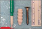
We conducted a study to biomechanically evaluate 3 methods of tibial-sided fixation for anterior cruciate ligament reconstruction: fully threaded...
Article
Dilute Betadine Lavage Reduces Implant-Related Bacterial Burden in a Rabbit Knee Prosthetic Infection Model
Treatment of acute postoperative arthroplasty infection with polyethylene exchange and retention of components has a limited success rate,...