Diagnostic puzzler: Acute eyelid edema

Article Type
Changed
Mon, 01/14/2019 - 11:36
Display Headline
Diagnostic puzzler: Acute eyelid edema

A 68-year-old man sought care in our emergency department for unilateral ptosis following superior and inferior right eyelid edema. The patient said that the edema had developed 3 days earlier and was getting worse each day; the ptosis was impairing his vision. The patient indicated that the edema was accompanied by mild burning in the right periocular region. His medical history included arterial hypertension, which was under control, and bilateral cataract surgery 5 years ago.

On examination, we noted superior and inferior nontender painless eyelid edema on the right eye, with no signs of acute inflammation (FIGURE). The patient also had right supraciliary folliculitis that was improving; the folliculitis had been treated 3 days earlier at a primary care facility.

We performed a complete ocular examination, including visual acuity (20/20 in both eyes) and found no other significant problems. Nor did the patient have a fever or any other systemic symptoms.


FIGURE 1
Edema of the upper and lower eyelid


The swelling was not accompanied by inflammation or fever, but it did impair the patient’s vision.

WHAT IS YOUR DIAGNOSIS?
HOW WOULD YOU TREAT THIS PATIENT?

Diagnosis: Noninflammatory eyelid edema

The fact that the patient’s eyelid was edematous (but not tender or red) and that he’d been scratching the area following a case of folliculitis prompted us to diagnose noninflammatory eyelid edema.

Noninflammatory eyelid edema—also called noninflammatory palpebral edema—is a relatively common disorder that usually occurs after some local irritation or microtrauma. In its early phase, patients start out with nonerythematous, painless edema of the upper eyelid, which eventually affects the lower eyelid.


As was the case with our patient, this disorder is not associated with fever or any other systemic sign or symptom. Complete blood count and C-reactive protein will be negative, as no infection or systemic alteration is present.

Consider these infectious alternatives
In cases like this one, be sure to differentiate between noninflammatory palpebral edema and other common but less benign ocular conditions. Among the infectious causes of unilateral edema to rule out:

Preseptal cellulitis is an infection that is localized between the skin and the orbital septum. This disorder usually appears as a tender erythematous eyelid edema with no proptosis and no pain with eye movement.1 It is usually accompanied by an elevated white blood cell (WBC) count. Blood cultures are rarely positive, but when they are, Haemophilus influenzae is the most frequent pathogen.2 Preseptal cellulitis is treated with broad-spectrum oral antibiotics (amoxicillin/ clavulanate) for at least 7 days.

Orbital cellulitis is an infection that affects the orbital extraocular structures. It can lead to blindness, so prompt diagnosis is critical.

Orbital cellulitis usually develops when a sinus infection spreads into the orbit.3 Suspect it in a patient with proptosis, orbital pain, tenderness, conjunctival chemosis, decreased vision, elevated intraocular pressure, and pain on eye movement. Also look for an elevated WBC count. Perform blood cultures to confirm the presence of a pathogen.

If you suspect orbital cellulitis, order an orbital computed tomography scan with contrast infusion, including axial and coronal views; it may reveal an infection of the soft tissue behind the orbital septum. Treat this condition with broad-spectrum intravenous antibiotics (ceftriaxone) for 1 week, followed by 2 weeks of oral antibiotics, completing a total of 21 days of antibiotics.

Hordeolum is a localized infection that occurs when a meibomian gland becomes blocked. It usually appears as a palpable subcutaneous nodule within the eyelid and requires treatment with warm compresses and a combination of antibiotics and steroid topical ointment (tobramycin and dexamethasone ophthalmic ointment 4 times a day for a week).4

Acute dacryoadenitis is a lacrimal gland infection that usually causes unilateral, painful swelling of the outer third of the upper lid. You may also see tearing and discharge.5 Look for an elevated WBC count; however, blood cultures are rarely positive. These patients must be referred to an ophthalmologist as treatment varies from symptomatic measures in viral cases (warm compresses and a nonsteroidal anti-inflammatory drug, such as ibuprofen) to hospitalization and oral or intravenous antibiotics (eg, amoxicillin/ clavulanate for at least 7 days), depending on the severity and origin of the infection.6

Ruling out other noninfectious disorders
Noninfectious inflammatory diseases should also be part of the differential diagnosis, including:

Idiopathic orbital inflammation. Also known as orbital pseudotumor, this disorder is a nongranulomatous acute-to-subacute inflammatory disease with no systemic manifestations.7 It frequently involves the lacrimal gland and can cause abrupt pain, conjunctival edema, and lid edema. Other common signs are proptosis and ocular motility alterations, which the patient may report as diplopia and visual impairment.


This disorder can result from optic neuropathy, exudative retinal detachment, or uveitis. It is a diagnosis of exclusion and is often arrived at by seeing the patient respond to systemic corticosteroids. The specific drug and dose will vary based on disease severity.

Graves’ ophthalmopathy. This bilateral, asymmetric immunological disorder affects the conjunctiva, eyelids,8 extraocular muscles, lacrimal gland, and optic nerve. Although it is typically diagnosed clinically, thyroid hormone abnormalities (increased T4 and a drop in thyroid-stimulating hormone) help support the diagnosis. In the acute phase, it has to be treated with systemic steroids; if the condition becomes chronic, the complications may require surgery. Treatment of the underlying hyperthyroidism may, in some cases, worsen the ophthalmopathy.

One more consideration. Finally, consider a local allergic reaction in the eyelids,9 which can also cause painless palpebral edema. Expect to see erythematous edema and intense itching a few hours after exposure to the allergen. A cosmetic product is often the culprit. As you might expect, initial treatment calls for withdrawal of the offending product. Instruct the patient to apply a topical steroid ointment on the eyelid if the edema is intense.


Good news for our patient
Diagnosing noninflammatory eyelid edema requires keen observational skills and a knowledge of several alternative diagnoses. Fortunately for our patient, this benign condition did not require any local or systemic treatment. The edema resolved on its own in 6 days.

CORRESPONDENCE
Omar Rayward, MD, Calle Profesor Martín Lagos, Madrid, Spain 28040; orayward@yahoo.com

References


1. Bilyk JR. Periocular infection. Curr Opin Ophthalmol. 2007;18:414-423.

2. Jackson K, Baker SR. Periorbital cellulitis. Head Neck Surg. 1987;9:227-234.

3. Adam R, Gupta V, Harvey J. Question: can you identify this condition? This condition is orbital cellulitis. Can Fam Physician. 2009;55:1097.-

4. Lindsley K, Nichols JJ, Dickersin K. Interventions for acute internal hordeolum. Cochrane Database Syst Rev. 2010;(9):CD007742.-

5. Colegrove JA. Localized orbital inflammation: a case of dacryoadenitis. Optom Vis Sci. 2000;77:121-124.

6. Durand ML. Periocular infections. In: Mandell GL, Bennett JE, Dolin R, eds. Mandell, Douglas, and Bennett’s Principles and Practice of Infectious Diseases. 7th ed. Philadelphia, Pa: Elsevier Churchill Livingstone; 2009:1569–1575.

7. Cooney NL. Orbital pseudotumor. Int J Emerg Med. 2009;2:263.-

8. Imaizumi M. Recurrent upper eyelid edema as first sign of Graves’ disease. Thyroid. 2006;16:95-96.

9. Friedlaender MH. Objective measurement of allergic reactions in the eye. Curr Opin Allergy Clin Immunol. 2004;4:447-453.

Article PDF
Author and Disclosure Information


Omar Rayward, MD, PhD
Hospital Clinico San Carlos, Madrid, Spain
orayward@yahoo.com

Jose Luis Vallejo-Garcia, MD
Humanitas Clinical and Research Center, Milan, Italy

Paula Moreno-Martin, MD
Hospital del Henares, Coslada, Madrid

Sergio Vano-Galvan, MD, PhD
Hospital Ramón y Cajal, Madrid

The authors reported no potential conflict of interest relevant to this article.

Issue
The Journal of Family Practice - 62(5)
Publications
Topics
Page Number
E1-E3
Legacy Keywords
Omar Rayward; MD; PhD; Jose Luis Vallejo-Garcia; MD; Paula Moreno-Martin; MD; Sergio Vano-Galvan; MD; PhD; supraciliary folliculitis; noninflammatory eyelid edema; C-reactive protein; preseptal cellulitis; orbital cellulitus; optic neuropathy; exudative retinal detachment; PURLs
Sections
Author and Disclosure Information


Omar Rayward, MD, PhD
Hospital Clinico San Carlos, Madrid, Spain
orayward@yahoo.com

Jose Luis Vallejo-Garcia, MD
Humanitas Clinical and Research Center, Milan, Italy

Paula Moreno-Martin, MD
Hospital del Henares, Coslada, Madrid

Sergio Vano-Galvan, MD, PhD
Hospital Ramón y Cajal, Madrid

The authors reported no potential conflict of interest relevant to this article.

Article PDF
Article PDF

A 68-year-old man sought care in our emergency department for unilateral ptosis following superior and inferior right eyelid edema. The patient said that the edema had developed 3 days earlier and was getting worse each day; the ptosis was impairing his vision. The patient indicated that the edema was accompanied by mild burning in the right periocular region. His medical history included arterial hypertension, which was under control, and bilateral cataract surgery 5 years ago.

On examination, we noted superior and inferior nontender painless eyelid edema on the right eye, with no signs of acute inflammation (FIGURE). The patient also had right supraciliary folliculitis that was improving; the folliculitis had been treated 3 days earlier at a primary care facility.

We performed a complete ocular examination, including visual acuity (20/20 in both eyes) and found no other significant problems. Nor did the patient have a fever or any other systemic symptoms.


FIGURE 1
Edema of the upper and lower eyelid


The swelling was not accompanied by inflammation or fever, but it did impair the patient’s vision.

WHAT IS YOUR DIAGNOSIS?
HOW WOULD YOU TREAT THIS PATIENT?

Diagnosis: Noninflammatory eyelid edema

The fact that the patient’s eyelid was edematous (but not tender or red) and that he’d been scratching the area following a case of folliculitis prompted us to diagnose noninflammatory eyelid edema.

Noninflammatory eyelid edema—also called noninflammatory palpebral edema—is a relatively common disorder that usually occurs after some local irritation or microtrauma. In its early phase, patients start out with nonerythematous, painless edema of the upper eyelid, which eventually affects the lower eyelid.


As was the case with our patient, this disorder is not associated with fever or any other systemic sign or symptom. Complete blood count and C-reactive protein will be negative, as no infection or systemic alteration is present.

Consider these infectious alternatives
In cases like this one, be sure to differentiate between noninflammatory palpebral edema and other common but less benign ocular conditions. Among the infectious causes of unilateral edema to rule out:

Preseptal cellulitis is an infection that is localized between the skin and the orbital septum. This disorder usually appears as a tender erythematous eyelid edema with no proptosis and no pain with eye movement.1 It is usually accompanied by an elevated white blood cell (WBC) count. Blood cultures are rarely positive, but when they are, Haemophilus influenzae is the most frequent pathogen.2 Preseptal cellulitis is treated with broad-spectrum oral antibiotics (amoxicillin/ clavulanate) for at least 7 days.

Orbital cellulitis is an infection that affects the orbital extraocular structures. It can lead to blindness, so prompt diagnosis is critical.

Orbital cellulitis usually develops when a sinus infection spreads into the orbit.3 Suspect it in a patient with proptosis, orbital pain, tenderness, conjunctival chemosis, decreased vision, elevated intraocular pressure, and pain on eye movement. Also look for an elevated WBC count. Perform blood cultures to confirm the presence of a pathogen.

If you suspect orbital cellulitis, order an orbital computed tomography scan with contrast infusion, including axial and coronal views; it may reveal an infection of the soft tissue behind the orbital septum. Treat this condition with broad-spectrum intravenous antibiotics (ceftriaxone) for 1 week, followed by 2 weeks of oral antibiotics, completing a total of 21 days of antibiotics.

Hordeolum is a localized infection that occurs when a meibomian gland becomes blocked. It usually appears as a palpable subcutaneous nodule within the eyelid and requires treatment with warm compresses and a combination of antibiotics and steroid topical ointment (tobramycin and dexamethasone ophthalmic ointment 4 times a day for a week).4

Acute dacryoadenitis is a lacrimal gland infection that usually causes unilateral, painful swelling of the outer third of the upper lid. You may also see tearing and discharge.5 Look for an elevated WBC count; however, blood cultures are rarely positive. These patients must be referred to an ophthalmologist as treatment varies from symptomatic measures in viral cases (warm compresses and a nonsteroidal anti-inflammatory drug, such as ibuprofen) to hospitalization and oral or intravenous antibiotics (eg, amoxicillin/ clavulanate for at least 7 days), depending on the severity and origin of the infection.6

Ruling out other noninfectious disorders
Noninfectious inflammatory diseases should also be part of the differential diagnosis, including:

Idiopathic orbital inflammation. Also known as orbital pseudotumor, this disorder is a nongranulomatous acute-to-subacute inflammatory disease with no systemic manifestations.7 It frequently involves the lacrimal gland and can cause abrupt pain, conjunctival edema, and lid edema. Other common signs are proptosis and ocular motility alterations, which the patient may report as diplopia and visual impairment.


This disorder can result from optic neuropathy, exudative retinal detachment, or uveitis. It is a diagnosis of exclusion and is often arrived at by seeing the patient respond to systemic corticosteroids. The specific drug and dose will vary based on disease severity.

Graves’ ophthalmopathy. This bilateral, asymmetric immunological disorder affects the conjunctiva, eyelids,8 extraocular muscles, lacrimal gland, and optic nerve. Although it is typically diagnosed clinically, thyroid hormone abnormalities (increased T4 and a drop in thyroid-stimulating hormone) help support the diagnosis. In the acute phase, it has to be treated with systemic steroids; if the condition becomes chronic, the complications may require surgery. Treatment of the underlying hyperthyroidism may, in some cases, worsen the ophthalmopathy.

One more consideration. Finally, consider a local allergic reaction in the eyelids,9 which can also cause painless palpebral edema. Expect to see erythematous edema and intense itching a few hours after exposure to the allergen. A cosmetic product is often the culprit. As you might expect, initial treatment calls for withdrawal of the offending product. Instruct the patient to apply a topical steroid ointment on the eyelid if the edema is intense.


Good news for our patient
Diagnosing noninflammatory eyelid edema requires keen observational skills and a knowledge of several alternative diagnoses. Fortunately for our patient, this benign condition did not require any local or systemic treatment. The edema resolved on its own in 6 days.

CORRESPONDENCE
Omar Rayward, MD, Calle Profesor Martín Lagos, Madrid, Spain 28040; orayward@yahoo.com

References


1. Bilyk JR. Periocular infection. Curr Opin Ophthalmol. 2007;18:414-423.

2. Jackson K, Baker SR. Periorbital cellulitis. Head Neck Surg. 1987;9:227-234.

3. Adam R, Gupta V, Harvey J. Question: can you identify this condition? This condition is orbital cellulitis. Can Fam Physician. 2009;55:1097.-

4. Lindsley K, Nichols JJ, Dickersin K. Interventions for acute internal hordeolum. Cochrane Database Syst Rev. 2010;(9):CD007742.-

5. Colegrove JA. Localized orbital inflammation: a case of dacryoadenitis. Optom Vis Sci. 2000;77:121-124.

6. Durand ML. Periocular infections. In: Mandell GL, Bennett JE, Dolin R, eds. Mandell, Douglas, and Bennett’s Principles and Practice of Infectious Diseases. 7th ed. Philadelphia, Pa: Elsevier Churchill Livingstone; 2009:1569–1575.

7. Cooney NL. Orbital pseudotumor. Int J Emerg Med. 2009;2:263.-

8. Imaizumi M. Recurrent upper eyelid edema as first sign of Graves’ disease. Thyroid. 2006;16:95-96.

9. Friedlaender MH. Objective measurement of allergic reactions in the eye. Curr Opin Allergy Clin Immunol. 2004;4:447-453.

Issue
The Journal of Family Practice - 62(5)
Issue
The Journal of Family Practice - 62(5)
Page Number
E1-E3
Page Number
E1-E3
Publications
Publications
Topics
Article Type
Display Headline
Diagnostic puzzler: Acute eyelid edema
Display Headline
Diagnostic puzzler: Acute eyelid edema
Legacy Keywords
Omar Rayward; MD; PhD; Jose Luis Vallejo-Garcia; MD; Paula Moreno-Martin; MD; Sergio Vano-Galvan; MD; PhD; supraciliary folliculitis; noninflammatory eyelid edema; C-reactive protein; preseptal cellulitis; orbital cellulitus; optic neuropathy; exudative retinal detachment; PURLs
Legacy Keywords
Omar Rayward; MD; PhD; Jose Luis Vallejo-Garcia; MD; Paula Moreno-Martin; MD; Sergio Vano-Galvan; MD; PhD; supraciliary folliculitis; noninflammatory eyelid edema; C-reactive protein; preseptal cellulitis; orbital cellulitus; optic neuropathy; exudative retinal detachment; PURLs
Sections
Disallow All Ads
Alternative CME
Article PDF Media

A persistently swollen lip

Article Type
Changed
Wed, 01/31/2018 - 09:13
Display Headline
A persistently swollen lip

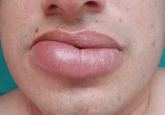
A 44-year-old man is referred for evaluation of asymptomatic swelling of the lower lip that has persisted for 10 months. He has been treated unsuccessfully with oral antihistamines for suspected chronic angioedema. He has no other symptoms and appears to be well otherwise. He has no history of applied irritants or local trauma, and his medical history is unremarkable.

Figure 1.
Physical examination shows a swollen and indurated lower lip (Figure 1). The tongue and gums are normal, and the ophthalmologic evaluation reveals no abnormalities. No facial paralysis is observed.

Results of the laboratory evaluation, including serum angiotensin-converting enzyme level, are normal. Patch tests to detect contact sensitivity to food additives are negative. Biopsy of the affected lip reveals dense infiltrate of the submucosal connective tissue with focal nonnecrotizing granulomas. Imaging and endoscopic studies show no evidence of sarcoidosis or Crohn disease.

Q: Given what we know so far, which of the following is the most likely diagnosis of the persistent lip swelling?

  • Melkersson-Rosenthal syndrome
  • Amyloidosis
  • Quincke edema
  • Cheilitis granulomatosa
  • Cutaneous tuberculosis

A: From what we know so far, the correct answer is cheilitis granulomatosa. While this rare condition may be a feature of Melkersson-Rosenthal syndrome and amyloidosis, at this point in the evaluation these have not been confirmed. Quincke edema (ie, angioedema) is unlikely, given the ineffectiveness of previous treatment with oral antihistamines. Cutaneous tuberculosis usually presents as “lupus vulgaris,” which is characterized by solitary, small, sharply marginated, red-brown papules of gelatinous consistency (“apple-jelly nodules”), mainly on the head and neck.

Cheilitis granulomatosa is a rare inflammatory disorder1 that primarily affects young adults. Its key feature is recurrent or persistent painless swelling of one or both lips. It may occur without other signs of disease, but it is also a manifestation of Melkersson-Rosenthal syndrome and it may be a presenting symptom of Crohn disease or, rarely, sarcoidosis.2 The term “orofacial granulomatosis” was introduced to encompass the broad spectrum of nonnecrotizing granulomatous inflammation in the orofacial region, including cheilitis granulomatosa, the complete Melkersson-Rosenthal syndrome, sarcoidosis, Crohn disease, and infectious disorders such as tuberculosis.1

The cause of cheilitis granulomatosa is unknown. Specific T-cell clonality has been identified in several patients with orofacial granulomatosis, suggesting a delayed hypersensitivity response. Moreover, the HLA haplotypes HLA-A2 and HLA-A11 have been found in 25% of patients with orofacial granulomatosis, suggesting a viral etiology. A genetic predisposition may exist in Melkersson- Rosenthal syndrome: siblings have been affected, and otherwise unaffected relatives may have a fissured tongue (lingua plicata).

Melkersson-Rosenthal syndrome, a rare condition, is characterized by a classic triad of recurrent swelling of the lips or face (or both), fissured tongue, and relapsing peripheral facial nerve paralysis. It is an unusual cause of facial swelling that can be confused with angioedema.3 This syndrome can be ruled out in this patient because he has only one of the three classic signs. Contact antigens are sometimes implicated.

DIFFERENTIAL DIAGNOSIS OF CHEILITIS GRANULOMATOSA

The differential diagnosis of cheilitis granulomatosa is extensive and includes amyloidosis, cheilitis glandularis, sarcoidosis, Crohn disease, actinic cheilitis, neoplasms, and infections, such as tuberculosis, syphilis, and leprosy.1

As many as 11% of patients with Crohn disease may develop mucocutaneous lesions. Oral lesions of Crohn disease include apthae, cobblestoning of the buccal mucosa, swelling of one or both lips (soft or rubbery), vertical clefts of the lips, or hypertrophic gingivitis. Only 5% of patients with Crohn disease ever develop cheilitis granulomatosa, though most cases occur in children.

Ultimately, the diagnosis of cheilitis granulomatosa is made by correlating the patient’s history and clinical features, usually supported by histopathologic findings of nonnecrotic granulomas extending into the deep dermis, composed of histiocytes and giant cells and associated with a lymphomonocytic infiltrate.

TREATMENT

Treatment of cheilitis granulomatosa is difficult because the cause is unknown and the rate of recurrence is high. Response to treatment is often late and unpredictable. Corticosteroids, clofazimine (Lamprene), and surgical intervention such as cheiloplasty have been described as treatment options. Other treatment options include thalidomide (Thalomid), sulfasalazine (Sulfazine), erythromycin, azathioprine (Imuran), and cyclosporine (Sandimmune). Infliximab (Remicade) has been recently reported as a new alternative treatment, in particular for Melkersson-Rosenthal syndrome.4

In our patient, twice-monthly injections of 1 mL of triamcinolone acetonide 10 mg/mL into the affected lip brought acceptable improvement at 3 months. The patient is on maintenance treatment with twice-monthly triamcinolone injections and has had no relapses after 2 years.

References
  1. van der Waal RI, Schulten EA, van de Scheur MR, Wauters IM, Starink TM, van der Waal I. Cheilitis granulomatosa. J Eur Acad Dermatol Venereol 2001; 15:519523.
  2. van der Waal RI, Schulten EA, van der Meij EH, van de Scheur MR, Starink TM, van der Waal I. Cheilitis granulomatosa: overview of 13 patients with long-term follow-up—results of management. Int J Dermatol 2002; 41:225229.
  3. Kakimoto C, Sparks C, White AA. Melkersson-Rosenthal syndrome: a form of pseudoangioedema. Ann Allergy Asthma Immunol 2007; 99:185189.
  4. Ratzinger G, Sepp N, Vogetseder W, Tilg H. Cheilitis granulomatosa and Melkersson-Rosenthal syndrome: evaluation of gastrointestinal involvement and therapeutic regimens in a series of 14 patients. J Eur Acad Dermatol Venereol 2007; 21:10651070.
Article PDF
Author and Disclosure Information

Sergio Vañó-Galván, MD
Department of Dermatology, Ramón y Cajal Hospital, University of Alcalá, Madrid, Spain

Paula Moreno-Martin, MD
Department of Ophthalmology, La Princesa University Hospital, Madrid, Spain

José-María Arrazola, MD
Department of Dermatology, Ramón y Cajal Hospital, University of Alcalá, Madrid, Spain

Pedro Jaén, PhD
Department of Dermatology, Ramón y Cajal Hospital, University of Alcalá, Madrid, Spain

Address: Sergio Vañó-Galván, MD, Department of Dermatology, Ramón y Cajal Hospital, University of Alcalá, Carretera de Colmenar Viejo, km 9.100, 28034 Madrid, Spain; e-mail sergiovano@yahoo.es

Issue
Cleveland Clinic Journal of Medicine - 76(1)
Publications
Topics
Page Number
12, 15
Sections
Author and Disclosure Information

Sergio Vañó-Galván, MD
Department of Dermatology, Ramón y Cajal Hospital, University of Alcalá, Madrid, Spain

Paula Moreno-Martin, MD
Department of Ophthalmology, La Princesa University Hospital, Madrid, Spain

José-María Arrazola, MD
Department of Dermatology, Ramón y Cajal Hospital, University of Alcalá, Madrid, Spain

Pedro Jaén, PhD
Department of Dermatology, Ramón y Cajal Hospital, University of Alcalá, Madrid, Spain

Address: Sergio Vañó-Galván, MD, Department of Dermatology, Ramón y Cajal Hospital, University of Alcalá, Carretera de Colmenar Viejo, km 9.100, 28034 Madrid, Spain; e-mail sergiovano@yahoo.es

Article PDF
Article PDF

A 44-year-old man is referred for evaluation of asymptomatic swelling of the lower lip that has persisted for 10 months. He has been treated unsuccessfully with oral antihistamines for suspected chronic angioedema. He has no other symptoms and appears to be well otherwise. He has no history of applied irritants or local trauma, and his medical history is unremarkable.

Figure 1.
Physical examination shows a swollen and indurated lower lip (Figure 1). The tongue and gums are normal, and the ophthalmologic evaluation reveals no abnormalities. No facial paralysis is observed.

Results of the laboratory evaluation, including serum angiotensin-converting enzyme level, are normal. Patch tests to detect contact sensitivity to food additives are negative. Biopsy of the affected lip reveals dense infiltrate of the submucosal connective tissue with focal nonnecrotizing granulomas. Imaging and endoscopic studies show no evidence of sarcoidosis or Crohn disease.

Q: Given what we know so far, which of the following is the most likely diagnosis of the persistent lip swelling?

  • Melkersson-Rosenthal syndrome
  • Amyloidosis
  • Quincke edema
  • Cheilitis granulomatosa
  • Cutaneous tuberculosis

A: From what we know so far, the correct answer is cheilitis granulomatosa. While this rare condition may be a feature of Melkersson-Rosenthal syndrome and amyloidosis, at this point in the evaluation these have not been confirmed. Quincke edema (ie, angioedema) is unlikely, given the ineffectiveness of previous treatment with oral antihistamines. Cutaneous tuberculosis usually presents as “lupus vulgaris,” which is characterized by solitary, small, sharply marginated, red-brown papules of gelatinous consistency (“apple-jelly nodules”), mainly on the head and neck.

Cheilitis granulomatosa is a rare inflammatory disorder1 that primarily affects young adults. Its key feature is recurrent or persistent painless swelling of one or both lips. It may occur without other signs of disease, but it is also a manifestation of Melkersson-Rosenthal syndrome and it may be a presenting symptom of Crohn disease or, rarely, sarcoidosis.2 The term “orofacial granulomatosis” was introduced to encompass the broad spectrum of nonnecrotizing granulomatous inflammation in the orofacial region, including cheilitis granulomatosa, the complete Melkersson-Rosenthal syndrome, sarcoidosis, Crohn disease, and infectious disorders such as tuberculosis.1

The cause of cheilitis granulomatosa is unknown. Specific T-cell clonality has been identified in several patients with orofacial granulomatosis, suggesting a delayed hypersensitivity response. Moreover, the HLA haplotypes HLA-A2 and HLA-A11 have been found in 25% of patients with orofacial granulomatosis, suggesting a viral etiology. A genetic predisposition may exist in Melkersson- Rosenthal syndrome: siblings have been affected, and otherwise unaffected relatives may have a fissured tongue (lingua plicata).

Melkersson-Rosenthal syndrome, a rare condition, is characterized by a classic triad of recurrent swelling of the lips or face (or both), fissured tongue, and relapsing peripheral facial nerve paralysis. It is an unusual cause of facial swelling that can be confused with angioedema.3 This syndrome can be ruled out in this patient because he has only one of the three classic signs. Contact antigens are sometimes implicated.

DIFFERENTIAL DIAGNOSIS OF CHEILITIS GRANULOMATOSA

The differential diagnosis of cheilitis granulomatosa is extensive and includes amyloidosis, cheilitis glandularis, sarcoidosis, Crohn disease, actinic cheilitis, neoplasms, and infections, such as tuberculosis, syphilis, and leprosy.1

As many as 11% of patients with Crohn disease may develop mucocutaneous lesions. Oral lesions of Crohn disease include apthae, cobblestoning of the buccal mucosa, swelling of one or both lips (soft or rubbery), vertical clefts of the lips, or hypertrophic gingivitis. Only 5% of patients with Crohn disease ever develop cheilitis granulomatosa, though most cases occur in children.

Ultimately, the diagnosis of cheilitis granulomatosa is made by correlating the patient’s history and clinical features, usually supported by histopathologic findings of nonnecrotic granulomas extending into the deep dermis, composed of histiocytes and giant cells and associated with a lymphomonocytic infiltrate.

TREATMENT

Treatment of cheilitis granulomatosa is difficult because the cause is unknown and the rate of recurrence is high. Response to treatment is often late and unpredictable. Corticosteroids, clofazimine (Lamprene), and surgical intervention such as cheiloplasty have been described as treatment options. Other treatment options include thalidomide (Thalomid), sulfasalazine (Sulfazine), erythromycin, azathioprine (Imuran), and cyclosporine (Sandimmune). Infliximab (Remicade) has been recently reported as a new alternative treatment, in particular for Melkersson-Rosenthal syndrome.4

In our patient, twice-monthly injections of 1 mL of triamcinolone acetonide 10 mg/mL into the affected lip brought acceptable improvement at 3 months. The patient is on maintenance treatment with twice-monthly triamcinolone injections and has had no relapses after 2 years.

References
  1. van der Waal RI, Schulten EA, van de Scheur MR, Wauters IM, Starink TM, van der Waal I. Cheilitis granulomatosa. J Eur Acad Dermatol Venereol 2001; 15:519523.
  2. van der Waal RI, Schulten EA, van der Meij EH, van de Scheur MR, Starink TM, van der Waal I. Cheilitis granulomatosa: overview of 13 patients with long-term follow-up—results of management. Int J Dermatol 2002; 41:225229.
  3. Kakimoto C, Sparks C, White AA. Melkersson-Rosenthal syndrome: a form of pseudoangioedema. Ann Allergy Asthma Immunol 2007; 99:185189.
  4. Ratzinger G, Sepp N, Vogetseder W, Tilg H. Cheilitis granulomatosa and Melkersson-Rosenthal syndrome: evaluation of gastrointestinal involvement and therapeutic regimens in a series of 14 patients. J Eur Acad Dermatol Venereol 2007; 21:10651070.
Issue
Cleveland Clinic Journal of Medicine - 76(1)
Issue
Cleveland Clinic Journal of Medicine - 76(1)
Page Number
12, 15
Page Number
12, 15
Publications
Publications
Topics
Article Type
Display Headline
A persistently swollen lip
Display Headline
A persistently swollen lip
Sections
Disallow All Ads
Alternative CME
Article PDF Media

Generalized pruritus after a beach vacation

Article Type
Changed
Mon, 04/23/2018 - 09:00
Display Headline
Generalized pruritus after a beach vacation

A 25-year-old man presents with a 2-month history of generalized itching that began 5 weeks after returning from a trip to a beach in Brazil, during which he had sexual relations without protection. He has already been treated with topical steroids and antihistamines, with no improvement.

Figure 1.
On physical examination, he has papules and excoriations on the abdomen (Figure 1A) and buttocks (Figure 1B), a linear grayish threadlike elevation on the flexor surface of the left wrist (Figure 1C), and multiple red nodules on the penis and scrotum (Figure 1D).

Q: What is the most likely diagnosis?

  • Herpes simplex type 2
  • Acute trypanosomiasis
  • Syphilis
  • Scabies
  • Papular urticaria

A: The most likely diagnosis is scabies, an intensely pruritic skin infestation caused by the host-specific mite Sarcoptes scabiei var hominis.1 Herpes simplex type 2 infection presents with clustered vesicles, not nodules, on an erythematous base in the genital area, without generalized pruritus. Acute trypanosomiasis involves malaise, fever, vomiting, diarrhea, anorexia, rash, tachycardia, and even generalized lymphadenopathy and meningeal irritation, but generalized itching is not typical. The location and morphology of the patient’s lesions are not consistent with syphilis. Topical steroids and oral antihistamines should have improved the pruritus of papular urticaria.

A GLOBAL PUBLIC HEALTH PROBLEM

Scabies is a worldwide public health problem, affecting people of all ages, races, and socioeconomic groups.1 Overcrowding, delayed diagnosis and treatment, and poor public education contribute to the prevalence of scabies in both industrialized and nonindustrialized nations.2 Prevalence rates are higher in children and people who are sexually active.3

Sexual transmission is by close skin-to-skin contact. Poor sensory perception in conditions such as leprosy and compromised immunity due to organ transplantation, human immunodeficiency virus infection, or old age increase the risk for the crusted variant of scabies.2 Patients with the crusted variant tend to present with clinically atypical lesions, and because of this they are often misdiag-nosed, thus delaying treatment and elevating the risk of local epidemics.

CLINICAL ASPECTS

Scabies can mimic a broad range of skin diseases. Patients present with intense itching that is worse at night. The face and neck are rarely affected. The pathognomonic signs of scabies are burrows, erythematous papules, and generalized pruritus (also on non-infested skin) with nocturnal predominance.3 Reddish to brownish extremely pruritic nodules of 2 to 20 mm in diameter may be also present on the genitalia (more commonly in males than in females), buttocks, groin, and axillary regions. Patients usually have secondary papules, pustules, vesicles, and excoriations.

DIAGNOSIS

Every patient with intense pruritus should be suspected of having scabies, but especially if a family member reports similar symptoms.3 A diagnosis can be made clinically if a burrow is detected at a typical predilection site and if the lesion itches severely. In this case, even a single burrow is pathognomonic.2 The diagnosis is confirmed by light-microscopic identification of mites, larvae, ova, or scybala (fecal pellets) in skin scrapings.1

TREATMENT

Treatment includes a scabicidal agent, an antipruritic agent such as a sedating antihistamine, and an appropriate antimicrobial agent in cases of secondary infection. Permethrin (Acticin), a 5% synthetic pyrethroid cream, is an excellent scabicide and is the preferred treatment.1 All family members and close contacts must be evaluated and treated, even if they do not have symptoms.1

References
  1. Chosidow O. Clinical practices. Scabies. N Engl J Med 2006; 354:17181727.
  2. Heukelbach J, Feldmeier H. Scabies. Lancet 2006; 367:17671774.
  3. Hengge UR, Currie BJ, Jäger G, Lupi O, Schwartz RA. Scabies: a ubiquitous neglected skin disease. Lancet Infect Dis 2006; 6:769779.
Article PDF
Author and Disclosure Information

Sergio Vañó-Galván, MD
Department of Dermatology, Ramón y Cajal Hospital, University of Alcalá, Madrid, Spain

Paula Moreno-Martin, MD
La Princesa University Hospital, Madrid, Spain

Address: Sergio Vañó-Galván, MD, Department of Dermatology, Ramón y Cajal University Hospital, Carretera de Colmenar Viejo km 9.100, 28034 Madrid, Spain; e-mail sergiovano@yahoo.es.

Issue
Cleveland Clinic Journal of Medicine - 75(7)
Publications
Topics
Page Number
474, 478
Sections
Author and Disclosure Information

Sergio Vañó-Galván, MD
Department of Dermatology, Ramón y Cajal Hospital, University of Alcalá, Madrid, Spain

Paula Moreno-Martin, MD
La Princesa University Hospital, Madrid, Spain

Address: Sergio Vañó-Galván, MD, Department of Dermatology, Ramón y Cajal University Hospital, Carretera de Colmenar Viejo km 9.100, 28034 Madrid, Spain; e-mail sergiovano@yahoo.es.

Article PDF
Article PDF

A 25-year-old man presents with a 2-month history of generalized itching that began 5 weeks after returning from a trip to a beach in Brazil, during which he had sexual relations without protection. He has already been treated with topical steroids and antihistamines, with no improvement.

Figure 1.
On physical examination, he has papules and excoriations on the abdomen (Figure 1A) and buttocks (Figure 1B), a linear grayish threadlike elevation on the flexor surface of the left wrist (Figure 1C), and multiple red nodules on the penis and scrotum (Figure 1D).

Q: What is the most likely diagnosis?

  • Herpes simplex type 2
  • Acute trypanosomiasis
  • Syphilis
  • Scabies
  • Papular urticaria

A: The most likely diagnosis is scabies, an intensely pruritic skin infestation caused by the host-specific mite Sarcoptes scabiei var hominis.1 Herpes simplex type 2 infection presents with clustered vesicles, not nodules, on an erythematous base in the genital area, without generalized pruritus. Acute trypanosomiasis involves malaise, fever, vomiting, diarrhea, anorexia, rash, tachycardia, and even generalized lymphadenopathy and meningeal irritation, but generalized itching is not typical. The location and morphology of the patient’s lesions are not consistent with syphilis. Topical steroids and oral antihistamines should have improved the pruritus of papular urticaria.

A GLOBAL PUBLIC HEALTH PROBLEM

Scabies is a worldwide public health problem, affecting people of all ages, races, and socioeconomic groups.1 Overcrowding, delayed diagnosis and treatment, and poor public education contribute to the prevalence of scabies in both industrialized and nonindustrialized nations.2 Prevalence rates are higher in children and people who are sexually active.3

Sexual transmission is by close skin-to-skin contact. Poor sensory perception in conditions such as leprosy and compromised immunity due to organ transplantation, human immunodeficiency virus infection, or old age increase the risk for the crusted variant of scabies.2 Patients with the crusted variant tend to present with clinically atypical lesions, and because of this they are often misdiag-nosed, thus delaying treatment and elevating the risk of local epidemics.

CLINICAL ASPECTS

Scabies can mimic a broad range of skin diseases. Patients present with intense itching that is worse at night. The face and neck are rarely affected. The pathognomonic signs of scabies are burrows, erythematous papules, and generalized pruritus (also on non-infested skin) with nocturnal predominance.3 Reddish to brownish extremely pruritic nodules of 2 to 20 mm in diameter may be also present on the genitalia (more commonly in males than in females), buttocks, groin, and axillary regions. Patients usually have secondary papules, pustules, vesicles, and excoriations.

DIAGNOSIS

Every patient with intense pruritus should be suspected of having scabies, but especially if a family member reports similar symptoms.3 A diagnosis can be made clinically if a burrow is detected at a typical predilection site and if the lesion itches severely. In this case, even a single burrow is pathognomonic.2 The diagnosis is confirmed by light-microscopic identification of mites, larvae, ova, or scybala (fecal pellets) in skin scrapings.1

TREATMENT

Treatment includes a scabicidal agent, an antipruritic agent such as a sedating antihistamine, and an appropriate antimicrobial agent in cases of secondary infection. Permethrin (Acticin), a 5% synthetic pyrethroid cream, is an excellent scabicide and is the preferred treatment.1 All family members and close contacts must be evaluated and treated, even if they do not have symptoms.1

References
  1. Chosidow O. Clinical practices. Scabies. N Engl J Med 2006; 354:17181727.
  2. Heukelbach J, Feldmeier H. Scabies. Lancet 2006; 367:17671774.
  3. Hengge UR, Currie BJ, Jäger G, Lupi O, Schwartz RA. Scabies: a ubiquitous neglected skin disease. Lancet Infect Dis 2006; 6:769779.
References
  1. Chosidow O. Clinical practices. Scabies. N Engl J Med 2006; 354:17181727.
  2. Heukelbach J, Feldmeier H. Scabies. Lancet 2006; 367:17671774.
  3. Hengge UR, Currie BJ, Jäger G, Lupi O, Schwartz RA. Scabies: a ubiquitous neglected skin disease. Lancet Infect Dis 2006; 6:769779.
Issue
Cleveland Clinic Journal of Medicine - 75(7)
Issue
Cleveland Clinic Journal of Medicine - 75(7)
Page Number
474, 478
Page Number
474, 478
Publications
Publications
Topics
Article Type
Display Headline
Generalized pruritus after a beach vacation
Display Headline
Generalized pruritus after a beach vacation
Sections
Disallow All Ads
Alternative CME
Use ProPublica
Article PDF Media