Article

Diverticulitis: A Primer for Primary Care Providers
Although accreditation for this CE/CME activity has expired, and the posttest is no longer available, you can still read the full article.
...
Article
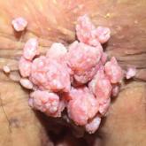
Anorectal Evaluations: Diagnosing & Treating Benign Conditions
Although accreditation for this CE/CME activity has expired, and the posttest is no longer available, you can still read the full article.
...