Article
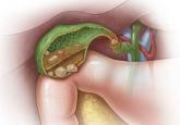
Gallstones: Watch and wait, or intervene?
Consider laparoscopic cholecystectomy for symptomatic cholelithiasis, expectant management for asymptomatic cases.
Article
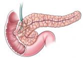
Total pancreatectomy and islet cell autotransplantation: Definitive treatment for chronic pancreatitis
Most patients report less pain after this procedure, and many avoid overt diabetes.