Article
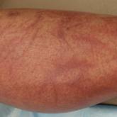
Flagellate Shiitake Mushroom Reaction With Histologic Features of Acute Generalized Exanthematous Pustulosis
Ingestion of shiitake mushrooms and bleomycin is associated with flagellate dermatitis.
Article
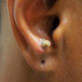
Cutaneous Sarcoidosis Presenting as a Cutaneous Horn
Biopsy of a cutaneous horn should be deep enough to capture the neoplastic or inflammatory process at the base of the lesion. Cutaneous...