User login
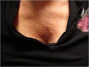
"Peach Pit" Décolleté Defect Can Be Repaired With Fillers
SANTA MONICA, CALIF. — The volume loss and wrinkled skin that appears in the décolleté area of some older women can be significantly softened with filler followed by fractionated laser treatments, Dr. Joel L. Cohen said at a cosmetic dermatology seminar sponsored by Skin Disease Education Foundation.
This cosmetic defect, which Dr. Cohen has dubbed "the peach pit," responds well to injections of hyaluronic acid fillers along with fractionated laser sessions.
"Over the course of many years, I've noticed that many patients become concerned about their décolleté area," said Dr. Cohen, director of AboutSkin Dermatology and DermSurgery, Englewood, Colo., in an interview. "I've tried botulinum toxin [type A]. And despite some reports of efficacy, I haven't been able to actually see this type of efficacy myself. People have topically treated it with retinoids and although that can be helpful, there are patients who have more severe crumpling of the skin."
Instead, he uses Juvéderm (Allergan) or Restylane (Medicis). With such hyaluronic acid fillers, "If you do get lumps or bumps, or the patients aren't happy with it, you could inject the enzyme hyaluronidase and make it go away," he said. "It's easier to mold out little contour irregularities with these types of agents as well."
Using only topical anesthesia, Dr. Cohen injects two or sometimes three syringes of the hyaluronic acid product, after which he asks a female assistant to massage it in. He follows up a few weeks later with two or more treatments with a fractionated laser, often combined with a light erbium laser peel for more texture improvement.
Pharmaceuticals Inc. Skin Disease Education Foundation (SDEF) and this news organization are owned by Elsevier.
Frequent, Lower Botox Dosing Best for Some Areas
Lower doses of botulinum toxin type A delivered more frequently yield better results in certain areas of the face, according to Dr. Joel L. Cohen.
While crow's feet and the glabella still do better with a higher dose and a 3- to 4-month dosing interval, there are several areas that do better with half the typical dose and half the dosing interval.

The forehead is one such area. "We use lighter doses in the forehead because we really want things to be more natural in terms of still having movement," Dr. Cohen said at a cosmetic dermatology seminar sponsored by Skin Disease Education Foundation in Santa Monica, Calif. "Sometimes these patients need to be dosed in the forehead every 8-10 weeks, whereas in the glabella and the crow's feet, if you're using regular doses, you might only need to inject them every 3.5 or 4 months. Sometimes they're in a cycle where they come in for Botox or Dysport in the forehead every 8 or 10 weeks and every 16 weeks or 18 weeks for the glabella and the crow's feet."
Brow lifts also do better with lower doses at shorter intervals. But Dr. Cohen, director of AboutSkin Dermatology and DermSurgery, Englewood, Colo., cautioned that brow lifts with botulinum toxin should not be attempted in patients who have a lot of sagging in the lateral brow. These patients require surgery; botulinum toxin should be reserved for patients with mild redundancy between the brow and the eyelid. Occasionally, patients with moderate sagging can benefit as well.
Around the mouth, Dr. Cohen uses 5-7 units of botulinum toxin to treat the vertical muscle columns of the upper lip and to prevent etched-in lines. At the same time, he uses 2-3 units in the lower lip, "so it doesn't feel funny." Once again, this needs to be repeated every 8-10 weeks, he said.
There is another class of patients in which low doses of perioral botulinum toxin may be beneficial. Studies have shown that healing after resurfacing procedures tends to be better after the area has previously been immobilized with botulinum toxin.
"There's decreasing contraction across the wound, so you're not imprinting lines where you already have lines," Dr. Cohen said. "And the second thing is there is decreased inflammation because there's less mobility of the area."
For one-time erbium or CO2 laser resurfacing, Dr. Cohen recommends the patient be treated with botulinum toxin a week to 10 days before. "Or you can do pretreatment a week or two before you do a fractionated ablative laser where the plan is to do three, four, or five treatments. By treating the patient with the fractionated laser every 3 weeks, you may really be able to get a session of three treatments in for every one pretreatment with Botox. The overall goal is less movement across what you're trying to heal."
Dr. Cohen acknowledged participating in clinical trials and serving as a consultant for Allergan, Medicis, Johnson and Johnson/Mentor, and Merz. SDEF and this news organization are owned by Elsevier.
Lower doses of botulinum toxin type A delivered more frequently yield better results in certain areas of the face, according to Dr. Joel L. Cohen.
While crow's feet and the glabella still do better with a higher dose and a 3- to 4-month dosing interval, there are several areas that do better with half the typical dose and half the dosing interval.
The forehead is one such area. "We use lighter doses in the forehead because we really want things to be more natural in terms of still having movement," Dr. Cohen said at a cosmetic dermatology seminar sponsored by Skin Disease Education Foundation in Santa Monica, Calif. "Sometimes these patients need to be dosed in the forehead every 8-10 weeks, whereas in the glabella and the crow's feet, if you're using regular doses, you might only need to inject them every 3.5 or 4 months. Sometimes they're in a cycle where they come in for Botox or Dysport in the forehead every 8 or 10 weeks and every 16 weeks or 18 weeks for the glabella and the crow's feet."
Brow lifts also do better with lower doses at shorter intervals. But Dr. Cohen, director of AboutSkin Dermatology and DermSurgery, Englewood, Colo., cautioned that brow lifts with botulinum toxin should not be attempted in patients who have a lot of sagging in the lateral brow. These patients require surgery; botulinum toxin should be reserved for patients with mild redundancy between the brow and the eyelid. Occasionally, patients with moderate sagging can benefit as well.
Around the mouth, Dr. Cohen uses 5-7 units of botulinum toxin to treat the vertical muscle columns of the upper lip and to prevent etched-in lines. At the same time, he uses 2-3 units in the lower lip, "so it doesn't feel funny." Once again, this needs to be repeated every 8-10 weeks, he said.
There is another class of patients in which low doses of perioral botulinum toxin may be beneficial. Studies have shown that healing after resurfacing procedures tends to be better after the area has previously been immobilized with botulinum toxin.
"There's decreasing contraction across the wound, so you're not imprinting lines where you already have lines," Dr. Cohen said. "And the second thing is there is decreased inflammation because there's less mobility of the area."
For one-time erbium or CO2 laser resurfacing, Dr. Cohen recommends the patient be treated with botulinum toxin a week to 10 days before. "Or you can do pretreatment a week or two before you do a fractionated ablative laser where the plan is to do three, four, or five treatments. By treating the patient with the fractionated laser every 3 weeks, you may really be able to get a session of three treatments in for every one pretreatment with Botox. The overall goal is less movement across what you're trying to heal."
Dr. Cohen acknowledged participating in clinical trials and serving as a consultant for Allergan, Medicis, Johnson and Johnson/Mentor, and Merz. SDEF and this news organization are owned by Elsevier.
Bone Loss in Teens on DMPA Tied to Vitamin D
Major Finding: In a substudy of 15 adolescent girls with significant bone loss while using depot medroxy-progesterone acetate, only 1 participant had a “sufficient” serum vitamin D level of greater than 30 ng/mL.
Data Source: Subset of a prospective study of 181 adolescent girls on depot medroxyprogesterone acetate.
Disclosures: The study was sponsored by Pfizer/Pharmacia, and one of the investigators was employed by that company. Dr. Harel disclosed financial relationships with Merck, Teva/Duramed, Ortho-McNeil, GlaxoSmithKline, Novartis, and Warner Chilcott.
LAS VEGAS — Abnormally low levels of vitamin D were seen in a subset of 15 adolescent girls who had substantial losses in bone mineral density while using depot medroxyprogesterone acetate for contraception, according to preliminary results from a prospective study presented at the annual meeting of the North American Society for Pediatric and Adolescent Gynecology.
The girls were among 181 adolescents using depot medroxyprogesterone acetate (Depo-Provera, Pfizer) in a prospective study. Bone mineral density (BMD) losses of 5% or more were seen at the lumbar spine in 25% and at the hip in 50% of the study participants.
The relative estrogen deficiency associated with depot medroxyprogesterone acetate (DMPA) did not correlate with the magnitude of BMD loss, according to Dr. Zeev Harel of Brown University in Providence, R.I.
Moreover, serum estradiol remained above 40-50 pg/mL in almost all participants, Dr. Harel said. This level is considered to be sufficient to conserve bone in elderly women.
Dr. Harel and his colleagues examined a subset of 15 young women who lost at least 5% of BMD from baseline. Their average age was 17 years, and they were an average of 61 months postmenarche. Their BMIs were within the normal range, and none was obese. Their ethnicity was diverse and they resided in various U.S. locations.
Investigators noted BMD losses in the majority of the 15 girls after two or three DMPA injections, but some participants did not exhibit BMD losses until after their 10th or 13th injection.
Serum 25-hydroxyvitamin D (25[OH]D) levels were available for 14 of the 15 girls, and all but 1 had low levels of vitamin D. Levels above 30 ng/mL are considered sufficient, levels between 20-30 ng/mL are referred to as “insufficient,” and levels below 20 ng/mL are referred to as “deficient.” Seven of the 14 participants (50%) were vitamin D insufficient, 6 (43%) were vitamin D deficient, and 1 (7%) had normal levels of vitamin D. The mean serum 25(OH)D level among the participants was about 25 ng/mL, in the insufficient range.
Mean levels of parathyroid hormone, on the other hand, were in the normal range.
In an interview, Dr. Harel expressed surprise at these results. “I was expecting probably less than 30% [of the participants would have low levels of vitamin D],” he said
“We were surprised specifically because when we drew the blood we did it at the end of the summer. Typically we absorb vitamin D from the sun. Also, most of the patients were Caucasian. We know that vitamin D deficiency is common in African Americans and Hispanics. Also, they were not extremely obese. We know we can find vitamin D deficiency in obesity. And we also had representatives from states that were really sunny, California for example.”
Dr. Harel emphasized at the meeting that the results were preliminary. Additional studies would require a comparison group of young women on depot medroxyprogesterone acetate who did not experience declines in BMD. And he said that it would be important to study whether vitamin D supplementation would reverse the decline in BMD.
Still, Dr. Harel found results sufficiently worrisome to recommend close monitoring of young women on depot medroxy-progesterone acetate. “We know in the adult elderly population that many are aware of their vitamin D status. In adolescents still we are in the beginning,” he said. He recommended that total 25(OH)D status be measured in all adolescent girls using depot medroxy-progesterone acetate. “And if it's low—deficient or insufficient—treat it accordingly.
“The current recommendation if we have a patient with vitamin D deficiency is to take 50,000 IU once a week for 8 weeks and then repeat the total 25(OH)D. Those who have insufficient vitamin D, we typically treat with 800 IU of vitamin D a day and we do it for 3 months, and then again we repeat the total 25(OH)D,” Dr. Harel said at the meeting.
'We were surprised … because when we drew the blood we did it at the end of the summer.'
Source DR. HAREL
Pearls for Avoiding Filler Injection Danger Zones
Most dermatologists are well aware of the danger of damaging nerves during cold-steel surgery, but may be less aware of the danger zones involved in filler injection, said Dr. Howard K. Steinman at a cosmetic dermatology seminar sponsored by Skin Disease Education Foundation in Santa Monica, Calif.
"Unlike the risk with a scalpel, where you're going to damage a nerve, the risk with fillers is that you may compress or occlude an artery," Dr. Steinman said in an interview. "You will get necrosis and scarring and disfigurement."

Dr. Steinman, director of dermatologic and skin cancer surgery at Texas A&M College of Medicine in College Station, offered some tips for avoiding the problem and suggestions on what to do if an occlusion occurs.
"The one everybody knows about from all the way back to the days of collagen is in the glabella," he said. If collagen is injected into the frown line, it is possible to damage the supratrochlear artery. If compressed or occluded, this artery can get a band-like area of damage extending up the forehead.
A second danger zone is at the nasolabial fold, where the angular artery runs close to the surface. Interrupting the blood flow here can cause damage to skin at the nasolabial fold and the side and tip of the nose. Although this is a relatively rare complication, the nasolabial fold is a popular site for filler injections, and physicians need to be mindful of this side effect, Dr. Steinman noted.
The third danger zone comes at the lips, where injections can interrupt flow in the labial artery.
In all cases, the symptoms are similar: There will be blanching, erythema, or edema, followed by necrosis unless the occlusion is treated promptly.
To avoid occlusion, he recommended injecting the filler perpendicular to the direction of the artery and remaining superficial but also pointed out that an artery can be indirectly occluded. According to one theory, simply injecting too much filler can compress the artery to the point of interrupting the flow.
Occlusions and compressions should be treated as soon as detected. The first step is an aggressive tapping and massaging of the area to try to break up the mass, Dr. Steinman said.
If that does not work, and the filler being used is hyaluronic acid-based, the next step is an injection of hyaluronidase, an enzyme that dissolves hyaluronic acid.
A third possibility is to apply nitroglycerin paste to the skin directly above the affected area. Typically used to keep the arteries dilated, the paste can improve flow in the case of occlusion or compression.
Unfortunately, the symptoms of an occluded artery do not always become evident immediately. "In some cases the patient won't notice this for hours after you have done the procedure," Dr. Steinman said. "They'll call you and they'll say, 'I'm having a problem.' You can certainly have them tap and massage as soon as possible, but you should meet them, even if it's at night, and start this protocol as soon as practical. It's not something you want to ignore. 'I'll see you in the morning,' is not the correct option for this."
Dr. Steinman stated that he had no conflicts of interest to disclose. SDEF and this news organization are owned by Elsevier.
Most dermatologists are well aware of the danger of damaging nerves during cold-steel surgery, but may be less aware of the danger zones involved in filler injection, said Dr. Howard K. Steinman at a cosmetic dermatology seminar sponsored by Skin Disease Education Foundation in Santa Monica, Calif.
"Unlike the risk with a scalpel, where you're going to damage a nerve, the risk with fillers is that you may compress or occlude an artery," Dr. Steinman said in an interview. "You will get necrosis and scarring and disfigurement."
Dr. Steinman, director of dermatologic and skin cancer surgery at Texas A&M College of Medicine in College Station, offered some tips for avoiding the problem and suggestions on what to do if an occlusion occurs.
"The one everybody knows about from all the way back to the days of collagen is in the glabella," he said. If collagen is injected into the frown line, it is possible to damage the supratrochlear artery. If compressed or occluded, this artery can get a band-like area of damage extending up the forehead.
A second danger zone is at the nasolabial fold, where the angular artery runs close to the surface. Interrupting the blood flow here can cause damage to skin at the nasolabial fold and the side and tip of the nose. Although this is a relatively rare complication, the nasolabial fold is a popular site for filler injections, and physicians need to be mindful of this side effect, Dr. Steinman noted.
The third danger zone comes at the lips, where injections can interrupt flow in the labial artery.
In all cases, the symptoms are similar: There will be blanching, erythema, or edema, followed by necrosis unless the occlusion is treated promptly.
To avoid occlusion, he recommended injecting the filler perpendicular to the direction of the artery and remaining superficial but also pointed out that an artery can be indirectly occluded. According to one theory, simply injecting too much filler can compress the artery to the point of interrupting the flow.
Occlusions and compressions should be treated as soon as detected. The first step is an aggressive tapping and massaging of the area to try to break up the mass, Dr. Steinman said.
If that does not work, and the filler being used is hyaluronic acid-based, the next step is an injection of hyaluronidase, an enzyme that dissolves hyaluronic acid.
A third possibility is to apply nitroglycerin paste to the skin directly above the affected area. Typically used to keep the arteries dilated, the paste can improve flow in the case of occlusion or compression.
Unfortunately, the symptoms of an occluded artery do not always become evident immediately. "In some cases the patient won't notice this for hours after you have done the procedure," Dr. Steinman said. "They'll call you and they'll say, 'I'm having a problem.' You can certainly have them tap and massage as soon as possible, but you should meet them, even if it's at night, and start this protocol as soon as practical. It's not something you want to ignore. 'I'll see you in the morning,' is not the correct option for this."
Dr. Steinman stated that he had no conflicts of interest to disclose. SDEF and this news organization are owned by Elsevier.
Bone Loss in Adolescents on DMPA Tied to Vitamin D
Major Finding: In a substudy of 15 adolescent girls with significant bone loss while using depot medroxyprogesterone acetate, only 1 participant had a “sufficient” serum vitamin D level of greater than 30 ng/mL.
Data Source: Subset of a prospective study of 181 adolescent girls on depot medroxyprogesterone acetate.
Disclosures: The study was sponsored by Pfizer/Pharmacia, and one of the investigators was employed by that company. Dr. Harel disclosed financial relationships with Merck, Teva/Duramed, Ortho-McNeil, GlaxoSmithKline, Novartis, and Warner Chilcott.
LAS VEGAS — Abnormally low levels of vitamin D were seen in a subset of 15 adolescent girls who had substantial losses in bone mineral density while using depot medroxyprogesterone acetate for contraception, according to preliminary results from a prospective study presented at the annual meeting of the North American Society for Pediatric and Adolescent Gynecology.
The girls were among 181 adolescents using depot medroxyprogesterone acetate (Depo-Provera, Pfizer) in a prospective study. Bone mineral density (BMD) losses of 5% or more were seen at the lumbar spine in 25% and at the hip in 50% of the study participants.
The relative estrogen deficiency associated with depot medroxyprogesterone acetate (DMPA) did not correlate with the magnitude of BMD loss, according to Dr. Zeev Harel of Brown University in Providence, R.I.
Moreover, serum estradiol remained above 40–50 pg/mL in almost all participants, Dr. Harel said. This level is considered to be sufficient to conserve bone in elderly women.
Dr. Harel and his colleagues examined a subset of 15 young women who lost at least 5% of BMD from baseline. Their average age was 17 years, and they were an average of 61 months post menarche. Body mass index was within the normal range, and none of the women was obese. Their ethnicity was diverse and they resided in various U.S. locations.
Investigators noted BMD losses in the majority of the 15 girls after two or three DMPA injections, but some participants did not exhibit BMD losses until after their 10th or 13th injection.
Serum 25-hydroxyvitamin D (25[OH]D) levels were available for 14 of the 15 girls, and all but 1 had low levels of vitamin D. Levels above 30 ng/mL are considered sufficient, levels between 20 and 30 ng/mL are referred to as “insufficient,” and levels below 20 ng/mL are referred to as “deficient.” Seven of the 14 participants (50%) were vitamin D insufficient, 6 (43%) were vitamin D deficient, and 1 (7%) had normal levels of vitamin D.
The mean serum 25(OH)D level among the participants was about 25 ng/mL, in the insufficient range. Mean levels of parathyroid hormone, on the other hand, were in the normal range.
In an interview, Dr. Harel expressed surprise at these results. “I was expecting probably less than 30% [of the participants would have low levels of vitamin D],” he said “We were surprised specifically because when we drew the blood we did it at the end of the summer. Typically we absorb vitamin D from the sun. Also, most of the patients were [white]. We know that vitamin D deficiency is common in African Americans and Hispanics. Also, they were not extremely obese. We know we can find vitamin D deficiency in obesity. And we also had representatives from states that were really sunny, California for example.”
Dr. Harel emphasized that the results were preliminary. Additional studies would require a comparison group of young women on depot medroxyprogesterone acetate who did not experience declines in BMD. And he said that it would be important to study whether vitamin D supplementation would reverse the decline in BMD.
Still, Dr. Harel found results sufficiently worrisome to recommend close monitoring of young women on depot medroxyprogesterone acetate. “We know in the adult elderly population that many are aware of their vitamin D status. In adolescents still we are in the beginning,” he said.
He recommended that total 25(OH)D status be measured in all adolescent girls using depot medroxyprogesterone acetate.
“And if it's low—deficient or insufficient—treat it accordingly. The current recommendation if we have a patient with vitamin D deficiency is to take 50,000 IU once a week for 8 weeks and then repeat the total 25(OH)D. Those who have insufficient vitamin D, we typically treat with 800 IU of vitamin D a day and we do it for 3 months, and then again we repeat the total 25(OH)D,” he said.
The results were surprising, given that the cohort comprised mostly white, nonobese women who had sun exposure.
Source DR. HAREL
After Many Complaints, FDA to Regulate Infusion Pumps
The Food and Drug Administration will regulate the design and manufacture of infusion pumps in the wake of thousands of adverse-event reports, the agency announced in a teleconference.
Over the past 5 years the FDA has received more than 56,000 reports of adverse events, including more than 500 deaths, related to the pumps. During that period there have been 87 recalls of infusion pumps undertaken to address identified safety concerns.
“Infusion pumps rank among the top of most frequently recalled devices for this 5-year period, and they are one of the top categories of devices for reporting of adverse events,” Dr. Jeffrey Shuren, director of the FDA's Center for Devices and Radiological Health, said during the teleconference. “There have been problems with every kind of infusion pump on the market across the entire industry…. In some cases, pumps have actually exploded in a patient's room.”
The problems have ranged from manufacturing defects to software bugs to user error. Dr. Shuren described one case in which a woman who was taking the blood thinner heparin accidentally gave herself 10 times the correct dose. The culprit was “key bounce,” in which someone trying to enter 20, for example, will enter 200 by mistake. The woman died.
The FDA is taking several steps to address the devices' problems at the level of manufacture.
In the interim, the agency is advising clinicians to use several strategies to reduce risk when using infusion pumps: Have a plan in place and be prepared to respond to pump failures; prevent errors by labeling infusion pump channels and tubing; check settings of infusion pumps and check patients for signs of under- or overinfusion; prevent and respond to pump problems by using available resources; and promptly report adverse events to the FDA.
The agency published draft guidance on April 23 recommending that manufacturers of infusion pumps start providing additional information on design and engineering to the agency during premarket review.
The draft guidance document, available on the FDA's Web site, is open for public comment, and the agency plans to hold a public infusion pump workshop on May 25-26.
Dr. Shuren said that the agency plans to move quickly in turning the draft guidance document into an actual set of regulations.
Among other things, the FDA recommends that each premarket submission include a structured, evidence-based discussion of all steps the manufacturer has taken to mitigate risk at each stage of the device's life cycle. That includes device design, manufacture, servicing, maintenance, and use in clinical or home settings.
FDA inspectors will visit production facilities for the first time to examine manufacturing practices.
In addition, manufacturers will need to show that they have tested their pump in the environment in which it is intended to be used and with the types of clinicians and patients who are expected to use it.
The agency offered to help manufacturers with the task of checking the software controlling these devices. Even before premarket review, manufacturers may submit software code to the FDA, whose experts will conduct “static analysis,” an automated diagnostic technique that can detect software problems early in the development process.
Dr. Shuren described the CDRH action as “a marked departure” in the way it has handled such cases in the past.
In response to a reporter's question, he noted that other medical devices have been the subject of large numbers of adverse event reports, citing implantable cardiac defibrillators as an example.
The FDA has established a Web site with detailed information on infusion pump problems and the agency's proposed solutions at www.fda.gov/MedicalDevices/ProductsandMedicalProcedures/GeneralHospitalDevicesandSupplies/InfusionPumps/default.htm
A white paper on the Infusion Pump Improvement Initiative can be found at www.fda.gov/MedicalDevices/ProductsandMedicalProcedures/GeneralHospitalDevicesandSupplies/InfusionPumps/ucm205424.htm
Common Lab Tests Can Predict Thiopurine Response in IBD
Algorithms based on common laboratory values outperformed expensive metabolite testing in determining which patients with inflammatory bowel disease are likely to respond to thiopurine therapy, Dr. Akbar K. Waljee and his colleagues reported.
Thiopurines are known to be effective immunomodulators in patients with inflammatory bowel disease (IBD) who have failed 5-aminosalicylic acid therapy. The problem is that thiopurines have a narrow therapeutic index, and individuals vary widely in how they metabolize these agents.
Experienced clinicians can use inexpensive complete blood count and standard blood chemistry values to balance efficacy and risk in individual patients, but this takes expert judgment, and there are no established algorithms.
A more reproducible approach is to measure the metabolites 6-thioguanine (6-TGN) and 6 methylmercaptopurine (6-MMP). Unfortunately, monitoring these metabolites is expensive, and the sensitivity and specificity of this approach are only 62% and 72%, respectively.
In an effort to resolve this dilemma, Dr. Waljee and his colleagues from the University of Michigan, Ann Arbor, used a machine learning technique to tease out the most accurate algorithms based on CBC and blood chemistries (Clin. Gastroenterol. Hepatol. 2010 [doi:10.1016/j.cgh.2009.09.031]).
The investigators used data collected in 774 cases from 346 individuals who were seen at the University of Michigan between May 2004 and August 2006. To be included in the study, the patients had to have had thiopurine metabolite analysis, CBC, and a comprehensive chemistry panel within the same 24-hour period.
Using a randomly selected 70% of the cases, investigators used a statistical technique called the “random forest” method to derive the most accurate algorithms based on data from the CBC and chemistry panels. They then tested that algorithm on the remaining 30% of the cases, comparing the accuracy to that of thiopurine metabolite analysis.
Their primary outcome measure was the area under the receiver operating characteristic curve (AuROC), a standard measure of accuracy.
The random forest algorithm differentiated clinical response from nonresponse with an AuROC of 0.856, compared with 0.594, for 6-TGN levels, a difference that was highly statistically significant.
The most important independent variables in differentiating responders from nonresponders were neutrophil count, alkaline phosphatase, red-cell distribution width, age, and white blood cell count.
The investigators also derived a random forest algorithm that would predict patient nonadherence, and another that would predict which patients were likely to have unfavorable pharmacodynamic responses to thiopurine therapy. Both of those algorithms proved to be significantly better than thiopurine metabolite analysis.
They also developed a simple prediction rule that was reasonably accurate at differentiating responders from nonresponders. Patients with a ratio of mean corpuscular volume (MCV) to white blood cell count (WBC) of 12 or more had a 67% likelihood of having a clinical response, while those with a ratio less than 12 had a 35% likelihood of having a clinical response. This simplified algorithm was significantly worse than the more complex algorithm, but it was still significantly better than metabolite analysis.
The investigators hypothesized that the algorithms based on common laboratory tests serve as surrogate markers for immune system changes induced by effective thiopurine therapy.
“It seems likely that patients with inadequate responses in these parameters are underdosed and that some patients, even at high doses, will not achieve these physiologic changes, due to differences in pharmacodynamics,” the investigators wrote. “We speculate that the eventual clinical utility of this machine learning approach, depending on a positive outcome of a prospective clinical trial, will be in guiding thiopurine dosing and timely changes to a different class of therapy.”
Disclosures: The investigators disclosed that the Regents of the University of Michigan, along with several of the study's coauthors, have applied for a patent on the application of machine learning to the prediction of clinical response to thiopurines.
'Metabolically Healthy' Obesity Not So Healthy
Major Finding: Men who were obese at age 50 but who did not have metabolic syndrome or insulin resistance had 10–15 times the chance of developing type 2 diabetes as did normal-weight men over 10 and 20 years.
Data Source: Longitudinal study of 934 men.
Disclosures: None reported.
SAN FRANCISCO — Men who are obese but “metabolically healthy” have no protection against developing type 2 diabetes, according to a large longitudinal study.
Compared with men of normal weight, men who have a body mass index greater than 30 kg/m
Other studies have shown that 25%-30% of individuals who are obese do not meet criteria for metabolic syndrome or insulin resistance. Earlier studies appeared to indicate that those individuals were unlikely to develop diabetes or cardiovascular disease. But those studies were hampered by relatively short period of follow-up.
“Our conclusion from this study … is that metabolically healthy obesity is not very healthy, that it is not a benign condition,” said Dr. Lars Lind of the University of Uppsala, Sweden, the study's lead investigator.
The investigators used data from the Uppsala Longitudinal Study of Adult Men, a cohort of men born during 1920–1924. At age 50 years 1,758 of these men were available for study; that number declined to 1,420 at age 60 and 934 at age 70.
The investigators defined normal weight as a BMI less than 25 kg/m
During 10 years of follow-up, 124 of the men developed diabetes, and that number increased to 169 after 20 years.
After correction for age, smoking, and LDL cholesterol level, obese men with and without metabolic syndrome had 10 times the risk of developing diabetes as did normal-weight men after 10 years. Overweight men without metabolic syndrome had a threefold increase in risk. All those increases were statistically significant. After 20 years, obese men without metabolic syndrome had 15 times the risk of developing diabetes and overweight men had 4 times the risk.
The situation with men who were not insulin resistant was similar. After 10 years, obese men with and without insulin resistance had a 15-fold increase in the risk of developing diabetes, and overweight men had a 3-fold increase in risk.
After 20 years, a statistically significant difference in risk appeared between obese men with and without insulin resistance. Men without insulin resistance were 15 times as likely to develop diabetes, while men with insulin resistance had a 30-fold increase in risk. But the 15-fold increase in risk among men without insulin resistance at baseline was still significantly elevated.
© Obese men without metabolic syndrome had a high risk of developing diabetes.
Source geronimo/Fotolia.com
Neighborhood Factors May Predict Cardiac Risks
Major Finding: Women in the highest quartile of neighborhood deprivation had about 2.5-fold the risk of coronary artery calcification as women in the lowest quartile.
Data Source: The CARDIA study of 2,974 adults.
Disclosures: Research supported by a grant from the John D. and Catherine T. MacArthur Foundation Research Network on Socioeconomic Status and Health.
SAN FRANCISCO — Women who live in economically deprived neighborhoods or neighborhoods that lack social cohesion have higher rates of coronary artery calcification than other women.
In men, however, the association with coronary artery calcification is statistically significant only for those who live in neighborhoods characterized both by economic deprivation and lack of social cohesion.
“Neighborhood deprivation and low cohesion may predict very early coronary heart disease in younger, asymptomatic adults,” Dr. Daniel Kim said at a conference sponsored by the American Heart Association. “These results are consistent with neighborhood deprivation and low cohesion as fundamental or root causes of coronary heart disease.”
Women in the highest quartile of neighborhood deprivation had about a 2.5-fold risk of coronary artery calcification (CAC), compared with women in the lowest quartile. Similarly, women in the lowest quartile of neighborhood cohesion had about twice the risk of CAC as women in the highest quartile, Dr. Kim reported.
Neither neighborhood cohesion nor neighborhood deprivation alone was significantly associated with CAC in men, but those living in the lowest three quartiles of neighborhood cohesion who were also living in deprived neighborhoods were about three times as likely to have CAC as those living in deprived neighborhoods with the highest levels of cohesion.
Data from the study came from 2,974 adults participating in the Coronary Artery Risk Development in Young Adults (CARDIA) study, said Dr. Kim of the Harvard School of Public Health, Boston.
CARDIA is a prospective cohort study of young adults in four urban areas—Birmingham, Ala.; Chicago; Minneapolis; and Oakland, Calif.—who have been followed since 1985. Included in this study were those participants who had undergone measures of CAC in 2005 when they were 37–50 years old.
The investigators assessed neighborhood deprivation by combining six indicators: median household income; median value of housing units; percentage of households receiving interest, dividend, or rental income; percentage of adults who had completed high school; percentage of adults who had completed college; and percentage employed in executive, managerial, or specialty occupations.
They assessed perceived neighborhood cohesion from participants' answers to five survey items: people's willingness to help their neighbors, whether the neighborhood is “close-knit,” whether people in the neighborhood can be trusted, whether they get along with each other, and whether they share the same values.
Investigators adjusted their results.
My Take
Access to High-Quality Food Is Key
Dr. Kim's study is important because we don't pay enough attention to neighborhoods and their role in encouraging or discouraging good dietary practices. It's well known that economically deprived neighborhoods tend to have lots of fast-food emporia and not much in the way of first-class supermarkets.
I'm reminded that back in the 1940s two great congressmen, Sen. Lister Hill (1894–1984, D-Ala.) and Sen. Harold Burton (1888–1964, R-Ohio), were successful in passing the landmark Hospital Survey and Construction Act. This financed the construction and renovation of more than 9,200 medical facilities, many of which were in low-income communities.
The modern version of the law would be an act by Congress to finance the construction of first-class supermarkets in more communities. These supermarkets could stock high-quality food and make it available at low prices. This would allow people living in deprived communities to improve their diets and overcome their unfavorable cardiovascular risk status.
JEREMIAH STAMLER, M.D., is professor emeritus in the department of preventive medicine at Northwestern University, Chicago.
Vitals
'Metabolically Healthy' Obesity Is … Unhealthy
Major Finding: Men who were obese at age 50 years but did not have metabolic syndrome or insulin resistance had 10-15 times the risk of type 2 diabetes as did normal-weight men after 10 and 20 years.
Data Source: Longitudinal study of 934 men.
Disclosures: The investigators said they had no relevant financial ties.
SAN FRANCISCO — Men who are obese but “metabolically healthy” have no protection against developing type 2 diabetes, according to a large longitudinal study.
Compared with men of normal weight, men who have a body mass index greater than 30 kg/m
Other studies have shown that 25%-30% of individuals who are obese do not meet criteria for metabolic syndrome or insulin resistance. Earlier studies appeared to indicate that those individuals were unlikely to develop diabetes or cardiovascular disease. But those studies were hampered by a relatively short period of follow-up.
“Our conclusion from this study … is that metabolically healthy obesity is not very healthy, that it is not a benign condition,” said Dr. Lars Lind of the University of Uppsala, Sweden, the study's lead investigator.
The investigators used data from the Uppsala Longitudinal Study of Adult Men, a cohort of men who were born during 1920-1924. At age 50 years 1,758 of these men were available for study; that number declined to 1,420 at age 60 and 934 at age 70.
The investigators defined normal weight as a BMI less than 25 kg/m
During 10 years of follow-up, 124 of the men developed diabetes, and that number increased to 169 after 20 years.
After correction for age, smoking, and LDL cholesterol level, obese men with and without metabolic syndrome had 10 times the risk of developing diabetes as did normal-weight men after 10 years. Overweight men without metabolic syndrome had a threefold increase in risk. All those increases in risk were statistically significant, Dr. Lind reported.
After 20 years, obese men without metabolic syndrome had 15 times the risk of developing diabetes and overweight men had 4 times the risk.
The situation with men who were not insulin resistant was similar. After 10 years, obese men with and without insulin resistance had a 15-fold increase in the risk of developing diabetes, and overweight men had a 3-fold increase in risk.
After 20 years, a statistically significant difference in risk appeared between obese men with and without insulin resistance. Men without insulin resistance were 15 times as likely to develop diabetes, while men with insulin resistance had a 30-fold increase in risk. But the 15-fold increase in risk among men without insulin resistance at baseline was still significantly elevated.
Obese men without metabolic syndrome had a high risk of developing diabetes.
Source ©geronimo/Fotolia.com