Article
Hospital Dermatology: Review of Research in 2023-2024
Inpatient consultative dermatologists play a critical role in the care of hospitalized patients with skin disease. Our review of the 2023-2024...
Article
Inpatient Management of Hidradenitis Suppurativa: A Delphi Consensus Study
These recommendations serve as an important resource for providers caring for inpatients with hidradenitis suppurativa (HS) and represent a...
Article
Hospital Dermatology: Review of Research in 2022-2023
In this review, we highlight 4 areas of focus from the published literature in 2022-2023—severe cutaneous adverse reactions, supportive...
Article
Cutaneous Manifestations of COVID-19: Characteristics, Pathogenesis, and the Role of Dermatology in the Pandemic
Cutaneous manifestations of COVID-19 may reflect the range of host immunologic responses to SARS-CoV-2.
Article
Nutritional Dermatoses in the Hospitalized Patient
This review details the risk factors for nutritional deficiency, illustrates the presentations of cutaneous disease, reviews diagnostic workups,...
Article
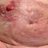
Erythematous Plaques and Nodules on the Abdomen and Groin
An 82-year-old man presented with acute abdominal pain and distension as well as an abdominal rash of 4 months' duration that was expanding...

Article
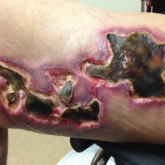
Update on Calciphylaxis Etiopathogenesis, Diagnosis, and Management
Calciphylaxis is associated with potentially severe complications and high mortality rates. This article highlights the challenges faced in...
Article
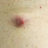
Enlarging Red Papulonodule on the Chest
A man in his 60s presented with a subcutaneous nodule on the right side of the chest. Due to impaired mental status, he was unable to describe the...