User login
Stains and Smears: Resident Guide to Bedside Diagnostic Testing
Dermatologists are fortunate to specialize in the organ that is most accessible to evaluation. Although we use the physical examination to formulate the initial differential diagnosis, at times we must rely on ancillary tests to narrow down the diagnosis. Various bedside testing modalities—potassium hydroxide (KOH) preparation, Tzanck smear, mineral oil preparation, and Gram stain—are most useful in diagnosing infectious causes of cutaneous disease. This guide serves as a useful reference for residents on how to perform these tests and which conditions they can help diagnose. Several of these procedures have no standard protocol for performing them; the literature is littered with various methodologies, fixatives, and stains, and as such, this article will attempt to describe a technique that is convenient and quick to perform with readily available materials while still offering high diagnostic utility.
KOH Preparation
A standard in the armamentarium of a dermatologist, the KOH preparation is invaluable to diagnose fungal and yeast infections. Although there are many available preparations including varying concentrations of KOH, dimethyl sulfoxide, and various inks, the procedure is similar for all of them.1 The first step involves collecting the specimen, which can be scale from an active border of suspected cutaneous dermatophyte or Malassezia infection, debris from suspected candidiasis, or hair shafts plucked from an area of alopecia of presumed tinea capitis. A no. 15 blade can be used to scrape the specimen onto a microscope slide, though a second microscope slide can be used in lieu of a blade in patients who will not remain still, and then a coverslip is placed. Two drops of the KOH solution of your choice are then placed on opposite ends of the coverslip, allowing capillary action to spread the stain evenly. A paper towel can be folded in half and pushed down on the surface of the coverslip to spread the stain and soak up any excess, and this pressure also can help the KOH solution digest the keratin in the specimen. Briefly heating the underside of the slide (below boiling point) will help digest the keratin; this step is not necessary when you are using a KOH preparation with dimethyl sulfoxide. Although many dermatologists view the slide almost immediately, ideally at least 5 minutes should pass before it is read. Particularly thick specimens may require additional digestion time, so setting them aside for later review may help visualize infectious agents. In a busy clinic where an immediate diagnosis may not be requisite and a prescription can be called in pending the result, waiting to review the slide may be feasible.
Tzanck Smear
The Tzanck smear is a useful cytopathologic test in the rapid diagnosis of herpetic lesions, though it cannot differentiate between herpes simplex virus type 1, herpes simplex virus type 2, and varicella-zoster virus. It also has shown utility for rapid diagnosis of protean other dermatologic conditions including autoimmune blistering disorders, cutaneous malignancies, and other infectious processes, though it has been superseded by histopathology in most cases.2 An ideal sample is collected by scraping the base of a fresh blister with a no. 15 blade or a second microscope slide. The scrapings then are smeared onto another microscope slide and allowed to air-dry briefly. Then, Wright-Giemsa stain is dispensed to cover the sample and allowed to sit for 15 minutes before being washed off with sterile water. After air-drying, the sample is examined for the presence of clumped multinucleated giant cells, a feature that confirms herpetic infection and allows rapid initiation of antiviral medication.3
Mineral Oil Preparation
A mineral oil preparation has utility in diagnosing ectoparasitic infestation. In the case of scabies, a positive microscopic examination is diagnostic and requires no further testing, allowing for rapid initiation of therapy. This technique also is useful in diagnosing rosacea related to Demodex, which requires a treatment algorithm that differs from the classic papulopustular rosacea which it mimics.4
Mineral oil preparations can be rapidly performed and interpreted. Several drops of mineral oil are placed onto a microscope slide and a no. 15 blade is dipped into this oil prior to scraping the sample lesion. For scabies, a burrow is scraped repeatedly with the blade, and the debris is collected in the mineral oil. Occasionally, the mite can be dermoscopically visualized as a jet plane or arrowhead at the leading edge of a burrow; scraping should be focused in the vicinity of the mite.5 A coverslip is applied to the microscope slide and examination for the mite, egg casings, and scybala can be performed with microscopy.6 For Demodex infestation, a facial pustule can be expressed or several eyelash hairs can be plucked and suspended in mineral oil. Examination of this specimen is identical to scabies.
Gram Stain
The Gram stain is invaluable in classifying bacteria, and a properly performed test can narrow the identification of a causative organism based on cellular morphology. Although it is more technically complex than other bedside diagnostic maneuvers, it can be rapidly performed once the sequence of stains is mastered. The collected sample is smeared onto a glass slide and then briefly passed over a flame several times to heat-fix the specimen. Caution should be taken to avoid direct or prolonged flame contact with the underside of the slide. After fixation, the staining can be performed. First, crystal violet is instilled onto the slide and remains on for 30 seconds before being rinsed off with sink water. Then, Gram iodine is used for 30 seconds, followed by another rinse in water. Next, pour the decolorizer solution over the slide until the runoff is clear, and then rinse in water. Finally, flood with safranin counterstain for 30 seconds and give the slide a final rinse. After air-drying, it is ready to be interpreted.7
Final Thoughts
Although the modern dermatologist has access to biopsies, cultures, and sophisticated diagnostic techniques, it is important to remember these useful bedside tests. The ability to rapidly pin a diagnosis is particularly useful on the consultative service where critically ill patients can benefit from identification of a causative pathogen sooner rather than later. Residents should master these stains in their training, as this knowledge may prove to be invaluable in their careers.
1. Trozak DJ, Tennenhouse DJ, Russell JJ. Dermatology Skills for Primary Care: An Illustrated Guide. Totowa, NJ: Humana Press; 2006.
2. Kelly B, Shimoni T. Reintroducing the Tzanck smear. Am J Clin Dermatol. 2009;10:141-152.
3. Singhi M, Gupta L. Tzanck smear: a useful diagnostic tool. Indian J Dermatol Venereol Leprol. 2005;71:295.
4. Elston DM. Demodex mites: facts and controversies. Clin Dermatol. 2010;28:502-504.
5. Dupuy A, Dehen L, Bourrat E, et al. Accuracy of standard dermoscopy for diagnosing scabies. J Am Acad Dermatol. 2007;56:53-62.
6. Bolognia J, Schaffer J, Duncan K, et al. Dermatology Essentials. St. Louis, MO: Saunders Elsevier; 2014.
7. Ruocco E, Baroni A, Donnarumma G, et al. Diagnostic procedures in dermatology. Clin Dermatol. 2011;29:548-556.
Dermatologists are fortunate to specialize in the organ that is most accessible to evaluation. Although we use the physical examination to formulate the initial differential diagnosis, at times we must rely on ancillary tests to narrow down the diagnosis. Various bedside testing modalities—potassium hydroxide (KOH) preparation, Tzanck smear, mineral oil preparation, and Gram stain—are most useful in diagnosing infectious causes of cutaneous disease. This guide serves as a useful reference for residents on how to perform these tests and which conditions they can help diagnose. Several of these procedures have no standard protocol for performing them; the literature is littered with various methodologies, fixatives, and stains, and as such, this article will attempt to describe a technique that is convenient and quick to perform with readily available materials while still offering high diagnostic utility.
KOH Preparation
A standard in the armamentarium of a dermatologist, the KOH preparation is invaluable to diagnose fungal and yeast infections. Although there are many available preparations including varying concentrations of KOH, dimethyl sulfoxide, and various inks, the procedure is similar for all of them.1 The first step involves collecting the specimen, which can be scale from an active border of suspected cutaneous dermatophyte or Malassezia infection, debris from suspected candidiasis, or hair shafts plucked from an area of alopecia of presumed tinea capitis. A no. 15 blade can be used to scrape the specimen onto a microscope slide, though a second microscope slide can be used in lieu of a blade in patients who will not remain still, and then a coverslip is placed. Two drops of the KOH solution of your choice are then placed on opposite ends of the coverslip, allowing capillary action to spread the stain evenly. A paper towel can be folded in half and pushed down on the surface of the coverslip to spread the stain and soak up any excess, and this pressure also can help the KOH solution digest the keratin in the specimen. Briefly heating the underside of the slide (below boiling point) will help digest the keratin; this step is not necessary when you are using a KOH preparation with dimethyl sulfoxide. Although many dermatologists view the slide almost immediately, ideally at least 5 minutes should pass before it is read. Particularly thick specimens may require additional digestion time, so setting them aside for later review may help visualize infectious agents. In a busy clinic where an immediate diagnosis may not be requisite and a prescription can be called in pending the result, waiting to review the slide may be feasible.
Tzanck Smear
The Tzanck smear is a useful cytopathologic test in the rapid diagnosis of herpetic lesions, though it cannot differentiate between herpes simplex virus type 1, herpes simplex virus type 2, and varicella-zoster virus. It also has shown utility for rapid diagnosis of protean other dermatologic conditions including autoimmune blistering disorders, cutaneous malignancies, and other infectious processes, though it has been superseded by histopathology in most cases.2 An ideal sample is collected by scraping the base of a fresh blister with a no. 15 blade or a second microscope slide. The scrapings then are smeared onto another microscope slide and allowed to air-dry briefly. Then, Wright-Giemsa stain is dispensed to cover the sample and allowed to sit for 15 minutes before being washed off with sterile water. After air-drying, the sample is examined for the presence of clumped multinucleated giant cells, a feature that confirms herpetic infection and allows rapid initiation of antiviral medication.3
Mineral Oil Preparation
A mineral oil preparation has utility in diagnosing ectoparasitic infestation. In the case of scabies, a positive microscopic examination is diagnostic and requires no further testing, allowing for rapid initiation of therapy. This technique also is useful in diagnosing rosacea related to Demodex, which requires a treatment algorithm that differs from the classic papulopustular rosacea which it mimics.4
Mineral oil preparations can be rapidly performed and interpreted. Several drops of mineral oil are placed onto a microscope slide and a no. 15 blade is dipped into this oil prior to scraping the sample lesion. For scabies, a burrow is scraped repeatedly with the blade, and the debris is collected in the mineral oil. Occasionally, the mite can be dermoscopically visualized as a jet plane or arrowhead at the leading edge of a burrow; scraping should be focused in the vicinity of the mite.5 A coverslip is applied to the microscope slide and examination for the mite, egg casings, and scybala can be performed with microscopy.6 For Demodex infestation, a facial pustule can be expressed or several eyelash hairs can be plucked and suspended in mineral oil. Examination of this specimen is identical to scabies.
Gram Stain
The Gram stain is invaluable in classifying bacteria, and a properly performed test can narrow the identification of a causative organism based on cellular morphology. Although it is more technically complex than other bedside diagnostic maneuvers, it can be rapidly performed once the sequence of stains is mastered. The collected sample is smeared onto a glass slide and then briefly passed over a flame several times to heat-fix the specimen. Caution should be taken to avoid direct or prolonged flame contact with the underside of the slide. After fixation, the staining can be performed. First, crystal violet is instilled onto the slide and remains on for 30 seconds before being rinsed off with sink water. Then, Gram iodine is used for 30 seconds, followed by another rinse in water. Next, pour the decolorizer solution over the slide until the runoff is clear, and then rinse in water. Finally, flood with safranin counterstain for 30 seconds and give the slide a final rinse. After air-drying, it is ready to be interpreted.7
Final Thoughts
Although the modern dermatologist has access to biopsies, cultures, and sophisticated diagnostic techniques, it is important to remember these useful bedside tests. The ability to rapidly pin a diagnosis is particularly useful on the consultative service where critically ill patients can benefit from identification of a causative pathogen sooner rather than later. Residents should master these stains in their training, as this knowledge may prove to be invaluable in their careers.
1. Trozak DJ, Tennenhouse DJ, Russell JJ. Dermatology Skills for Primary Care: An Illustrated Guide. Totowa, NJ: Humana Press; 2006.
2. Kelly B, Shimoni T. Reintroducing the Tzanck smear. Am J Clin Dermatol. 2009;10:141-152.
3. Singhi M, Gupta L. Tzanck smear: a useful diagnostic tool. Indian J Dermatol Venereol Leprol. 2005;71:295.
4. Elston DM. Demodex mites: facts and controversies. Clin Dermatol. 2010;28:502-504.
5. Dupuy A, Dehen L, Bourrat E, et al. Accuracy of standard dermoscopy for diagnosing scabies. J Am Acad Dermatol. 2007;56:53-62.
6. Bolognia J, Schaffer J, Duncan K, et al. Dermatology Essentials. St. Louis, MO: Saunders Elsevier; 2014.
7. Ruocco E, Baroni A, Donnarumma G, et al. Diagnostic procedures in dermatology. Clin Dermatol. 2011;29:548-556.

Learning Dermatopathology in the Digital Age
As in the study of clinical dermatology, establishing a strong fund of knowledge regarding dermatopathology requires visual exposure to countless representative cases. In the not-so-distant past, textbooks relied on grayscale representations to illustrate these diagnoses, but residents today enjoy full-color images; however, textbooks lack the plasticity of digital media, which allow for more immersive interaction with the content. With technological advances in whole-slide imaging, teaching cases can be saved and shared, and rare diagnoses can be studied by individuals who are far removed from the original specimen.1 Even more exciting is that many of the applications (apps) that facilitate digital learning of dermatopathology are available free of charge. In this article, I will review some of the available apps, focusing on their usability, content, and utility as a learning resource for dermatologists at all stages of training. They are discussed in the order of their utility to students of dermatopathology. I have no financial ties to any of the products reviewed, and my recommendations reflect my opinions and observations after real-world use.
Winner: Clearpath
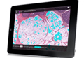
The Clearpath app (http://www.dermpathlab.com/clearpath/) is a fantastic representation of well-executed digital pathology software. Initially released for $50.00 in 2013, the app has since become free while maintaining a steady stream of updates and expanded content. The app is incredibly intuitive and easy to use, made possible by its modern user interface and versatile search function (Figure). For those just beginning to learn dermatopathology, the glossary contains well-written definitions as well as images, which have highlighting that can be toggled on and off to show an area of interest; for instance, if you cannot wrap your mind around the concept of a “grenz zone,” the app can highlight and focus your attention on the respective area in a related image. The app’s library contains more than 250 diagnoses; by clicking on a diagnosis, you are first shown several images displaying features of the pathology identified with highlighting. Then you can study a digital slide as if your tablet was a microscope stage, panning and zooming as you choose. When you are comfortable with the slides, the integrated quiz mode allows for board review with up to 25 answer choices per slide. Although Clearpath’s image-intensive program does require a wireless connection, it also offers the ability to download slides for offline review.
The app has few notable shortcomings related mostly to compatibility, as it is only available for download from the Apple App Store for iPad. Additionally, in comparison to other programs, there is a relative paucity of pathology images to look at, though new diagnoses frequently are added. Regardless, for those with iPads, it is the most refined introduction to a digital dermatopathology product, and a must-have download.
Runner-up: myDermPath
In my February 2014 column,2 I interviewed Dirk M. Elston, MD, and we briefly discussed the myDermPath app (http://mydermpath.com/), which had just recently been made available for free. The myDermPath app excels in the sheer volume of diagnoses it presents—more than 1000 in all—including more unusual pathologic entities. Physicians looking for images of barnyard pox or inflammatory myofibroblastic tumor, for example, do not need to go any further. The pathologic images presented are accompanied by coherent descriptions of clinical features and usually are supplemented with clinical photographs. Furthermore, the app includes a video primer on normal histology narrated by Dr. Elston, a step-by-step algorithm for arriving at a diagnosis, and detailed descriptions of immunofluorescence studies and stains. These additional features make myDermPath a more comprehensive application and a more useful reference source. Its universal compatibility on a range of digital devices makes access to myDermPath convenient for users on any platform (ie, iOS, Android, Web).
The app’s most notable limitation is that, at the time of this writing, it feels somewhat less polished, especially compared to the Clearpath app. This antiquated feel also is evident in the app’s apparent instability on my smartphone, as it frequently stops responding while I am navigating through the menus or looking at histology and often makes it cumbersome to use. This stability issue is not evident on the Web-based version. The app also does not fully support the larger screen sizes of some of the newer smartphones, and therefore the display includes wasted dead space. These faults aside, the volume of material presented and the app’s comprehensive content still make myDermPath a useful addition to your digital dermatopathology repertoire.
Honorable Mention: Derm In-Review
Derm In-Review (http://dermatologyinreview.com/Merz) is sponsored by Merz Pharma and is well known as a broad-reaching resource that reviews the entire breadth of our field for those preparing for in-service or the boards examination. To learn dermatopathology, there are 2 ways to access the digital images: through the Web-based interface and via the mobile app (compatible with iOS and Android). The slides are not categorized but rather are presented in a random order to facilitate quiz taking. The slide images are only photographs of individual features and are not meant to be manipulated as true digital slides; however, the images are good representations of diagnoses, and short descriptions help with learning histologic features. Currently, Aurora Diagnostics (Woodbury, New York) is funding the dermatopathology portion of Derm In-Review, and the online application has already seen a face-lift. With the addition of more content, an updated mobile app, and possibly digital slides, this app will become a more useful tool for learning dermatopathology. Access to Derm In-Review is free with registration on the Web site.
Honorable Mention: Dermpath University
Dermpath University (http://www.dermpathdiagnostics.com/university/digitaldermpath) is a Web-based educational resource of Dermpath Diagnostics that houses a large collection of digital slides. These slides are categorized and can be viewed as unknown cases or with the diagnoses revealed. The images are of high quality and the software is intuitive; however, aside from the diagnosis, slides are not labeled with histologic features or comments about them. The best way to think of this collection is to imagine it is a digital version of the organized slide boxes many residency programs have for teaching purposes. Access to Dermpath University is free with registration on the Web site. Dermpath University also is home to weekly live teledermatology sessions; the schedule can be found on the Web site.
Online Courses: DermpathMD and MDlive
Although structured differently than the other apps described in this article, DermpathMD (http://www.dermpathmd.com) and MDlive (http://www.mdlive.net/dermpath_sch.htm) offer free online dermatopathology courses that are also valuable resources. Rather than discrete apps or digital slides, the courses available from these sources are presented in a lecture-based format to provide overviews on specific topics in dermatopathology. DermpathMD has lectures available as PDFs to download, while MDlive has narrated presentations. Both of these resources are good supplements to a dermatopathology textbook and can be used to obtain a basic foothold on the subject matter before more detailed study.
Conclusion
Learning dermatopathology is no longer done exclusively behind a microscope. The resources presented here bring the experience of learning and reviewing histology slides to your fingertips, sharpening your ability to hone in on the correct features to make an accurate diagnosis. Studying from these digital resources is convenient, comprehensive, and generally free of charge. I hope that you enjoy experimenting with these programs to find a combination that suits your educational needs.
1. Pantanowitz L, Valenstein PN, Evans AJ, et al. Review of the current state of whole slide imaging in pathology. J Pathol Inform. 2011;2:36.
2. Bronfenbrener R. Learning from a leader: an interview with Dirk M. Elston, MD. Cutis. 2014;93:E7-E9.
As in the study of clinical dermatology, establishing a strong fund of knowledge regarding dermatopathology requires visual exposure to countless representative cases. In the not-so-distant past, textbooks relied on grayscale representations to illustrate these diagnoses, but residents today enjoy full-color images; however, textbooks lack the plasticity of digital media, which allow for more immersive interaction with the content. With technological advances in whole-slide imaging, teaching cases can be saved and shared, and rare diagnoses can be studied by individuals who are far removed from the original specimen.1 Even more exciting is that many of the applications (apps) that facilitate digital learning of dermatopathology are available free of charge. In this article, I will review some of the available apps, focusing on their usability, content, and utility as a learning resource for dermatologists at all stages of training. They are discussed in the order of their utility to students of dermatopathology. I have no financial ties to any of the products reviewed, and my recommendations reflect my opinions and observations after real-world use.
Winner: Clearpath
The Clearpath app (http://www.dermpathlab.com/clearpath/) is a fantastic representation of well-executed digital pathology software. Initially released for $50.00 in 2013, the app has since become free while maintaining a steady stream of updates and expanded content. The app is incredibly intuitive and easy to use, made possible by its modern user interface and versatile search function (Figure). For those just beginning to learn dermatopathology, the glossary contains well-written definitions as well as images, which have highlighting that can be toggled on and off to show an area of interest; for instance, if you cannot wrap your mind around the concept of a “grenz zone,” the app can highlight and focus your attention on the respective area in a related image. The app’s library contains more than 250 diagnoses; by clicking on a diagnosis, you are first shown several images displaying features of the pathology identified with highlighting. Then you can study a digital slide as if your tablet was a microscope stage, panning and zooming as you choose. When you are comfortable with the slides, the integrated quiz mode allows for board review with up to 25 answer choices per slide. Although Clearpath’s image-intensive program does require a wireless connection, it also offers the ability to download slides for offline review.
The app has few notable shortcomings related mostly to compatibility, as it is only available for download from the Apple App Store for iPad. Additionally, in comparison to other programs, there is a relative paucity of pathology images to look at, though new diagnoses frequently are added. Regardless, for those with iPads, it is the most refined introduction to a digital dermatopathology product, and a must-have download.
Runner-up: myDermPath
In my February 2014 column,2 I interviewed Dirk M. Elston, MD, and we briefly discussed the myDermPath app (http://mydermpath.com/), which had just recently been made available for free. The myDermPath app excels in the sheer volume of diagnoses it presents—more than 1000 in all—including more unusual pathologic entities. Physicians looking for images of barnyard pox or inflammatory myofibroblastic tumor, for example, do not need to go any further. The pathologic images presented are accompanied by coherent descriptions of clinical features and usually are supplemented with clinical photographs. Furthermore, the app includes a video primer on normal histology narrated by Dr. Elston, a step-by-step algorithm for arriving at a diagnosis, and detailed descriptions of immunofluorescence studies and stains. These additional features make myDermPath a more comprehensive application and a more useful reference source. Its universal compatibility on a range of digital devices makes access to myDermPath convenient for users on any platform (ie, iOS, Android, Web).
The app’s most notable limitation is that, at the time of this writing, it feels somewhat less polished, especially compared to the Clearpath app. This antiquated feel also is evident in the app’s apparent instability on my smartphone, as it frequently stops responding while I am navigating through the menus or looking at histology and often makes it cumbersome to use. This stability issue is not evident on the Web-based version. The app also does not fully support the larger screen sizes of some of the newer smartphones, and therefore the display includes wasted dead space. These faults aside, the volume of material presented and the app’s comprehensive content still make myDermPath a useful addition to your digital dermatopathology repertoire.
Honorable Mention: Derm In-Review
Derm In-Review (http://dermatologyinreview.com/Merz) is sponsored by Merz Pharma and is well known as a broad-reaching resource that reviews the entire breadth of our field for those preparing for in-service or the boards examination. To learn dermatopathology, there are 2 ways to access the digital images: through the Web-based interface and via the mobile app (compatible with iOS and Android). The slides are not categorized but rather are presented in a random order to facilitate quiz taking. The slide images are only photographs of individual features and are not meant to be manipulated as true digital slides; however, the images are good representations of diagnoses, and short descriptions help with learning histologic features. Currently, Aurora Diagnostics (Woodbury, New York) is funding the dermatopathology portion of Derm In-Review, and the online application has already seen a face-lift. With the addition of more content, an updated mobile app, and possibly digital slides, this app will become a more useful tool for learning dermatopathology. Access to Derm In-Review is free with registration on the Web site.
Honorable Mention: Dermpath University
Dermpath University (http://www.dermpathdiagnostics.com/university/digitaldermpath) is a Web-based educational resource of Dermpath Diagnostics that houses a large collection of digital slides. These slides are categorized and can be viewed as unknown cases or with the diagnoses revealed. The images are of high quality and the software is intuitive; however, aside from the diagnosis, slides are not labeled with histologic features or comments about them. The best way to think of this collection is to imagine it is a digital version of the organized slide boxes many residency programs have for teaching purposes. Access to Dermpath University is free with registration on the Web site. Dermpath University also is home to weekly live teledermatology sessions; the schedule can be found on the Web site.
Online Courses: DermpathMD and MDlive
Although structured differently than the other apps described in this article, DermpathMD (http://www.dermpathmd.com) and MDlive (http://www.mdlive.net/dermpath_sch.htm) offer free online dermatopathology courses that are also valuable resources. Rather than discrete apps or digital slides, the courses available from these sources are presented in a lecture-based format to provide overviews on specific topics in dermatopathology. DermpathMD has lectures available as PDFs to download, while MDlive has narrated presentations. Both of these resources are good supplements to a dermatopathology textbook and can be used to obtain a basic foothold on the subject matter before more detailed study.
Conclusion
Learning dermatopathology is no longer done exclusively behind a microscope. The resources presented here bring the experience of learning and reviewing histology slides to your fingertips, sharpening your ability to hone in on the correct features to make an accurate diagnosis. Studying from these digital resources is convenient, comprehensive, and generally free of charge. I hope that you enjoy experimenting with these programs to find a combination that suits your educational needs.
1. Pantanowitz L, Valenstein PN, Evans AJ, et al. Review of the current state of whole slide imaging in pathology. J Pathol Inform. 2011;2:36.
2. Bronfenbrener R. Learning from a leader: an interview with Dirk M. Elston, MD. Cutis. 2014;93:E7-E9.
1. Pantanowitz L, Valenstein PN, Evans AJ, et al. Review of the current state of whole slide imaging in pathology. J Pathol Inform. 2011;2:36.
2. Bronfenbrener R. Learning from a leader: an interview with Dirk M. Elston, MD. Cutis. 2014;93:E7-E9.

Learning From a Leader: An Interview With Jean L. Bolognia, MD
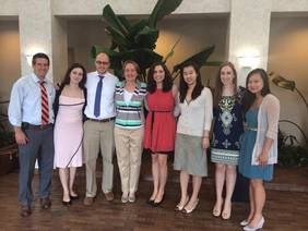
As senior editor of the current seminal textbook in dermatology, Dermatology, Jean L. Bolognia, MD, has a special place in the hearts and minds of dermatology residents, both past and present. Early on in our training, we learn that a surefire way to quickly win a debate is to start your counterargument with, “Well, Bolognia says. . . .” This respect is well garnered, as Dr. Bolognia’s book has helped elucidate difficult diagnostic and therapeutic concepts to dermatology trainees with coherent diagrams and clearly written text. It is a must-read during one’s formative education. Dr. Bolognia currently serves as professor in dermatology and vice chair of clinical affairs at Yale University in New Haven, Connecticut. During her visit to the State University of New York at Stony Brook (Figure), I sat down with Dr. Bolognia for an interview.
BRONFENBRENER: The dermatology textbook is broad and expansive. What inspired you to start this project?
BOLOGNIA: I’ll divide my answer into 2 parts: why I thought a new textbook was needed and how it came to be. At the time, I felt that many of the current comprehensive textbooks were too daunting for a first-year resident. I felt that in order to teach skin biology, for example, you needed to simplify it. The goal was simplification, at least in terms of how the content was explained to the novice reader, without sacrificing sophistication. I do believe that you need to understand basic science in order to understand the breakthroughs that will occur during your career. However, if the content is confusing or too many details are presented without schematics, at the end of the reading you are not learning. So, I wanted to teach concepts à la Scientific American, where an initial review of the schematics then makes the text much easier to understand. I thought that was a very important way to teach skin biology.
As for my early involvement in the book, I had previously organized a Web-based curriculum for medical students for the American Academy of Dermatology, which was divided into approximately 20 disease chapters, each 2 to 3 pages in length. I made sure the templates were simple and straightforward so that the authors could complete the assigned task within 2 to 3 hours. My invitation to be an editor came from the publisher based upon recommendations from some of those authors. Of note, that medical student curriculum has since been revised and is now much more refined than the original—we were version 1.0!
A few months earlier, Ron (Ronald) Rapini had approached [the publisher] with an outline for a comprehensive dermatology textbook and the publisher became interested in pursuing such a project. Joe (Joseph) Jorizzo was then asked to be the third editor. However, there was stiff competition, as we were going up against the established Fitzpatrick’s textbook. It was important to have a different approach and I was lucky that Julie Schaffer was a dermatology resident in our program at that time and she provided great critiques of the chapters.
BRONFENBRENER: When did you first realize the book was a hit?
BOLOGNIA: Well, it was fairly easy; I knew the sales numbers! That said, I think people were looking for something new and fresh. I remember one of the early reviewers likening us to USA Today! I think the Key Features were very important in making the book a success because they give first-year residents a sense of control when reading about a disease they have never heard about before. The first 6 months of dermatology residency are incredibly difficult because you need to incorporate words you never used before to talk about diseases you’ve never heard of before. I think one of my most meaningful experiences was when I visited South Africa and a young resident approached me who had photocopied the entire book and put it into individual binders so as not to desecrate his pristine original copy by writing in the margins, or underlining and highlighting. Something else I am proud of is that even if you are in a resource-poor country, you can read the book and can be on equal footing with any dermatologist from countries around the world, including ours. In other words, it is an educational equalizer.
BRONFENBRENER: What first inspired you to go into dermatology?
BOLOGNIA: I was a third-year medical student and had just finished a rotation in the neonatal intensive care unit, which was particularly demanding; I think you had more clinical responsibilities back then. I was sitting at dinner with some classmates looking for an elective to do, and they all pushed me towards dermatology. It was just what I needed, they said; you only really worked 4 days a week, and the hours were so civilized! My first Friday on the rotation I borrowed Irwin Braverman’s book Skin Signs of Systemic Disease from the clinic and read a good portion of it over the weekend. It struck a chord with me because of its multiple links to internal medicine. The next week I was in dermatology Grand Rounds, and back then the medical students were the first to present their differential diagnoses, prior to and without talking with the residents. It was a bit of sport to hear the rather imaginative diagnoses generated by the students. So when called upon, I stated that I thought my patient had histiocytosis X, and less likely dermatitis. The attending running Grand Rounds questioned me: What could possibly make me think that it was this rare entity histiocytosis X? I told him it looked just like the picture in Dr. Braverman’s book! Everyone got a chuckle out of my answer, but believe it or not, the patient actually had histiocytosis X. Nowadays I would never put histiocytosis above more common entities, but talk about beginner’s luck. I was hooked! The fact that I got the diagnosis right reflects how important a well-written book is for trainees, and I’m honored to still see patients in the same pod as Dr. Braverman.
BRONFENBRENER: What pearls would you give graduating residents now?
BOLOGNIA: I think you have to reflect on what you like to do, what resonates with you, whether you like to take care of people with contact dermatitis, or you like cutting cancer off patients’ faces, or you want to practice phototherapy. Think about where your heart is and that’s what you should do, not what your professors, or your parents, or your chairman thinks you should do. You’re going to be engaged in these activities for at least 30 years, so find something you are passionate about and build on that. Analyze your talents as well as your strengths and go with it. Another important point is to find an area—or 2, or even 3—where you do a lot of reading and then let others know of your interests. Before long you will be able to build upon your greater knowledge and clinical experience and become an expert within a few years. It doesn’t matter if you’re in an academic or private practice; there will always be a niche that you can fill and patients you can help because of your expertise.
BRONFENBRENER: What areas do you find particularly fascinating in dermatology?
BOLOGNIA: Oh, there’s so much! Patients with monoclonal gammopathies, where they can have follicular spines composed of the monoclonal protein coming out of their nose, are fascinating. That’s the kind of dermatology I like: the “believe-it-or-not” side of dermatology. Once I met a patient at a dermatology conference who had fallen down on a farm and had sustained a traumatic brain injury, after which he could only sweat on one side of his body. He had Grover disease limited to the side of his body that could sweat, while the other side was totally clear. We know Grover disease comes from a combination of sweating and sun damage, but seeing that sharp cutoff on his midline drove the point home for me.
BRONFENBRENER: How do you learn best?
BOLOGNIA: I put notes in the margins. If I go to a lecture, I use address labels for my notes. Then I take those stickers and put them in the relevant part of the textbook. It is so much easier to peel off the stickers than it is to write the information. As a result, there is a greater likelihood that the information will actually be transferred.
BRONFENBRENER: What is your daily routine?
BOLOGNIA: Monday, Tuesday, Friday I see patients. Wednesday is Grand Rounds and electronic medical record cleanup time, the paperwork that is no longer on paper. Thursday is my academic day where I can work on projects, and I like this full-day approach because I work best in larger blocks of time. That’s why I recommend, for residents especially, to get a large chunk of their reading done on Saturday or Sunday morning in 2- to 3-hour blocks. I’m not someone who rises hours before everyone else; I’m a 6:00 am type of gal. If you wake up and get your work done during a planned block of time, then you feel like you’ve accomplished something and can enjoy the rest of your day. For me, I’m freshest in the morning, no question about it.
BRONFENBRENER: Where do you think dermatology is going?
BOLOGNIA: I don’t know exactly, and I don’t think anyone knows exactly. I think we need to work hard to maintain our unique body of knowledge and to remind others of this accomplishment. I am often struck in Grand Rounds that no other specialty would have a fighting chance at generating a differential diagnosis or appropriate treatment strategy for the patients presented there. I can’t answer whether if in 10 years most dermatologists will be employees at hospitals or will still have some degree of autonomy, and I think that is unsettling.
BRONFENBRENER: What’s on the horizon for you?
BOLOGNIA: I’m working on the fourth edition of Dermatology, slated to come out in 2017. Dermatology Essentials was just published and is meant to provide a quick overview if you’re scheduled to give a talk and want a 5-minute refresher or if you’re in between patients and would like to be reminded of additional entities in the differential diagnosis. These books are accessible via the Inkling app, so you can have the entire book correctly formatted on your phone or tablet. I was recently trying to provide a more specific site for a biopsy on the ear and just couldn’t remember the correct term, but a quick search and I had the entire external ear anatomy schematic up on my phone.
BRONFENBRENER: Any favorite quotations?
BOLOGNIA: I like this one by Benjamin Franklin: “Well done is better than well said.” I judge people on their actions; not on what they say they’re going to do but rather what they’ve done.
Final Thoughts
My interview provided a glimpse into the cooperative effort required to synthesize the vast quantity of information into a well-written textbook. Dr. Bolognia is passionate about the field, finds great pleasure in unraveling mysterious patients, and goes to great lengths to make teaching points. As impressive as her accomplishments are, I found myself most inspired by her intellectual curiosity for dermatology, a character trait that successful dermatology residents should also embody. I feel fortunate to have had the opportunity to interview her, and I hope that the resident community can also benefit from the advice she has shared.
Suggested Readings
Bolognia JL, Jorizzo JL, Schaffer JV, et al, eds. Dermatology. Phildelphia, PA: WB Saunders; 2012.
Bolognia JL, Schaffer JV, Duncan KO, et al, eds. Dermatology Essentials. Philadelphia, PA: WB Saunders; 2014.
Braverman IM. Skin Signs of Systemic Disease. 3rd ed. Philadelphia, PA: WB Saunders; 1998.
Goldsmith LA, Katz SI, Gilchrist BA, et al, eds. Fitzpatrick’s Dermatology in General Medicine. 8th ed. New York, NY: McGraw Hill; 2012.
As senior editor of the current seminal textbook in dermatology, Dermatology, Jean L. Bolognia, MD, has a special place in the hearts and minds of dermatology residents, both past and present. Early on in our training, we learn that a surefire way to quickly win a debate is to start your counterargument with, “Well, Bolognia says. . . .” This respect is well garnered, as Dr. Bolognia’s book has helped elucidate difficult diagnostic and therapeutic concepts to dermatology trainees with coherent diagrams and clearly written text. It is a must-read during one’s formative education. Dr. Bolognia currently serves as professor in dermatology and vice chair of clinical affairs at Yale University in New Haven, Connecticut. During her visit to the State University of New York at Stony Brook (Figure), I sat down with Dr. Bolognia for an interview.
BRONFENBRENER: The dermatology textbook is broad and expansive. What inspired you to start this project?
BOLOGNIA: I’ll divide my answer into 2 parts: why I thought a new textbook was needed and how it came to be. At the time, I felt that many of the current comprehensive textbooks were too daunting for a first-year resident. I felt that in order to teach skin biology, for example, you needed to simplify it. The goal was simplification, at least in terms of how the content was explained to the novice reader, without sacrificing sophistication. I do believe that you need to understand basic science in order to understand the breakthroughs that will occur during your career. However, if the content is confusing or too many details are presented without schematics, at the end of the reading you are not learning. So, I wanted to teach concepts à la Scientific American, where an initial review of the schematics then makes the text much easier to understand. I thought that was a very important way to teach skin biology.
As for my early involvement in the book, I had previously organized a Web-based curriculum for medical students for the American Academy of Dermatology, which was divided into approximately 20 disease chapters, each 2 to 3 pages in length. I made sure the templates were simple and straightforward so that the authors could complete the assigned task within 2 to 3 hours. My invitation to be an editor came from the publisher based upon recommendations from some of those authors. Of note, that medical student curriculum has since been revised and is now much more refined than the original—we were version 1.0!
A few months earlier, Ron (Ronald) Rapini had approached [the publisher] with an outline for a comprehensive dermatology textbook and the publisher became interested in pursuing such a project. Joe (Joseph) Jorizzo was then asked to be the third editor. However, there was stiff competition, as we were going up against the established Fitzpatrick’s textbook. It was important to have a different approach and I was lucky that Julie Schaffer was a dermatology resident in our program at that time and she provided great critiques of the chapters.
BRONFENBRENER: When did you first realize the book was a hit?
BOLOGNIA: Well, it was fairly easy; I knew the sales numbers! That said, I think people were looking for something new and fresh. I remember one of the early reviewers likening us to USA Today! I think the Key Features were very important in making the book a success because they give first-year residents a sense of control when reading about a disease they have never heard about before. The first 6 months of dermatology residency are incredibly difficult because you need to incorporate words you never used before to talk about diseases you’ve never heard of before. I think one of my most meaningful experiences was when I visited South Africa and a young resident approached me who had photocopied the entire book and put it into individual binders so as not to desecrate his pristine original copy by writing in the margins, or underlining and highlighting. Something else I am proud of is that even if you are in a resource-poor country, you can read the book and can be on equal footing with any dermatologist from countries around the world, including ours. In other words, it is an educational equalizer.
BRONFENBRENER: What first inspired you to go into dermatology?
BOLOGNIA: I was a third-year medical student and had just finished a rotation in the neonatal intensive care unit, which was particularly demanding; I think you had more clinical responsibilities back then. I was sitting at dinner with some classmates looking for an elective to do, and they all pushed me towards dermatology. It was just what I needed, they said; you only really worked 4 days a week, and the hours were so civilized! My first Friday on the rotation I borrowed Irwin Braverman’s book Skin Signs of Systemic Disease from the clinic and read a good portion of it over the weekend. It struck a chord with me because of its multiple links to internal medicine. The next week I was in dermatology Grand Rounds, and back then the medical students were the first to present their differential diagnoses, prior to and without talking with the residents. It was a bit of sport to hear the rather imaginative diagnoses generated by the students. So when called upon, I stated that I thought my patient had histiocytosis X, and less likely dermatitis. The attending running Grand Rounds questioned me: What could possibly make me think that it was this rare entity histiocytosis X? I told him it looked just like the picture in Dr. Braverman’s book! Everyone got a chuckle out of my answer, but believe it or not, the patient actually had histiocytosis X. Nowadays I would never put histiocytosis above more common entities, but talk about beginner’s luck. I was hooked! The fact that I got the diagnosis right reflects how important a well-written book is for trainees, and I’m honored to still see patients in the same pod as Dr. Braverman.
BRONFENBRENER: What pearls would you give graduating residents now?
BOLOGNIA: I think you have to reflect on what you like to do, what resonates with you, whether you like to take care of people with contact dermatitis, or you like cutting cancer off patients’ faces, or you want to practice phototherapy. Think about where your heart is and that’s what you should do, not what your professors, or your parents, or your chairman thinks you should do. You’re going to be engaged in these activities for at least 30 years, so find something you are passionate about and build on that. Analyze your talents as well as your strengths and go with it. Another important point is to find an area—or 2, or even 3—where you do a lot of reading and then let others know of your interests. Before long you will be able to build upon your greater knowledge and clinical experience and become an expert within a few years. It doesn’t matter if you’re in an academic or private practice; there will always be a niche that you can fill and patients you can help because of your expertise.
BRONFENBRENER: What areas do you find particularly fascinating in dermatology?
BOLOGNIA: Oh, there’s so much! Patients with monoclonal gammopathies, where they can have follicular spines composed of the monoclonal protein coming out of their nose, are fascinating. That’s the kind of dermatology I like: the “believe-it-or-not” side of dermatology. Once I met a patient at a dermatology conference who had fallen down on a farm and had sustained a traumatic brain injury, after which he could only sweat on one side of his body. He had Grover disease limited to the side of his body that could sweat, while the other side was totally clear. We know Grover disease comes from a combination of sweating and sun damage, but seeing that sharp cutoff on his midline drove the point home for me.
BRONFENBRENER: How do you learn best?
BOLOGNIA: I put notes in the margins. If I go to a lecture, I use address labels for my notes. Then I take those stickers and put them in the relevant part of the textbook. It is so much easier to peel off the stickers than it is to write the information. As a result, there is a greater likelihood that the information will actually be transferred.
BRONFENBRENER: What is your daily routine?
BOLOGNIA: Monday, Tuesday, Friday I see patients. Wednesday is Grand Rounds and electronic medical record cleanup time, the paperwork that is no longer on paper. Thursday is my academic day where I can work on projects, and I like this full-day approach because I work best in larger blocks of time. That’s why I recommend, for residents especially, to get a large chunk of their reading done on Saturday or Sunday morning in 2- to 3-hour blocks. I’m not someone who rises hours before everyone else; I’m a 6:00 am type of gal. If you wake up and get your work done during a planned block of time, then you feel like you’ve accomplished something and can enjoy the rest of your day. For me, I’m freshest in the morning, no question about it.
BRONFENBRENER: Where do you think dermatology is going?
BOLOGNIA: I don’t know exactly, and I don’t think anyone knows exactly. I think we need to work hard to maintain our unique body of knowledge and to remind others of this accomplishment. I am often struck in Grand Rounds that no other specialty would have a fighting chance at generating a differential diagnosis or appropriate treatment strategy for the patients presented there. I can’t answer whether if in 10 years most dermatologists will be employees at hospitals or will still have some degree of autonomy, and I think that is unsettling.
BRONFENBRENER: What’s on the horizon for you?
BOLOGNIA: I’m working on the fourth edition of Dermatology, slated to come out in 2017. Dermatology Essentials was just published and is meant to provide a quick overview if you’re scheduled to give a talk and want a 5-minute refresher or if you’re in between patients and would like to be reminded of additional entities in the differential diagnosis. These books are accessible via the Inkling app, so you can have the entire book correctly formatted on your phone or tablet. I was recently trying to provide a more specific site for a biopsy on the ear and just couldn’t remember the correct term, but a quick search and I had the entire external ear anatomy schematic up on my phone.
BRONFENBRENER: Any favorite quotations?
BOLOGNIA: I like this one by Benjamin Franklin: “Well done is better than well said.” I judge people on their actions; not on what they say they’re going to do but rather what they’ve done.
Final Thoughts
My interview provided a glimpse into the cooperative effort required to synthesize the vast quantity of information into a well-written textbook. Dr. Bolognia is passionate about the field, finds great pleasure in unraveling mysterious patients, and goes to great lengths to make teaching points. As impressive as her accomplishments are, I found myself most inspired by her intellectual curiosity for dermatology, a character trait that successful dermatology residents should also embody. I feel fortunate to have had the opportunity to interview her, and I hope that the resident community can also benefit from the advice she has shared.
Suggested Readings
Bolognia JL, Jorizzo JL, Schaffer JV, et al, eds. Dermatology. Phildelphia, PA: WB Saunders; 2012.
Bolognia JL, Schaffer JV, Duncan KO, et al, eds. Dermatology Essentials. Philadelphia, PA: WB Saunders; 2014.
Braverman IM. Skin Signs of Systemic Disease. 3rd ed. Philadelphia, PA: WB Saunders; 1998.
Goldsmith LA, Katz SI, Gilchrist BA, et al, eds. Fitzpatrick’s Dermatology in General Medicine. 8th ed. New York, NY: McGraw Hill; 2012.

New Sunscreen Guidelines: What Your Patients Need to Know
For more information, access Dr. Bronfenbrener's resident corner column, "Simplifying Sun Safety: A Guide to the New FDA Sunscreen Monograph."
For more information, access Dr. Bronfenbrener's resident corner column, "Simplifying Sun Safety: A Guide to the New FDA Sunscreen Monograph."
For more information, access Dr. Bronfenbrener's resident corner column, "Simplifying Sun Safety: A Guide to the New FDA Sunscreen Monograph."

Giving a Good Needle: Resident Guide to Decreasing Injection Pain
Think back to a busy day and try to recall how many biopsies you performed. Chances are during that time you administered several dozen injections for various indications. Although we have become accustomed to performing injections through repetition, it may be the focus of many patient visits and may be the basis on which a patient judges his/her physician. There are various techniques and products available that can help decrease the physical and psychological burden of injections for patients, some that should be incorporated into a resident’s repertoire to be perfected before becoming an attending.
Factors Related to Anesthetics
The most frequently used local anesthetic in dermatology clinics is lidocaine (1% or 2%) combined with epinephrine 1:100,000. This premixed formulation relieves the burden of mixing for nurses; however, its low pH (4.2) contributes to stinging and burning with infiltration.1 Buffering with sodium bicarbonate 8.4% in a 9:1 ratio (9 parts lidocaine-epinephrine to 1 part bicarbonate) more closely matches the neutral pH in human tissues and decreases injection pain.2 Alkalinizing the anesthetic mixture also decreases the time of onset of its effects, as higher pH solutions convert lidocaine into its active unionized form. However, buffering the anesthetic does have the drawback of decreasing its shelf life, and many clinics no longer store buffered solutions for fear of spoilage. It can be useful to prepare a freshly buffered mixture prior to injecting a particularly needlephobic patient or when injecting in a difficult anatomic location.
In keeping with the philosophy that infiltrating with a solution that closely mimics physiologic parameters minimizes discomfort, a recent meta-analysis found that warming the anesthetic prior to injection led to less pain.3 In my experience, I have found that rolling the syringe between my hands prior to injection also decreases the patient’s sensation of “feeling the anesthetic going in.”
Preinjection Preparation
Properly positioning the patient is paramount to safe injection. Murphy’s Law should be anticipated, not discovered. A few moments spent adjusting the chair and lighting can pay dividends if a patient suddenly has a vasovagal episode. Unfortunately, it is difficult to predict which patients are prone to such attacks, as even a patient who may spend hours playing football in the summer heat could collapse at the sight of a needle. Aside from proper positioning of the patient, the biopsy tray should not be in the patient’s direct line of sight. Even those who tolerate the anesthesia well may become distraught at the sight of bloody gauze.
There are several options for topical anesthesia to decrease injection pain. Cream or gel preparations (ie, eutectic mixture of lidocaine and prilocaine, lidocaine cream, tetracaine gel) generally are cumbersome in a busy clinic setting, as they require at least 30 minutes of contact before anesthesia is achieved; a longer duration of exposure provides further anesthetization and may improve patient outcomes.4 However, these formulations may be useful in planned procedures. I have found much utility in utilizing ethyl chloride vapocoolant spray as a numbing agent with an immediate onset of action, a feature that makes this product useful in busy clinics.5 Ice is another excellent local coolant and is readily available in most offices at a negligible cost. Placing the ice in aluminum foil instead of a glove delivers more rapid cooling, and the ice is safe to use on areas where vapocoolant spray may be inconvenient or contraindicated, such as around the eyes, nose, ears, or mouth. Holding the ice in place for approximately 10 seconds prior to injection numbs superficial nerve endings and facilitates painless needle insertion.6
Injection Technique
Injection technique is arguably the most important factor in minimizing pain for patients and ensuring effective anesthesia in the field. It also is the factor that a patient will either praise or blame, depending on their perception of the injection.
An important point is that the initial injection should be done perpendicular to the skin. The superficial skin has the highest concentration of nerve endings, which branch repeatedly from larger stems in the deeper dermis and subcutaneous fat. Tangential injections disrupt a relatively larger number of nerve endings as the needle tracks through more superficial skin. By injecting perpendicularly, you minimize damage done during the needle’s plunge.2
Anesthetic should initially be deposited into the subcutaneous fat and continued as the needle is withdrawn. Injection directly into the dense dermis leads to pain with hydrodissection, while deeper placement is less painful due to the malleability of fat and a decreased concentration of nerve endings.7
Subsequent injections should be strategically placed. Ideally, the initial injection should be the only one that the patient feels, with widening of the anesthetic field achieved by slowly infiltrating lidocaine through skin that is already numb. The needle should be inserted into the wheal and advanced slowly with continuous pressure on the plunger; special attention should be paid to avoid advancing the needle tip past the leading edge of the wheal and into skin with intact sensation (areas of skin that have not yet been numbed by anesthesia and therefore are still capable of sensing pain from injection).8 This method of delivering anesthesia with only one initial prick experienced by the patient has been coined the “hole-in-one” technique and has proven to be not only efficacious in minimizing injection discomfort but also easy to learn, even for amateur injectors.9
Distraction Techniques
Although patients should be made aware of the injection sites and reasonable expectations should be set, distraction techniques are easy to implement and can be valuable in directing the patient’s focus away from the anticipated injection. A patient’s clothing, jewelry, and body art; reading material; and the weather are examples of topics that can be discussed as “talkesthesia.” For younger patients, various distraction techniques have been developed, ranging from basic distraction with stickers or toys to increasingly sophisticated methods such as virtual reality glasses worn during the procedure.10 In actual practice, I prefer to use available technology that is inexpensive and already familiar to the child; for instance, allowing children to watch their favorite short Web videos or play a video game during the procedure, as long as the biopsy site allows it, is an ideal adjunct to proper topical anesthesia and good injection technique. For adults, quickly plugging in their favorite musical artist to an Internet radio application also can allevi-
ate anxiety.
Several distraction techniques also can be administered directly at the injection site based on the gate control theory of pain. Tactile stimuli delivered proximal to the injection site creates sensory “noise” at the level of the spinal cord, masking the noxious sensation carried on unmyelinated C fibers.7 If you have ever hit your head on an open cabinet, for instance, the instinctual desire to immediately rub the area to reduce the pain functions on this same premise. Various tactics may be employed, including stretching or pinching the skin, rubbing, and tapping; however, I find that the major limitation to many of these methods is performing them safely. An inadvertent stick may be more likely if your fingers are dancing in the path of the needle. Generally, I like to rub the skin about 3- to 5-cm proximal to the injection site with the index finger of the supporting hand as the needle is inserted and continue while the deposit of anesthetic is placed. A potentially safer option is a small handheld massager, though concerns about sterility and durability with autoclaving may limit its use to select patients.11
Special Patient Considerations
Although we do our best to control the operator-dependent aspects of injection pain, patient factors also can complicate the administration of adequate anesthesia and occasionally can lead to unintended surprises. Patients with red hair whose characteristic locks are the result of mutations in the melanocortin 1receptor have been identified as being resistant to the effects of local anesthesia, putatively related to the role of melanocortin 1 receptor in pain modulation.12 This discovery highlights the fact that there likely are numerous undiscovered mutations that cause resistance to anesthetic agents. It further underscores the need to ensure adequate numbing by testing the patient’s level of sensation prior to beginning any procedure.
Conclusion
By keeping your patient’s comfort in mind, you will not only enhance their confidence in you as their physician but will also encourage good follow-up care and adherence to any prescribed protocols. In the future, needleless injection devices and more rapid topical anesthesia may further decrease pain associated with dermatologic procedures.13
1. Frank SG, Lalonde DH. How acidic is the lidocaine we are injecting, and how much bicarbonate should we add? Can J Plast Surg. 2012;20:71-73.
2. Zilinsky I, Bar-Meir E, Zaslansky R, et al. Ten commandments for minimal pain during administration of local anesthetics. J Drugs Dermatol. 2005;4:212-216.
3. Hogan ME, vanderVaart S, Perampaladas K, et al. Systematic review and meta-analysis of the effect of warming local anesthetics on injection pain. Ann Emerg Med. 2011;58:86-98.
4. Smith DP, Gjellum M. The efficacy of LMX versus EMLA for pain relief in boys undergoing office meatotomy. J Urol. 2004;172:1760-1761.
5. Farion KJ, Splinter KL, Newhook K, et al. The effect of vapocoolant spray on pain due to intravenous cannulation in children: a randomized controlled trial. CMAJ. 2008;179:31-36.
6. Dixit S, Lowe P, Fischer G, et al. Ice anaesthesia in procedural dermatology. Australas J Dermatol. 2013;54:273-276.
7. Strazar AR, Leynes PG, Lalonde DH. Minimizing the pain of local anesthesia injection. Plast Reconstr Surg. 2013;132:675-684.
8. Lalonde DH. “Hole-in-one” local anesthesia for wide-awake carpal tunnel surgery. Plast Reconstr Surg. 2010;126:1642-1644.
9. Farhangkhoee H, Lalonde J, Lalonde DH. Teaching medical students and residents how to inject local anesthesia almost painlessly. Can J Plast Surg. 2012;20:169-172.
10. Koller D, Goldman RD. Distraction techniques for children undergoing procedures: a critical review of pediatric research. J Pediatr Nurs. 2012;27:652-681.
11. Nanitsos E, Vartuli R, Forte A, et al. The effect of vibration on pain during local anaesthesia injections. Aust Dent J. 2009;54:94-100.
12. Liem EB, Joiner TV, Tsueda K, et al. Increased sensitivity to thermal pain and reduced subcutaneous lidocaine efficacy in redheads. Anesthesiol. 2005;102:509-514.
13. Patakfalvi L, Benohanian A. Needle-free anesthesia, a promising option for the needle phobic patient. Br J Dermatol. 2014;170:1191-1192.
Think back to a busy day and try to recall how many biopsies you performed. Chances are during that time you administered several dozen injections for various indications. Although we have become accustomed to performing injections through repetition, it may be the focus of many patient visits and may be the basis on which a patient judges his/her physician. There are various techniques and products available that can help decrease the physical and psychological burden of injections for patients, some that should be incorporated into a resident’s repertoire to be perfected before becoming an attending.
Factors Related to Anesthetics
The most frequently used local anesthetic in dermatology clinics is lidocaine (1% or 2%) combined with epinephrine 1:100,000. This premixed formulation relieves the burden of mixing for nurses; however, its low pH (4.2) contributes to stinging and burning with infiltration.1 Buffering with sodium bicarbonate 8.4% in a 9:1 ratio (9 parts lidocaine-epinephrine to 1 part bicarbonate) more closely matches the neutral pH in human tissues and decreases injection pain.2 Alkalinizing the anesthetic mixture also decreases the time of onset of its effects, as higher pH solutions convert lidocaine into its active unionized form. However, buffering the anesthetic does have the drawback of decreasing its shelf life, and many clinics no longer store buffered solutions for fear of spoilage. It can be useful to prepare a freshly buffered mixture prior to injecting a particularly needlephobic patient or when injecting in a difficult anatomic location.
In keeping with the philosophy that infiltrating with a solution that closely mimics physiologic parameters minimizes discomfort, a recent meta-analysis found that warming the anesthetic prior to injection led to less pain.3 In my experience, I have found that rolling the syringe between my hands prior to injection also decreases the patient’s sensation of “feeling the anesthetic going in.”
Preinjection Preparation
Properly positioning the patient is paramount to safe injection. Murphy’s Law should be anticipated, not discovered. A few moments spent adjusting the chair and lighting can pay dividends if a patient suddenly has a vasovagal episode. Unfortunately, it is difficult to predict which patients are prone to such attacks, as even a patient who may spend hours playing football in the summer heat could collapse at the sight of a needle. Aside from proper positioning of the patient, the biopsy tray should not be in the patient’s direct line of sight. Even those who tolerate the anesthesia well may become distraught at the sight of bloody gauze.
There are several options for topical anesthesia to decrease injection pain. Cream or gel preparations (ie, eutectic mixture of lidocaine and prilocaine, lidocaine cream, tetracaine gel) generally are cumbersome in a busy clinic setting, as they require at least 30 minutes of contact before anesthesia is achieved; a longer duration of exposure provides further anesthetization and may improve patient outcomes.4 However, these formulations may be useful in planned procedures. I have found much utility in utilizing ethyl chloride vapocoolant spray as a numbing agent with an immediate onset of action, a feature that makes this product useful in busy clinics.5 Ice is another excellent local coolant and is readily available in most offices at a negligible cost. Placing the ice in aluminum foil instead of a glove delivers more rapid cooling, and the ice is safe to use on areas where vapocoolant spray may be inconvenient or contraindicated, such as around the eyes, nose, ears, or mouth. Holding the ice in place for approximately 10 seconds prior to injection numbs superficial nerve endings and facilitates painless needle insertion.6
Injection Technique
Injection technique is arguably the most important factor in minimizing pain for patients and ensuring effective anesthesia in the field. It also is the factor that a patient will either praise or blame, depending on their perception of the injection.
An important point is that the initial injection should be done perpendicular to the skin. The superficial skin has the highest concentration of nerve endings, which branch repeatedly from larger stems in the deeper dermis and subcutaneous fat. Tangential injections disrupt a relatively larger number of nerve endings as the needle tracks through more superficial skin. By injecting perpendicularly, you minimize damage done during the needle’s plunge.2
Anesthetic should initially be deposited into the subcutaneous fat and continued as the needle is withdrawn. Injection directly into the dense dermis leads to pain with hydrodissection, while deeper placement is less painful due to the malleability of fat and a decreased concentration of nerve endings.7
Subsequent injections should be strategically placed. Ideally, the initial injection should be the only one that the patient feels, with widening of the anesthetic field achieved by slowly infiltrating lidocaine through skin that is already numb. The needle should be inserted into the wheal and advanced slowly with continuous pressure on the plunger; special attention should be paid to avoid advancing the needle tip past the leading edge of the wheal and into skin with intact sensation (areas of skin that have not yet been numbed by anesthesia and therefore are still capable of sensing pain from injection).8 This method of delivering anesthesia with only one initial prick experienced by the patient has been coined the “hole-in-one” technique and has proven to be not only efficacious in minimizing injection discomfort but also easy to learn, even for amateur injectors.9
Distraction Techniques
Although patients should be made aware of the injection sites and reasonable expectations should be set, distraction techniques are easy to implement and can be valuable in directing the patient’s focus away from the anticipated injection. A patient’s clothing, jewelry, and body art; reading material; and the weather are examples of topics that can be discussed as “talkesthesia.” For younger patients, various distraction techniques have been developed, ranging from basic distraction with stickers or toys to increasingly sophisticated methods such as virtual reality glasses worn during the procedure.10 In actual practice, I prefer to use available technology that is inexpensive and already familiar to the child; for instance, allowing children to watch their favorite short Web videos or play a video game during the procedure, as long as the biopsy site allows it, is an ideal adjunct to proper topical anesthesia and good injection technique. For adults, quickly plugging in their favorite musical artist to an Internet radio application also can allevi-
ate anxiety.
Several distraction techniques also can be administered directly at the injection site based on the gate control theory of pain. Tactile stimuli delivered proximal to the injection site creates sensory “noise” at the level of the spinal cord, masking the noxious sensation carried on unmyelinated C fibers.7 If you have ever hit your head on an open cabinet, for instance, the instinctual desire to immediately rub the area to reduce the pain functions on this same premise. Various tactics may be employed, including stretching or pinching the skin, rubbing, and tapping; however, I find that the major limitation to many of these methods is performing them safely. An inadvertent stick may be more likely if your fingers are dancing in the path of the needle. Generally, I like to rub the skin about 3- to 5-cm proximal to the injection site with the index finger of the supporting hand as the needle is inserted and continue while the deposit of anesthetic is placed. A potentially safer option is a small handheld massager, though concerns about sterility and durability with autoclaving may limit its use to select patients.11
Special Patient Considerations
Although we do our best to control the operator-dependent aspects of injection pain, patient factors also can complicate the administration of adequate anesthesia and occasionally can lead to unintended surprises. Patients with red hair whose characteristic locks are the result of mutations in the melanocortin 1receptor have been identified as being resistant to the effects of local anesthesia, putatively related to the role of melanocortin 1 receptor in pain modulation.12 This discovery highlights the fact that there likely are numerous undiscovered mutations that cause resistance to anesthetic agents. It further underscores the need to ensure adequate numbing by testing the patient’s level of sensation prior to beginning any procedure.
Conclusion
By keeping your patient’s comfort in mind, you will not only enhance their confidence in you as their physician but will also encourage good follow-up care and adherence to any prescribed protocols. In the future, needleless injection devices and more rapid topical anesthesia may further decrease pain associated with dermatologic procedures.13
1. Frank SG, Lalonde DH. How acidic is the lidocaine we are injecting, and how much bicarbonate should we add? Can J Plast Surg. 2012;20:71-73.
2. Zilinsky I, Bar-Meir E, Zaslansky R, et al. Ten commandments for minimal pain during administration of local anesthetics. J Drugs Dermatol. 2005;4:212-216.
3. Hogan ME, vanderVaart S, Perampaladas K, et al. Systematic review and meta-analysis of the effect of warming local anesthetics on injection pain. Ann Emerg Med. 2011;58:86-98.
4. Smith DP, Gjellum M. The efficacy of LMX versus EMLA for pain relief in boys undergoing office meatotomy. J Urol. 2004;172:1760-1761.
5. Farion KJ, Splinter KL, Newhook K, et al. The effect of vapocoolant spray on pain due to intravenous cannulation in children: a randomized controlled trial. CMAJ. 2008;179:31-36.
6. Dixit S, Lowe P, Fischer G, et al. Ice anaesthesia in procedural dermatology. Australas J Dermatol. 2013;54:273-276.
7. Strazar AR, Leynes PG, Lalonde DH. Minimizing the pain of local anesthesia injection. Plast Reconstr Surg. 2013;132:675-684.
8. Lalonde DH. “Hole-in-one” local anesthesia for wide-awake carpal tunnel surgery. Plast Reconstr Surg. 2010;126:1642-1644.
9. Farhangkhoee H, Lalonde J, Lalonde DH. Teaching medical students and residents how to inject local anesthesia almost painlessly. Can J Plast Surg. 2012;20:169-172.
10. Koller D, Goldman RD. Distraction techniques for children undergoing procedures: a critical review of pediatric research. J Pediatr Nurs. 2012;27:652-681.
11. Nanitsos E, Vartuli R, Forte A, et al. The effect of vibration on pain during local anaesthesia injections. Aust Dent J. 2009;54:94-100.
12. Liem EB, Joiner TV, Tsueda K, et al. Increased sensitivity to thermal pain and reduced subcutaneous lidocaine efficacy in redheads. Anesthesiol. 2005;102:509-514.
13. Patakfalvi L, Benohanian A. Needle-free anesthesia, a promising option for the needle phobic patient. Br J Dermatol. 2014;170:1191-1192.

Denver AAD 2014

With the annual meeting of the American Academy of Dermatology (AAD) set in the “mile-high city” now behind us, we must begin to convert didactic sessions into improved outcomes in our daily patient encounters. The enormous variety of lectures and frequently overlapping schedules can make this event a whirlwind for unseasoned attendees. I aim to enlighten those attending future meetings about individual sessions of particularly high value to the dermatologist in training. As a disclaimer, my primary interests are in medical dermatology, so the content of the courses I recommend is by no means comprehensive; however, residents need to have a solid fund of medical knowledge to function in any practice setting and, perhaps more importantly, to pass the boards examination!
I think the session that takes the cake for utility and value for residents is “High Yield ‘Power Hour’ for Residents,” which was led by a group of education-oriented Harvard University dermatologists. The power “hour” in fact lasted 2 hours, during which there was a variety of material presented covering pediatric dermatology, allergic dermatitis, infectious disease dermatology, blistering disorders, and pharmacology. The presenters showed incredible enthusiasm for their respective topics, and their passion also was evident in the high-yield handouts that they provided that were jam-packed with tables, bullet points, and frequently tested material. I would recommend that attendees save or print the handouts and avoid taking extensive notes on them during the session. Sit back, relax, and just soak in the lectures; later on, review the handouts. Also, be sure to arrive early—this session fills up fast—and fill out your evaluation! These lecturers deserve credit for their presentations and deserve a much larger room to accommodate residents that are otherwise willing to sit on the floor, crowd against the walls, and peek in through the doorway to listen.
I also feel residents benefit from lectures that provide us with practical information regarding complicated medical problems. I greatly enjoyed the symposium “Biologics: Perils and Promise” led by Canadian Dermatology Association President Richard Langley, MD. This conglomerate of experts addressed the risk for infection and malignancy with the use of biologics as well as pediatric use. The symposium also touched on the cardiovascular risk inherent in psoriasis and new developments suggesting that the treatment of psoriasis decreases overall systemic inflammation and possibly even cardiovascular risk. Another symposium in the same vein was “Systemic Therapies for Dermatologists: A Comprehensive Review and Update,” which was divided into short lectures discussing agents from acitretin to Zelboraf (vemurafenib). Expert insight on the use of these drugs was invaluable for those of us who train in programs where we are not frequently exposed to these agents.
For those residents interested in an overview of dermatopathology, “Dermatopathology Made Simple” led by Christine Ko, MD, was a lightning-fast tour through the subject. Dr. Ko categorized diagnoses based on pattern and also compared similar pathology side-by-side, focusing on key differences to help cinch the correct diagnosis. Although this talk was only 2 hours, it covered surprising breadth, as more than 170 different cases were presented. The handout accompanying this talk was excellent and served as a condensed review of all the material she covered.
Any resident attending an AAD meeting should first review the schedule carefully and then find topics that would be most beneficial during training and later on during practice. The recommendations I have made summarize material that will benefit all trainees in dermatology. Even those of us who will be continuing on to a fellowship need to have a handle on these topics to remain current and better communicate with colleagues. I look forward to seeing you all at future meetings!