Article
Collagenase Enzymatic Fasciotomy for Dupuytren Contracture in Patients on Chronic Immunosuppression
Collagenase enzymatic fasciotomy is an accepted nonsurgical treatment for disabling hand contractures caused by Dupuytren disease. We conducted a...
Article
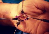
Open Carpal Tunnel Release With Use of a Nasal Turbinate Speculum
Incomplete release of the transverse carpal ligament (TCL) and median nerve injury are complications of carpal tunnel release (CTR). In this...