Article
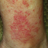
Exercise-Induced Vasculitis in a Patient With Negative Ultrasound Venous Reflux Study: A Mimic of Stasis Dermatitis
Exercise-induced vasculitis largely is documented in photographs or by history and may be misdiagnosed as stasis dermatitis due to its clinical...
Article
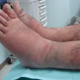
Severe Pretibial Myxedema Refractory to Systemic Immunosuppressants
Pretibial myxedema (PM) is a known manifestation of Graves disease that almost always occurs in the presence of Graves ophthalmopathy. The...
Article
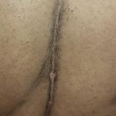
Linear Terra Firma–Forme Dermatosis of the Midline Back
Terra firma–forme dermatosis (TFFD) is a benign and likely underdiagnosed disorder with relatively few reports in the literature. A 46-year-old...
Article

The Use of Sodium Sulfacetamide in Dermatology
Sodium sulfacetamide is effective in the management of a variety of inflammatory facial dermatoses and often is used in combination with sulfur...