Article
Enhanced Care for Pediatric Patients With Generalized Lichen Planus: Diagnosis and Treatment Tips
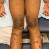
The rarity of generalized lichen planus (LP) in children often leads to misdiagnosis or delayed treatment, impacting the patient’...
The rarity of generalized lichen planus (LP) in children often leads to misdiagnosis or delayed treatment, impacting the patient’...