Article
Epidermolysis Bullosa Acquisita in Association With Mantle Cell Lymphoma
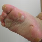
A 46-year-old man presented with multiple tense bullae and denuded patches on the palms (Figure 1A) and soles (Figure 1B). The blisters first...
Article
Rash on a Man's Hand; Itchy Lower-Body Lesions on a Woman
A 34-year-old man requests evaluation of a rash affecting his dorsal proximal right thumb and right wrist. A 35-year-old woman presents for...
Article
A Plantar Bullous Lesion, and a Case of Extranumerary Digits
What treatment recommendations can you offer for each patient?
Article
An Erythematous Plaque With a Clear Center, and a Case of Back Excoriations
Can you find the underlying cause for each of these conditions?
Article
Two Patients Have Erythematous Patches: Will the Diagnosis Differ?
Annular, erythematous patches and plaques prompt a woman with a family history of diabetes to seek a diagnosis. An 18-year-old has shiny...
Article
Foot Problems in Two Patients
A 23-year-old has eruptions on the soles of her feet and hands. An elderly man has a painful growth beneath his fifth toe. What is the diagnosis...
Article
An Eyelid Growth Affects Vision
A man presents with a 2-year history of a rash near his umbilicus; a woman requests removal of a growth on her left eyelid.
Article
Back-to-Back Dermatologic Challenges
Two women, two back lesions—what is the diagnosis in each case, and is one more worrisome?
Article
Crusted Nodule on Chin
An older woman presents with a crusted nodule on her chin; another woman has a large, linear rash affecting her right leg.
Article
Oozing and Enlarging Scalp Lesion
A man has an eruption on his nose; another man has an oozing, growing scalp lesion.
Article
Malodorous Rash
A woman has a malodorous rash; another woman has a large nodule on her forehead.
Article
A Young Man With a Concerning Mole
A young man is concerned about a mole that has been changing; a woman has a rash below her shoulder blades.
Article
Why Did Eating Ahi Cause Intense Erythema?
A man has intense upper-body erythema after eating ahi; another man’s skin has a fish-scale appearance.
Article
Painful Finger Lesion
A man in police custody has a burn on his thigh; a woman has a painful finger lesion.
Article
Eyelid With Vesicles
A boy’s facial rash is worsening; a woman’s eyelid has vesicles.