Article
Diagnosis at a Glance
A 56-year-old woman presents to the urgent care center with an ovoidshaped lump on the medial band of the plantar fascia; a 77-year-old man...
Article
Extensive Pruritic Rash; Itchy Lesion on the Ankle
A mother seeks consultation for her 3-year-old daughter, who presents with an extensive, mildly pruritic rash on her face, trunk, and extremities...
Article
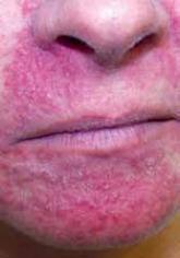
Penile Lesion; Papules on Forehead and Cheeks
A 46-year-old sexually active, divorced man presents to the ED shortly after midnight for evaluation of a sore on his penis. A 69-year-old man...
Article
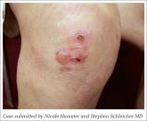
Rapidly Enlarging Lesion on Right Shoulder
A female patient in her early 20s has had a fluctuating rash on her neck and torso for seven years; another woman, 64, is concerned about an...
Article
Enlarging Scalp Lesion
A 67-year-old patient has an enlarging, dome-shaped nodule on his scalp; an infant is brought in with an underdeveloped auricle.
Article
Painful Bumps on Right Ankle
An 83-year-old patient displays firm, erythematous papules on her right ankle that she describes as persistent and painful; another elderly woman...