Article
Small renal masses: Toward more rational treatment
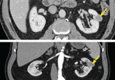
Although surgical removal of the whole kidney has long been the most common treatment, nephron-sparing approaches are now favored.
Although surgical removal of the whole kidney has long been the most common treatment, nephron-sparing approaches are now favored.