Article
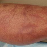
Flagellate Shiitake Mushroom Reaction With Histologic Features of Acute Generalized Exanthematous Pustulosis
Ingestion of shiitake mushrooms and bleomycin is associated with flagellate dermatitis.
Article
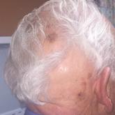
Remote-Onset Alopecia Areata Attributed to Ipilimumab
Cutaneous immune-related adverse effects (irAEs) are among the most common adverse effects of ipilimumab used to treat advanced-stage melanoma....