Article
Bullous Systemic Lupus Erythematosus Successfully Treated With Rituximab
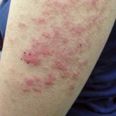
Bullous systemic lupus erythematosus can present with a waxing and waning course punctuated by flares. This case illustrates the role of rituximab...
Article
Heparin-Induced Bullous Hemorrhagic Dermatosis Confined to the Oral Mucosa
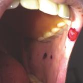
It is important for physicians to recognize the clinical appearance of cutaneous adverse reactions to heparin, including bullous hemorrhagic...