Article
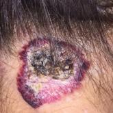
Large Hemorrhagic Plaque With Central Crusting
A 54-year-old woman presented with a large hemorrhagic plaque on the left side of the posterior neck with central brown-yellow crusting and a few...
A 54-year-old woman presented with a large hemorrhagic plaque on the left side of the posterior neck with central brown-yellow crusting and a few...