Article
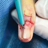
Customized Dermal Curette: An Alternative and Effective Shaving Tool in Nail Surgery
Surgical excision of the pigmented nail matrix followed by histopathologic examination is a common procedure aimed at managing longitudinal...
Article

Affixing a Scalp Dressing With Hairpins
We describe a method for using hairpins to affix a dressing after scalp surgery or trauma that is sturdy, does not compromise aesthetics, and does...