Article
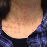
Rapid Development of Perifolliculitis Following Mesotherapy
Mesotherapy is a common procedure performed in both medical and nomedical settings for cosmetic rejuvenation. Complications can occur from...
Article
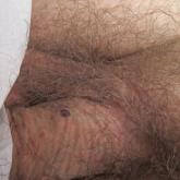
Multiple Eruptive Syringomas on the Penis
Penile syringoma can mimic sexually transmitted disease such as condyloma acuminatum or molluscum contagiosum. To prevent misdiagnosis and...