Opinion

Wave, surge, or tsunami
Predictive models for COVID-19 have strengths and weaknesses and none are perfect.
News

Doing things right vs. doing the right things
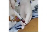
There has been inconsistent guidance in regard to the containment of COVID-19 within health care systems.
Article
A woman, age 35, with new-onset ascites
She also has jaundice, hepatomegaly, and multiple abnormal laboratory results. What is the next step?
News
How should asymptomatic hypertension be managed in the hospital?
The appropriate treatment of hypertension in the inpatient setting is suboptimal due to the lack of guidelines and inconsistent management.
Article
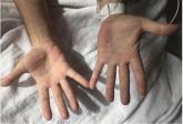
Methemoglobinemia in an HIV patient
He had restarted his home dapsone prophylaxis, but his dyspnea worsened and his urine became dark.