User login
LIVERPOOL, ENGLAND – A novel radiologic scoring system differentiated psoriatic arthritis (PsA) from nodal osteoarthritis (OA) of the hand in a pilot study.
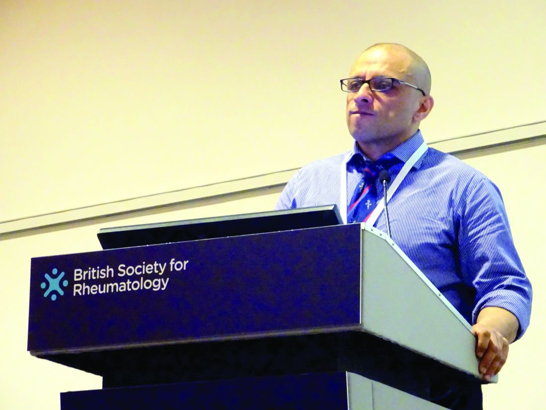
“It’s a dilemma that’s faced, perhaps every couple of weeks, by most [rheumatologists]: Is it osteoarthritis or is it early psoriatic arthritis?” said Sardar Bahadur, MD, at the British Society for Rheumatology annual conference.
Both conditions are seen in daily practice, although the prevalence of hand OA is less frequent than knee OA. Approximately one in five of all adults in the United Kingdom have OA and 1%-2% have psoriasis. Of these, the prevalence of hand OA is about 11% and 0.1%-0.3% have psoriatic arthritis.
Being able to differentiate between the two conditions has important consequences for treatment, Dr. Bahadur said.
“Getting the diagnosis wrong could have major implications,” he said. “If you miss psoriatic arthritis, then potentially you are going to find irreversible joint damage causing pain and disability, and the opposite is also true, with misdiagnosis of osteoarthritis, with overuse of immunosuppression and all the cost implications as well as medicolegal consequences.”
Dr. Bahadur of the department of rehabilitation medicine and rheumatology at the Defence Medical Rehabilitation Centre Headley Court, in Epsom, England, added: “So early diagnosis is very important, it means early treatment, it means better care, potentially preventing serious and irreversible damage.”
Together with researchers at Guy’s and St Thomas’ NHS Trust, London, Dr. Bahadur hypothesized that changes in hand x-rays were distinct and could be reliably used to differentiate between the two conditions. They developed a scoring system for hand radiographs that looked at the differences in the interphalangeal joints, soft tissue, and bone features of patients with known OA or PsA.
Dr. Bahadur noted that the aim was to focus on plain film radiographs of the hands because these were inexpensive, universally accessible, did not rely on radiologists’ interpretation, and changes in the hands were known to occur in both OA and PsA.
A total of 99 sets of hand x-rays taken between 2008 and 2016 from 50 patients with OA and 49 patients with PsA were obtained. These were anonymized and then analyzed by a musculoskeletal radiologist using the scoring system the team had developed. The radiologist was unaware of the patients’ clinical status. The results were then compared to the clinical diagnosis.
The novel method of scoring each x-ray was then taught to two rheumatology and one radiology trainee during a 1-hour training session and were then asked to score the same radiographs.
Dr. Bahadur reported that the radiologist reported normal hand radiographs in five patients and, of the remaining 94 sets of left- and right-hand radiographs, the scoring system correctly allocated 100% of images to either PsA, OA, or rheumatoid arthritis (RA).
Of note was that the radiologist correctly identified two patients with nodal hand OA who later developed PsA several years later, and one patient with RA who was initially thought to have PsA.
“The system could be successfully used by nonradiologists,” Dr. Bahadur proposed. There was good agreement between the scoring system results and the clinical diagnosis then used by the trainees, with 88% and 67% of the radiographs correctly matched to the clinical diagnosis by the rheumatology trainees, and 70% for the radiology trainee.
Dr. Bahadur noted that the features that were consistently identified as being different between hand OA and PsA patients were soft tissue changes, such as dactylitis, as well as erosions, new bone formation, and other features such as subchondral surface changes and cysts.
The results of this single-center study show that the novel radiologic scoring system of hand radiographs was effective at differentiating patients with PsA from nodal OA.
“The ambition is to make this usable by nonradiologists,” Dr. Bahadur said. A multicenter trial would be the next step to look at the use of the scoring system.
Dr. Bahadur had no conflicts of interest to disclose.
SOURCE: Bahadur S et al. Rheumatology. 2018;57[Suppl. 3]:key075.184
REPORTING FROM RHEUMATOLOGY 2018
Key clinical point:
Major finding: Using the scoring system, 100% of images were correctly allocated to PsA, OA, or RA.
Study details: Single center pilot study assessing 99 x-rays of both hands taken between 2008 and 2016 of patients with OA (n = 50) or PsA (n = 49).
Disclosures: Dr. Bahadur had no conflicts of interest to disclose.
Source: Bahadur S et al., Rheumatology. 2018;57[Suppl. 3]:key075.184

