User login
LAHAINA, HAWAII – Teledermatology and dermoscopy were made for each other, Trilokraj Tejasvi, MBBS, MD, declared at the Hawaii Dermatology Seminar provided by the Global Academy for Medical Education/Skin Disease Education Foundation.
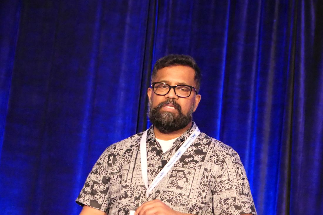
“If somebody is The dermatoscope manufacturers make teledermoscopy systems with attachments for iPhones, iPads, and Android devices,” said Dr. Tejasvi, who is director of teledermatology services and also director of the cutaneous lymphoma program at the University of Michigan, Ann Arbor.
To make his point, he presented slides of six standard unenhanced teledermatologic photos of ambiguous pigmented skin lesions. When he asked the large audience which ones they’d want to biopsy and which they were confident were benign, there was absolutely no consensus. But when he followed up with teledermoscopic photos of the same lesions, the dermoscopists in the audience quickly voiced agreement that four of the lesions were benign and two were obvious melanoma. Based on that information, instead of having to bring in all six patients for biopsy of their indeterminant suspicious lesions, only two of the patients would need to come in promptly for treatment of their malignancy.
“Dermoscopy changes the whole triage system and the teledermoscopy concept model, because dermoscopy remains the same: it’s going to stay [two-dimensional] whether you’re going to see the images in the clinic or in teledermatology. So using teledermoscopy images actually makes it far better for your teledermatology services,” explained Dr. Tejasvi, who is also chief of the dermatology service at the Ann Arbor Veterans Affairs Hospital.
Why get into teledermatology?
The benefits of teledermatology include earlier diagnosis and treatment of skin cancers as documented in a Spanish study of 43,677 patients. The Spanish dermatologists reported that teledermatologically detected melanomas had a thinner Breslow depth and lower tumor stage because they were diagnosed earlier. Teledermatology also brought a twofold increase in the basal cell carcinoma detection rate and – most importantly – a reduction in time to biopsy for what turned out to be skin cancers (JAMA Dermatol. 2015 Dec 1;151[12]:1289-90).
In addition, teledermatology is an effective triage tool for busy clinicians whose appointment calendars are booked weeks or months in advance.
“Let’s say you are the only dermatologist in the surrounding five counties. You can use teledermatology to see which patients actually need to come to your clinic,” Dr. Tejasvri said. Just make sure the referring primary care providers know to send photos taken with the dermatoscope attachment.
Internet-based teledermatology also provides a way to follow patients with chronic conditions, including psoriasis, atopic dermatitis, and venous ulcers, he noted.
Before getting started
Dr. Tejasvri emphasized the importance of visiting the American Academy of Dermatology Teledermatology Task Force website as well as the American Telemedicine Association’s Teledermatology Special Interest Group, which he chairs. These resources, he stressed, are invaluable.
The AAD site, open to all academy members, includes a tool kit for getting started in teledermatology. It’s individually tailored for the dermatologist in solo, small group, academic, or multispecialty practice. This highly practical tool kit includes a checklist that aids in determining whether a dermatologist’s practice is suited for teledermatology, as well as the suggested optimal teledermatology practice model for that individual, the nuts and bolts of equipment, relevant state laws, and how to navigate legal concerns, among the most critical of which is to get in writing the malpractice insurer’s verbal reassurance that the policy covers telemedicine.
The American Telemedicine Association Teledermatology Special Interest Group provides best-practice guidelines (Telemed J E Health. 2016 Dec;22[12]:981-90)
Teledermatology practice model options
The most common teledermatology model is called “store-and-forward.” It relies upon transmission of still images of skin lesions. Its advantages are that it’s not dependent upon internet speed and it accommodates physicians working in different time zones. Most commonly, this is a consult model in which a remote primary care provider takes the photos and transmits them to the dermatologist specialist. The referring provider retains responsibility for patient care.
The other model entails creation of a virtual clinic with real-time videoconference-based communication using a HIPAA-compliant high-speed broadband internet connection. The advantages are that reimbursement is good – indeed, the same as for a face-to-face office visit – and it’s possible to ask questions of the patient and referring physician, although that’s generally not necessary for the straightforward evaluation of suspicious pigmented or nonpigmented skin lesions. However, the video image quality isn’t as good as with still photos, the virtual clinic requires dedicated scheduling, and the quality of the experience is highly dependent upon internet speed.
“If you have a bad internet speed the whole process becomes choppy. When you ask a question, the answer you get is the one to your previous question,” Dr. Tejasvi said.
Reimbursement
Currently 38 states and Washington, D.C., have laws governing private payer telehealth reimbursement policy.
Under the 2019 Medicare physician fee schedule, code number 99446 – interprofessional telephone/internet consult lasting 5-10 minutes – pays $18.36. A 99447, lasting 11-20 minutes, pays $36.36, and a 99448, representing a 21-30 minute interprofessional consult, pays $54.72.
“Reimbursement is poor. It’s not a lot at all. If you spend 5-10 minutes on a consult you get paid about 20 bucks. But it’s better than nothing, and it used to be that patients had to pay out of pocket,” the dermatologist commented.
And of course, the improved timely and efficient patient access to dermatologist evaluation of potential skin cancer that’s afforded via teledermatology helps out with the profession’s workforce shortage and responds to the common criticism that dermatologists are geographically maldistributed and treatment delayed is treatment denied.
How accurate is teledermatology?
Numerous studies have reported diagnostic concordance rates between teledermatology and face-to-face clinical diagnosis of 72.5%-90% for melanoma, dropping off markedly to 31.2%-62% for lentigines. However, teledermoscopic images greatly improved the diagnostic accuracy.
In one recent study involving teledermatology versus face-to-face evaluation of 293 index lesions, the face-to-face dermatologist examination turned up 131 incidental skin lesions, including 6 incidental melanomas not suspected or photographed by the consulting primary care providers. That worked out to a 2.6% risk of incidental melanoma per consult, which Dr. Tejasvi called “kind of scary.”
“All six of the incidental melanomas were located on the back, chest, or abdomen, so a good teaching point is that, if you’re doing a teledermatology consult, ask the primary care provider who’s sending you this consult to do a careful waist-up exam to look for other lesions,” he advised.
He added that more and larger studies are needed in order to determine the diagnostic concordance rate for nonpigmented lesions.
Dr. Tejasvi reported having no financial conflicts regarding his presentation.
SDEF/Global Academy for Medical Education and this news organization are owned by the same parent company.
LAHAINA, HAWAII – Teledermatology and dermoscopy were made for each other, Trilokraj Tejasvi, MBBS, MD, declared at the Hawaii Dermatology Seminar provided by the Global Academy for Medical Education/Skin Disease Education Foundation.
“If somebody is The dermatoscope manufacturers make teledermoscopy systems with attachments for iPhones, iPads, and Android devices,” said Dr. Tejasvi, who is director of teledermatology services and also director of the cutaneous lymphoma program at the University of Michigan, Ann Arbor.
To make his point, he presented slides of six standard unenhanced teledermatologic photos of ambiguous pigmented skin lesions. When he asked the large audience which ones they’d want to biopsy and which they were confident were benign, there was absolutely no consensus. But when he followed up with teledermoscopic photos of the same lesions, the dermoscopists in the audience quickly voiced agreement that four of the lesions were benign and two were obvious melanoma. Based on that information, instead of having to bring in all six patients for biopsy of their indeterminant suspicious lesions, only two of the patients would need to come in promptly for treatment of their malignancy.
“Dermoscopy changes the whole triage system and the teledermoscopy concept model, because dermoscopy remains the same: it’s going to stay [two-dimensional] whether you’re going to see the images in the clinic or in teledermatology. So using teledermoscopy images actually makes it far better for your teledermatology services,” explained Dr. Tejasvi, who is also chief of the dermatology service at the Ann Arbor Veterans Affairs Hospital.
Why get into teledermatology?
The benefits of teledermatology include earlier diagnosis and treatment of skin cancers as documented in a Spanish study of 43,677 patients. The Spanish dermatologists reported that teledermatologically detected melanomas had a thinner Breslow depth and lower tumor stage because they were diagnosed earlier. Teledermatology also brought a twofold increase in the basal cell carcinoma detection rate and – most importantly – a reduction in time to biopsy for what turned out to be skin cancers (JAMA Dermatol. 2015 Dec 1;151[12]:1289-90).
In addition, teledermatology is an effective triage tool for busy clinicians whose appointment calendars are booked weeks or months in advance.
“Let’s say you are the only dermatologist in the surrounding five counties. You can use teledermatology to see which patients actually need to come to your clinic,” Dr. Tejasvri said. Just make sure the referring primary care providers know to send photos taken with the dermatoscope attachment.
Internet-based teledermatology also provides a way to follow patients with chronic conditions, including psoriasis, atopic dermatitis, and venous ulcers, he noted.
Before getting started
Dr. Tejasvri emphasized the importance of visiting the American Academy of Dermatology Teledermatology Task Force website as well as the American Telemedicine Association’s Teledermatology Special Interest Group, which he chairs. These resources, he stressed, are invaluable.
The AAD site, open to all academy members, includes a tool kit for getting started in teledermatology. It’s individually tailored for the dermatologist in solo, small group, academic, or multispecialty practice. This highly practical tool kit includes a checklist that aids in determining whether a dermatologist’s practice is suited for teledermatology, as well as the suggested optimal teledermatology practice model for that individual, the nuts and bolts of equipment, relevant state laws, and how to navigate legal concerns, among the most critical of which is to get in writing the malpractice insurer’s verbal reassurance that the policy covers telemedicine.
The American Telemedicine Association Teledermatology Special Interest Group provides best-practice guidelines (Telemed J E Health. 2016 Dec;22[12]:981-90)
Teledermatology practice model options
The most common teledermatology model is called “store-and-forward.” It relies upon transmission of still images of skin lesions. Its advantages are that it’s not dependent upon internet speed and it accommodates physicians working in different time zones. Most commonly, this is a consult model in which a remote primary care provider takes the photos and transmits them to the dermatologist specialist. The referring provider retains responsibility for patient care.
The other model entails creation of a virtual clinic with real-time videoconference-based communication using a HIPAA-compliant high-speed broadband internet connection. The advantages are that reimbursement is good – indeed, the same as for a face-to-face office visit – and it’s possible to ask questions of the patient and referring physician, although that’s generally not necessary for the straightforward evaluation of suspicious pigmented or nonpigmented skin lesions. However, the video image quality isn’t as good as with still photos, the virtual clinic requires dedicated scheduling, and the quality of the experience is highly dependent upon internet speed.
“If you have a bad internet speed the whole process becomes choppy. When you ask a question, the answer you get is the one to your previous question,” Dr. Tejasvi said.
Reimbursement
Currently 38 states and Washington, D.C., have laws governing private payer telehealth reimbursement policy.
Under the 2019 Medicare physician fee schedule, code number 99446 – interprofessional telephone/internet consult lasting 5-10 minutes – pays $18.36. A 99447, lasting 11-20 minutes, pays $36.36, and a 99448, representing a 21-30 minute interprofessional consult, pays $54.72.
“Reimbursement is poor. It’s not a lot at all. If you spend 5-10 minutes on a consult you get paid about 20 bucks. But it’s better than nothing, and it used to be that patients had to pay out of pocket,” the dermatologist commented.
And of course, the improved timely and efficient patient access to dermatologist evaluation of potential skin cancer that’s afforded via teledermatology helps out with the profession’s workforce shortage and responds to the common criticism that dermatologists are geographically maldistributed and treatment delayed is treatment denied.
How accurate is teledermatology?
Numerous studies have reported diagnostic concordance rates between teledermatology and face-to-face clinical diagnosis of 72.5%-90% for melanoma, dropping off markedly to 31.2%-62% for lentigines. However, teledermoscopic images greatly improved the diagnostic accuracy.
In one recent study involving teledermatology versus face-to-face evaluation of 293 index lesions, the face-to-face dermatologist examination turned up 131 incidental skin lesions, including 6 incidental melanomas not suspected or photographed by the consulting primary care providers. That worked out to a 2.6% risk of incidental melanoma per consult, which Dr. Tejasvi called “kind of scary.”
“All six of the incidental melanomas were located on the back, chest, or abdomen, so a good teaching point is that, if you’re doing a teledermatology consult, ask the primary care provider who’s sending you this consult to do a careful waist-up exam to look for other lesions,” he advised.
He added that more and larger studies are needed in order to determine the diagnostic concordance rate for nonpigmented lesions.
Dr. Tejasvi reported having no financial conflicts regarding his presentation.
SDEF/Global Academy for Medical Education and this news organization are owned by the same parent company.
EXPERT ANALYSIS FROM SDEF HAWAII DERMATOLOGY SEMINAR