User login
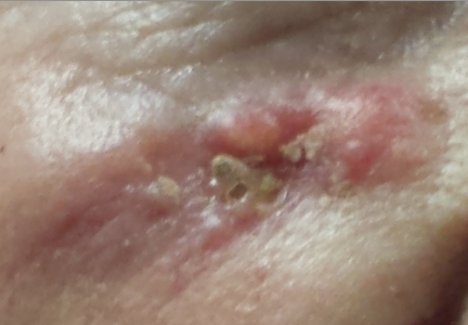
The lesion on the face of this 72-year-old woman has slowly grown over a period of years but has never caused pain. On several occasions, she has consulted primary care providers (PCPs), most of whom prescribed oral antibiotics for possible infection. However, these medications never helped.
She recently developed oral health problems and established care with a new PCP, who insisted she see a dermatologist about the facial lesion. The patient, who is “terrified” of needles, resisted this advice at first but finally agreed once her family got involved.
The patient reports a history of extensive sun exposure in childhood, when she worked in the fields, that continued into adulthood; she maintained a garden until recently. Her response to the sun is invariably a deep tan, which lasts all summer.
EXAMINATION
The lesion, almost 6 cm wide and more than 4 cm long, is located in the right infraorbital facial cheek area, well below the eyelid margin. It is focally eroded, peripherally erythematous, and quite firm to the touch. Fortunately, it is still mobile and not fixed to the underlying tissue. There are no palpable lymph nodes in the area and no change in sensation from one side of the face to the other.
A punch biopsy is performed under local infiltrate with 1% lidocaine with epinephrine. The 4-mm defect is closed with 2 x 5-0 nylon sutures and the specimen submitted to pathology. The report confirms the clinical suspicion of basal cell carcinoma (BCC).
Continue for Joe Monroe's discussion >>
DISCUSSION
When they are allowed to reach this size, BCCs can become a real problem. In extreme instances, they can erode into the face and even into bone. Patients can lose ears, noses, or even their entire face to this relentless but “safe” cancer. Given enough time and bad luck, BCC can metastasize to local lymph nodes and even to the brain or lung; it is even occasionally a cause of death.
Patients often ask why we have to remove BCCs if they’re usually “safe.” Without trying to scare them, we describe cases such as this one as the inevitable outcome of prolonged neglect and/or an aggressive tumor. BCCs almost never heal without treatment and almost always grow wider and deeper over time. Some are extremely indolent, taking 20 years to become noticeable, while others are more aggressive. In addition to the clinical behavior of any given BCC, there are also histologic clues to how aggressive a particular BCC might be.
This patient was referred to a Mohs surgeon, who will be able to do two things that require specialized training:
1. He’ll remove the biopsy-proven cancer with scalpel surgery, and while the patient is still present, check the margins for residual cancer. If the margins are positive, he’ll go back to the area and remove more, repeating this step until clear wide and deep microscopic margins are visualized by frozen section technique. (It’s important to note that the exact way the frozen specimen is processed and examined permits evaluation of the entire margin—top, bottom, sides—making it considerably different from ordinary frozen sections.)
2. Then, the Mohs surgeon will have the skill to close the defect in an acceptable way: usually by primary closure, sometimes with flaps or with grafts. All of this is typically done on an outpatient basis, on the same visit, although it may take most of a morning or afternoon.
Mohs surgery was pioneered in the 1930s by Frederic Mohs, MD, a general surgeon, as a way to address large and/or aggressive cancers located in difficult areas (eg, face or genitals). It is not indicated for ordinary, relatively small skin cancers on the arms, legs, or trunk.
Some BCCs and squamous cell carcinomas develop in especially difficult areas, such as the eyelids, or involve large areas of the ear. These lesions may require the attention of relevant surgical specialists, such as oculoplastic or head and neck surgeons.
This patient will undergo Mohs surgery in the near future, and her defect will probably be closed primarily. The depth and histologic markers indicating exceptional aggression may dictate a further step: Radiation therapy may be required to minimize the likelihood of recurrence. Her chances of a complete cure are 95% to 97% with Mohs surgery alone.
TAKE-HOME LEARNING POINTS
• Basal cell carcinomas (BCCs) are often mistaken for infection.
• BCCs almost never heal on their own, grow slowly but steadily, and can reach prodigious size and depth.
• BCCs can metastasize to local nodes or even to the brain and lung.
• Ordinary excision is adequate for most BCCs, but Mohs surgery is indicated for larger lesions located in difficult areas (eg, face, scalp or groin).
• Simple shave biopsy is adequate to diagnose most BCCs.