Case Letter

Verrucous Kaposi Sarcoma in an HIV-Positive Man
Verrucous Kaposi sarcoma (VKS) is an uncommon variant of Kaposi sarcoma that rarely is seen in clinical practice or reported in the literature.
Lisa M. Martorano, DO; Jonathan D. Cannella, MD; Jenifer R. Lloyd, DO
Dr. Martorano is from Heritage College of Osteopathic Medicine, Ohio University, Athens. Dr. Cannella is from the Department of Laboratory Medicine, Northside Medical Center/Valley Care Health System of Ohio, Youngstown. Dr. Lloyd is from the Department of Dermatology, University Hospital Richmond Medical Center, a Campus of UH Regional Hospitals, Cleveland, Ohio, and Lloyd Dermatology and Laser Center, Youngstown.
The authors report no conflict of interest.
Correspondence: Jenifer R. Lloyd, DO, Lloyd Dermatology and Laser Center, 8060 Market St, Youngstown, OH 44512 (jrl@lloyd-derm.com).

Kaposi sarcoma (KS) is a malignant proliferation of endothelial cells within the skin. The clinical presentation is characterized by clusters of violaceous macules and papules that often appear on the distal extremities or trunk with or without oral mucosal involvement. Mucocutaneous lesions are present at onset of diagnosis in a minority of cases. The lesions can evolve to include the mucous membranes of the gastric mucosa and the lungs. We present a unique case of KS in a 45-year-old, asymptomatic, human immunodeficiency virus (HIV)–positive man with mucocutaneous involvement to highlight the importance of recognizing KS in immunocompromised patients.
Practice Points
Case Report
A 45-year-old man presented with persistent swelling and “black-and-blue” lesions on the legs, feet, and toes of 6 months’ duration. The painless lesions first appeared on the left lower leg and then began to appear on the right leg in recent months. Three weeks prior to presentation, he developed swelling of the left lower leg during hospitalization for a lumbar laminectomy. A venous ultrasound was negative for a deep vein thrombosis. He denied trauma or history of bleeding diathesis. He did not report symptoms of dyspnea, angina, or claudication, and a review of systems was unremarkable.
The patient’s medical history included spinal stenosis, chronic back pain, osteoarthritis, and anxiety. His medications included oxycodone, zolpidem, and alprazolam. In addition to a recent lumbar laminectomy, he had undergone extensive dental work in the last 6 months. The patient denied the use of cigarettes, alcohol, or intravenous drugs.
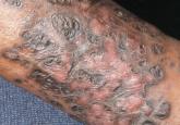
Physical examination revealed scattered, purple, segmented patches on the dorsal and plantar aspects of the feet, both calves, both heels, and toes (Figure 1). Mild nonpitting edema was present below the left knee along with edema on the dorsum of the left foot. The distribution of the lesions was initially suggestive of cholesterol embolization syndrome; however, both the femoral and posterior tibial pulses were symmetric and palpable (+2). Well-demarcated violaceous plaques with central clearing and a rustlike discoloration were noted on the hard and soft palates. Cervical lymphadenopathy was not present.
Figure 1. Scattered, purple, segmented patches along the dorsal surface of the toes. Figure 2. Punch biopsy specimen of the right fourth toe showed an acanthotic epidermis and focally lobulated proliferation of cells and small caliber vessels, representing the plaque stage of Kaposi sarcoma (H&E, original magnification ×40). Figure 3. Red blood cells were evident in the holes and in between the tumor cells. The promontory sign was present and the native vessels were seen protruding into the lumens of newly formed tumoral vessels (H&E, original magnification ×400). |
Laboratory tests including a repeat venous ultrasound of the left lower leg revealed no evidence of deep vein thrombosis. Ankle brachial index revealed no abnormalities and blood flow to the lower legs was adequate. Computed tomography scans of the chest, abdomen, and pelvis were unremarkable except for mild splenomegaly and moderate cardiomegaly. Lastly, human immunodeficiency virus (HIV) 1 and HIV-2 enzyme immunoassay was reactive.
Histopathologic examination of a punch biopsy from the right fourth toe was representative of the plaque stage of Kaposi sarcoma (KS) with a diffuse collection of extravasated erythrocytes and neoplastic vascular proliferation among a background of numerous plasma cells and hemosiderophages (Figure 2). Higher magnification illustrated the promontory sign, whereby native vessels encroach on neoplastic slitlike vascular spaces (Figure 3). A final diagnosis of AIDS-related KS was made. The patient was referred to an infectious disease specialist for evaluation of his CD4 levels and HIV management.
Comment
Kaposi sarcoma is a neoplastic proliferation of the blood vessels in the skin characterized by the formation of violaceous macules and papules that often appear on a single distal extremity, such as the foot. Over time the lesions can develop on the opposite extremity and coalesce into poorly demarcated plaques and nodules with accompanying stasis and lymphedema of the involved extremities.1 Evolution of the lesions depends on the KS subtype. The most common clinical variant of KS is the classic form, which primarily is seen in those of Mediterranean, Eastern European, or Ashkenazi Jewish descent, with a predilection for men and older adults.1,2 The endemic form of KS, or African KS, is more aggressive with rapid visceral involvement and rare skin lesions; it is common among prepubertal children in sub-Saharan Africa with no predilection for either sex.2 In the setting of severe immune suppression, organ transplantation, or chemotherapy, a third KS subtype known as iatrogenic KS can occur. The clinical course of iatrogenic KS may range from scattered cutaneous lesions to diffuse involvement secondary to increased dosages and long-term use of immunosuppressive agents.2
Our patient had AIDS-related or epidemic KS. AIDS-related KS is largely predominant among homosexual men, but due to the awareness of safe sexual practices and the introduction of highly active antiretroviral therapy (HAART), KS incidence in the United States has declined.1,2 However, despite recent advances in HIV therapy, AIDS-related KS is still the most common neoplasm seen in AIDS patients and is the presenting manifestation of AIDS in up to 30% of cases.3 Up to 22% of cases first appear on the gingiva, hard palate, and tongue, with concomitant dysphagia and airway obstruction in severe cases.4,5 More advanced cases of AIDS-related KS can present with initial symptoms such as abdominal pain, melena, dyspnea, lymphadenopathy, and weight loss, which suggests involvement of the gastrointestinal tract, lungs, and other organ systems.

Verrucous Kaposi sarcoma (VKS) is an uncommon variant of Kaposi sarcoma that rarely is seen in clinical practice or reported in the literature.

Kaposi sarcoma is a virally induced, low-grade angiosarcoma. It was initially described in 1872 and subsequently categorized into 4...
