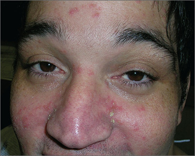
Taking into consideration the patient’s 2 predisposing risk factors (AIDS and dementia), the FP diagnosed seborrheic dermatitis. The FP also noted seborrheic dermatitis on the scalp. The scalp is frequently involved in cases of seborrheic dermatitis on the face.
Seborrheic dermatitis is a common, chronic, relapsing dermatitis affecting sebum-rich areas of the body. Its presentation may vary from mild erythema to greasy scale, and rarely, to erythroderma. Patients with seborrheic dermatitis may be colonized with certain species of lipophilic yeast of the genus Malassezia (also known as Pityrosporum). Malassezia, however, is considered normal skin flora because it is also found in unaffected people.
Some evidence suggests that an overgrowth of Malassezia may produce different irritants or metabolites on affected skin, leading to inflammation. It’s postulated that an overgrowth of Malassezia leads to the inflammatory dermatitis in seborrhea.
The goals of treatment are to diminish the fungal overgrowth and treat the inflammatory response. Thus, the mainstay of treatment is ongoing topical antifungals in shampoos and other preparations, along with a topical steroid to treat the inflammation.
The FP prescribed 2% ketoconazole shampoo to be used twice weekly and suggested that a selenium-containing shampoo be used on the other days of the week, as well as a 2.5% hydrocortisone cream to be applied twice daily to the face until the dermatitis resolved. The FP also prescribed a topical antifungal, cyclopirox 1% cream, to be applied twice daily on an ongoing basis to keep the Malassezia under control. At a one-month follow-up, the patient’s skin and scalp were cleared.
Photos and text for Photo Rounds Friday courtesy of Richard P. Usatine, MD. This case was adapted from: Hancock M, Bae Y, Usatine R. Seborrheic dermatitis. In: Usatine R, Smith M, Mayeaux EJ, et al, eds. Color Atlas of Family Medicine. 2nd ed. New York, NY: McGraw-Hill; 2013: 871-877.
To learn more about the Color Atlas of Family Medicine, see: www.amazon.com/Color-Family-Medicine-Richard-Usatine/dp/0071769641/
You can now get the second edition of the Color Atlas of Family Medicine as an app by clicking on this link: usatinemedia.com


