Diagnosis: Bullous pemphigoid
This patient had a severe and refractory case of bullous pemphigoid (BP), which was confirmed with a biopsy of the lesions.
BP is a rare autoimmune, blistering skin disease that typically occurs after age 60.1 The incidence rises with age, and is higher among women than men.1 The pathogenesis of BP involves development of autoantibodies against the subepidermal basement membrane. Deposition of immunoglobulin G (IgG) occurs, leading to immune-mediated destruction and subepidermal blistering.2
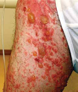
Patients will present with new-onset, widespread eruptions of bullous lesions and urticarial plaques (FIGURE 2).2 Bullae are frequent on flexural surfaces such as the groin and axillae. Urticarial plaques are often pruritic. Oral involvement occurs in a minority of cases.2 Nikolsky’s sign—exfoliation of the outermost layer of skin upon slight rubbing—is absent in BP.
FIGURE 2
Fluid-filled vesicles and bullae on right anterior thigh

Differential: Other autoimmune and blistering skin conditions
Two additional pemphigoid subtypes are part of the differential when a patient presents with a blistering skin condition: pemphigoid gestationis and mucous membrane pemphigoid.2
Pemphigoid gestationis occurs exclusively during pregnancy and the puerperium, and is self-limited.
Mucous membrane pemphigoid is pathophysiologically similar to BP, but distributes preferentially on mucosal surfaces.
Pemphigus vulgaris, another autoimmune blistering skin disease, is characterized by sparse intact bullae. The mucous membrane is frequently involved, and there is a positive Nikolsky’s sign.
Additional conditions to keep in mind include epidermolysis bullosa acquisita, dermatitis herpetiformis, bullous erythema multiforme, and bullous lupus erythematosus.

