The patient had tinea versicolor caused by Pityrosporum (Malassezia furfur), a lipophilic yeast that can be normal human cutaneous flora. Tinea versicolor starts when the yeast that normally colonizes the skin changes from the round form to the pathologic mycelial form and then invades the stratum corneum. Tinea versicolor tends to come in white, pink, and brown. While it is often associated with hypopigmentation, this case demonstrates the hyperpigmentation that can also occur (FIGURE 1). Pityrosporum thrives on sebum and moisture and tends to grow on the skin in areas where there are sebaceous follicles secreting sebum.
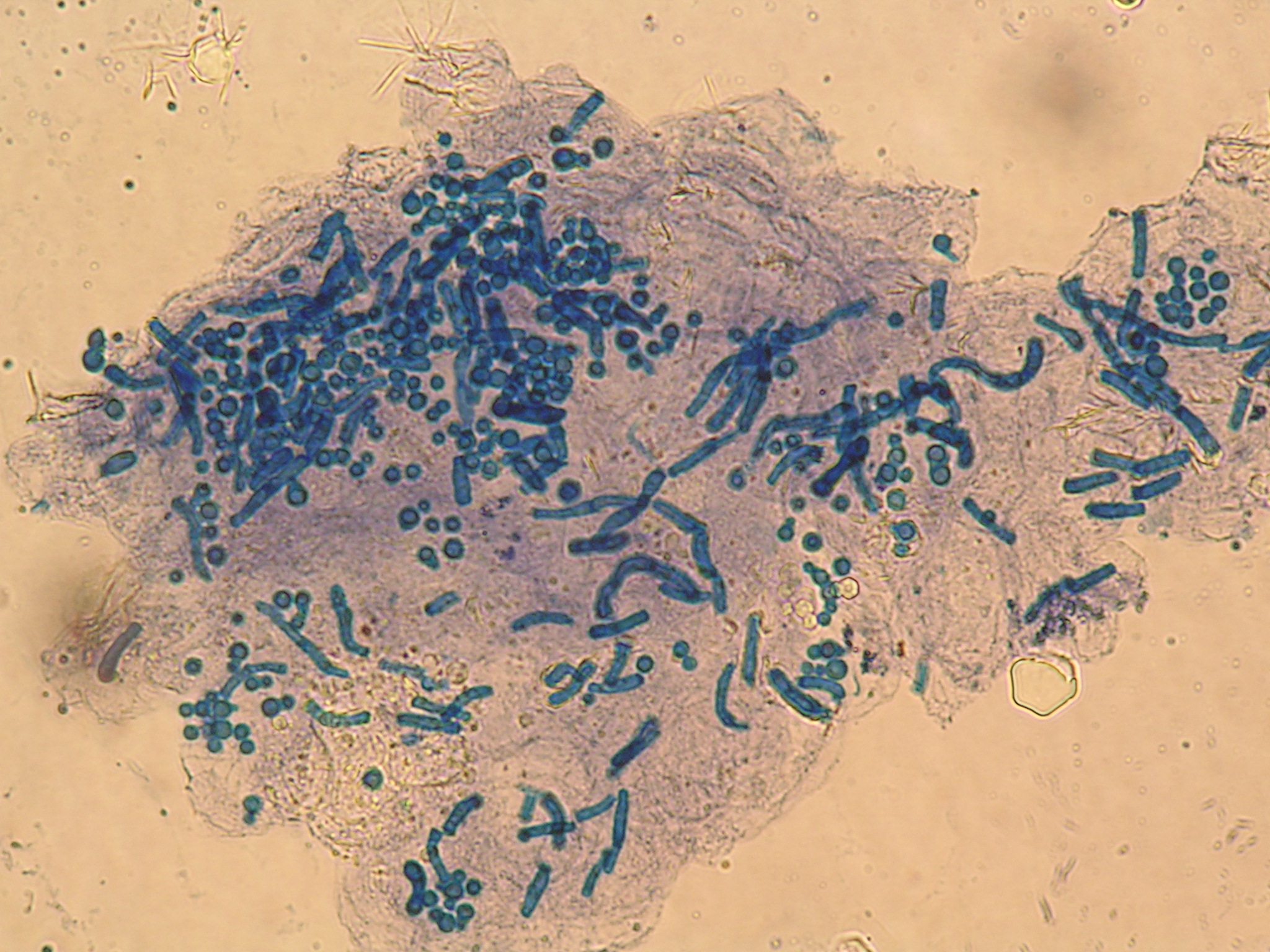
The physician did a scraping of the scale and applied Swartz-Lamkins stain (potassium hydroxide, dimethyl sulfoxide, and blue ink) to the slide. FIGURE 2 shows the “spaghetti and meatballs” pattern typical of tinea versicolor. The spaghetti or “ziti” is the short mycelial form and the meatballs are the round yeast of Pityrosporum ovale.
The patient received a single oral dose of 400 mg fluconazole. The condition cleared and the skin color returned to normal in the following months.
Photos and text for Photo Rounds Friday courtesy of Richard P. Usatine, MD. This case was adapted from: Usatine R. Tinea versicolor. In: Usatine R, Smith M, Mayeaux EJ, Chumley H, Tysinger J, eds. The Color Atlas of Family Medicine. New York, NY: McGraw-Hill; 2009:566-569.
To learn more about The Color Atlas of Family Medicine, see:
* http://www.amazon.com/Color-Atlas-Family-Medicine/dp/0071474641


