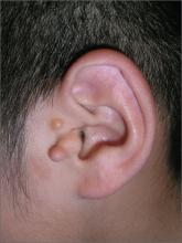
The FP explained to the patient’s mother that the boy had preauricular tags. These occur in approximately 1 out of 10,000 to 12,500 births, without predilection for gender or race. Ear malformations may occur in isolation, or as part of a constellation of abnormalities--often involving the renal system. Children with preauricular tags have a 5-fold increased risk of hearing impairment. Several chromosomal abnormalities (eg, Goldenhar syndrome) include preauricular tags as one of the phenotypic expressions.
Preauricular tags arise from remnants of supernumerary branchial hillocks. Early stage embryology involves the formation of several slit-like structures on the side of the head, the branchial clefts. The 3 hillocks between the first 4 clefts eventually form the structure of the outer ear. Preauricular tags are generally minor malformations arising from remnants of the hillocks. Preauricular tags can be left alone or surgically excised for cosmetic reasons.
In this case, the child had normal hearing and hadn’t had any urinary tract infections. The FP explained that the changing size of the tags was not worrisome and that they could be removed for cosmetic purposes, but her son would have to undergo general anesthesia for the surgery. The mother agreed with the FP that the benefits of the surgery did not outweigh the risks.
Photos and text for Photo Rounds Friday courtesy of Richard P. Usatine, MD. This case was adapted from: French L. Chondrodermatitis nodularis helicis and preauricular tags. In: Usatine R, Smith M, Mayeaux EJ, et al. Color Atlas of Family Medicine. 2nd ed. New York, NY: McGraw-Hill; 2013:189-192.
To learn more about the Color Atlas of Family Medicine, see: http://www.amazon.com/Color-Family-Medicine-Richard-Usatine/dp/0071769641/ref=dp_ob_title_bk
You can now get the second edition of the Color Atlas of Family Medicine as an app for mobile devices by clicking this link: http://usatinemedia.com/


