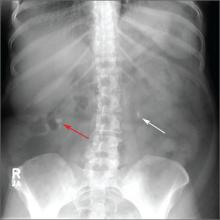
The family physician (FP) noted bilateral renal stones on the abdominal radiograph. A noncontrast CT scan confirmed multiple bilateral renal stones, with an obstructing right distal ureteral stone and enlargement of the right kidney.
A kidney stone is a solid mass that forms when minerals crystallize and collect in the urinary tract. Kidney stones can cause pain and hematuria, and may lead to complications such as urinary tract obstruction and infection.
Stones smaller than 5 mm are likely to pass spontaneously. About three-fourths of distal ureteral stones and about half of proximal ureteral stones will pass spontaneously. Medical expulsive therapy with α-adrenergic blockers (such as tamsulosin) or calcium-channel blockers can increase the chances of stone passage.
Effective pain control should be provided using nonsteroidal anti-inflammatory drugs (NSAIDs) and narcotics if needed. NSAIDs may need to be avoided if lithotripsy (see below) is planned because there is an increased risk of perinephric bleeding.
Stones that do not pass spontaneously or with medical expulsive therapy can be treated with lithotripsy or removed via ureteroscopy. Large stones may require percutaneous nephrolithotomy or open surgery.
In this case, the FP recommended adequate fluid intake, which consists of 2 to 3 L of water per day for most patients. The FP also did further work-up (in light of the multiple stones) and determined that the patient had hyperparathyroidism. The FP referred the patient to an endocrinologist for further evaluation and management.
Photo courtesy of Karl T. Rew, MD. Text for Photo Rounds Friday courtesy of Richard P. Usatine, MD. This case was adapted from: Rew K, Smith M. Kidney stones. In: Usatine R, Smith M, Mayeaux EJ, et al, eds. Color Atlas of Family Medicine. 2nd ed. New York, NY: McGraw-Hill; 2013:425-429.
To learn more about the Color Atlas of Family Medicine, see: http://www.amazon.com/Color-Family-Medicine-Richard-Usatine/dp/0071769641/
You can now get the second edition of the Color Atlas of Family Medicine as an app by clicking this link: http://usatinemedia.com/


