User login
Temporary tissue expanders optimize radiotherapy after mastectomy
LAS VEGAS – Radiation oncologists at the University of Texas MD Anderson Cancer Center, Houston, were able to complete 98% of their radiotherapy plans when women received temporary tissue expanders, instead of immediate reconstructions, at the time of skin-sparing mastectomy, in a series of 384 women, most with stage 2-3 breast cancer.
The expanders – saline-filled bags commonly used in plastic surgery to create new skin – were kept in place but deflated for radiotherapy, which allowed for optimal access to treatment fields; the final reconstruction, successful in 90% of women, came a median of 7 months following radiation.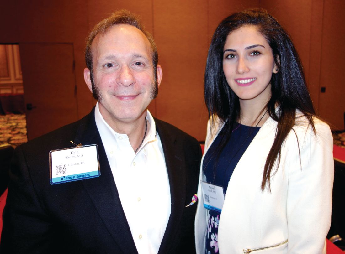
“The shape and volume of the reconstruction” – and the need to avoid damaging the new breast – “got in the way of putting radiation where we wanted it to be. We ended up having bad radiotherapy plans, patients not getting skin-sparing mastectomies, and high probabilities of radiation complications to the reconstruction,” said investigator Eric Strom, MD, professor of radiation oncology at MD Anderson.
Radiologists and plastic and oncologic surgeons collaborated to try tissue expanders instead. “We wanted the advantage of skin-sparing mastectomy without the disadvantages” of immediate reconstruction, Dr. Strom said at the American Society of Breast Surgeons annual meeting.
With the new approach, “radiotherapy is superior. We don’t have to compromise our plans. I can put radiation everywhere it needs to be, without frying the heart” and almost completely avoiding the lungs, he said.
The 5-year rates of locoregional control, disease-free survival, and overall survival were 99.2%, 86.1%, and 92.4%, respectively, which “is extraordinary” in patients with stage 2-3 breast cancer, and likely due at least in part to optimal radiotherapy, he said.
Tissue expanders also keep the skin envelope open so it’s able to receive a graft at final reconstruction; abdominal skin doesn’t have to brought up to recreate the breast.
“This approach lessens negative interactions between breast reconstruction and [radiotherapy] and offers patients what they most desire: a high probability of freedom from cancer and optimal final aesthetic outcome,” said Zeina Ayoub, MD, a radiation oncology fellow at Anderson who presented the findings.
The median age of the women was 44 years, and almost all were node positive. Radiation was delivered to the chest wall and regional lymphatics, including the internal mammary chain.
Fifty women (13.0%) required explantation after radiation but before reconstruction, most commonly because of cellulitis; even so, more than half went on to final reconstruction.
Abdominal autologous reconstruction was the most common type, followed by latissimus dorsi–based reconstruction, and exchange of the tissue expander with an implant.
Dr. Ayoub and Dr. Strom had no relevant disclosures.
AT ASBS 2017
Key clinical point:
Major finding: The 5-year rates of locoregional control, disease-free survival, and overall survival were 99.2%, 86.1%, and 92.4%, respectively, likely due at least in part to optimal radiotherapy.
Data source: Review of 384 patients.
Disclosures: The investigators said they had no relevant disclosures.
Breast cancer liquid biopsies don’t change outcomes....yet
LAS VEGAS – Although circulating tumor cells are prognostic in breast cancer, they aren’t likely to become a part of routine practice until they’ve been shown to improve outcomes, and that hasn’t happened yet, according to Anthony Lucci, MD, professor of breast surgical oncology at MD Anderson Cancer Center, Houston.
MD Anderson and other institutions have established that in breast cancer, from baseline through treatment follow-up, the presence of circulating tumor cells (CTCs) in the blood predicts worse outcomes in both metastatic and local disease, even among women who have a pathologic complete response to treatment. 
That knowledge has not yet translated into better outcomes. In a pivotal trial, survival was no better in metastatic breast cancer when women were switched to an alternative chemotherapy regimen after their CTC levels did not fall in response to first-line treatment, compared with women who remained on their initial agent despite persistently high CTCs (J Clin Oncol. 2014 Nov 1;32[31]:3483-9).
“When we tried to switch chemotherapy” based on CTCs, “it really didn’t make a difference, so no one is really quite sure yet what to do with this information. We can’t tell a patient that they are likely to have a much worse outcome, but there’s nothing we can do about it,” and “I don’t think payers will pay” for CTC testing in breast cancer until “we establish a predictive benefit, and show what agents will reduce CTCs and improve outcomes,” Dr. Lucci said at the American Society of Breast Surgeons annual meeting.
Even so, the promise of CTCs as a clinical tool is too great to abandon research, and work continues at MD Anderson and elsewhere. It’s thought that tumor cells in the blood aren’t simply a marker of disease, but rather microscopic disease in themselves that contributes to recurrence and progression. It’s possible that cells shed by tumors lie dormant in the bone marrow, then reactivate and reseed the original tumor site or give rise to distant metastases.
CTCs are similar to tumor cells that have been found in the bone marrow of women with apparently quiescent breast cancer, but finding them in the blood means “you don’t have to poke a needle in someone’s bone. You can just take a sample of their blood, and it tells you the same information,” Dr. Lucci said.
CTCs have been shown in breast cancer to rise and fall depending on tumor response. “In the future, I do think we will [use them] as a serial monitoring tool and a guide to therapy.” There might even be a role for breast cancer screening, he said.
Analyzing the blood for evidence of solid tumors – popularly called “liquid biopsy” – has shown benefits in a variety of cancers, particularly for identifying disease, and in some cases mutations, sooner than with conventional methods. Science is catching up to the old-school notion that cancer spreads through the blood.
Another approach is to look for circulating tumor DNA, which has been shown to be useful for early detection in gynecologic cancers. The idea is that a few tumor cells shatter and spill their genetic material into the blood early on. “In the majority of breast cancer patients,” however, “you don’t find actionable mutations” with circulating tumor DNA, especially in earlier-stage, nonmetastatic disease. “There’s not enough DNA released into the blood,” Dr. Lucci said.
But “we need to be monitoring the blood on a routine basis” for breast cancer clues. “That, I think, is the wave of the future. Just looking at x-rays and waiting for something to happen is too late,” he said.
Dr. Lucci had no disclosures related to his talk.
LAS VEGAS – Although circulating tumor cells are prognostic in breast cancer, they aren’t likely to become a part of routine practice until they’ve been shown to improve outcomes, and that hasn’t happened yet, according to Anthony Lucci, MD, professor of breast surgical oncology at MD Anderson Cancer Center, Houston.
MD Anderson and other institutions have established that in breast cancer, from baseline through treatment follow-up, the presence of circulating tumor cells (CTCs) in the blood predicts worse outcomes in both metastatic and local disease, even among women who have a pathologic complete response to treatment.
That knowledge has not yet translated into better outcomes. In a pivotal trial, survival was no better in metastatic breast cancer when women were switched to an alternative chemotherapy regimen after their CTC levels did not fall in response to first-line treatment, compared with women who remained on their initial agent despite persistently high CTCs (J Clin Oncol. 2014 Nov 1;32[31]:3483-9).
“When we tried to switch chemotherapy” based on CTCs, “it really didn’t make a difference, so no one is really quite sure yet what to do with this information. We can’t tell a patient that they are likely to have a much worse outcome, but there’s nothing we can do about it,” and “I don’t think payers will pay” for CTC testing in breast cancer until “we establish a predictive benefit, and show what agents will reduce CTCs and improve outcomes,” Dr. Lucci said at the American Society of Breast Surgeons annual meeting.
Even so, the promise of CTCs as a clinical tool is too great to abandon research, and work continues at MD Anderson and elsewhere. It’s thought that tumor cells in the blood aren’t simply a marker of disease, but rather microscopic disease in themselves that contributes to recurrence and progression. It’s possible that cells shed by tumors lie dormant in the bone marrow, then reactivate and reseed the original tumor site or give rise to distant metastases.
CTCs are similar to tumor cells that have been found in the bone marrow of women with apparently quiescent breast cancer, but finding them in the blood means “you don’t have to poke a needle in someone’s bone. You can just take a sample of their blood, and it tells you the same information,” Dr. Lucci said.
CTCs have been shown in breast cancer to rise and fall depending on tumor response. “In the future, I do think we will [use them] as a serial monitoring tool and a guide to therapy.” There might even be a role for breast cancer screening, he said.
Analyzing the blood for evidence of solid tumors – popularly called “liquid biopsy” – has shown benefits in a variety of cancers, particularly for identifying disease, and in some cases mutations, sooner than with conventional methods. Science is catching up to the old-school notion that cancer spreads through the blood.
Another approach is to look for circulating tumor DNA, which has been shown to be useful for early detection in gynecologic cancers. The idea is that a few tumor cells shatter and spill their genetic material into the blood early on. “In the majority of breast cancer patients,” however, “you don’t find actionable mutations” with circulating tumor DNA, especially in earlier-stage, nonmetastatic disease. “There’s not enough DNA released into the blood,” Dr. Lucci said.
But “we need to be monitoring the blood on a routine basis” for breast cancer clues. “That, I think, is the wave of the future. Just looking at x-rays and waiting for something to happen is too late,” he said.
Dr. Lucci had no disclosures related to his talk.
AT ASBS 2017
Intraoperative radiation looks good for DCIS
LAS VEGAS – Intraoperative radiation therapy (IORT) is effective in patients with ductal carcinoma in situ (DCIS), and bilateral digital mammography and breast MRI effectively predicted which patients would be most suited for the procedure, according to a nonrandomized study.
The TARGIT-A trial showed that IORT was noninferior to external beam radiation therapy in women with early-stage breast cancer (Lancet. 2014 Feb 15;383:603-13), but the technique hasn’t been tested in DCIS patients.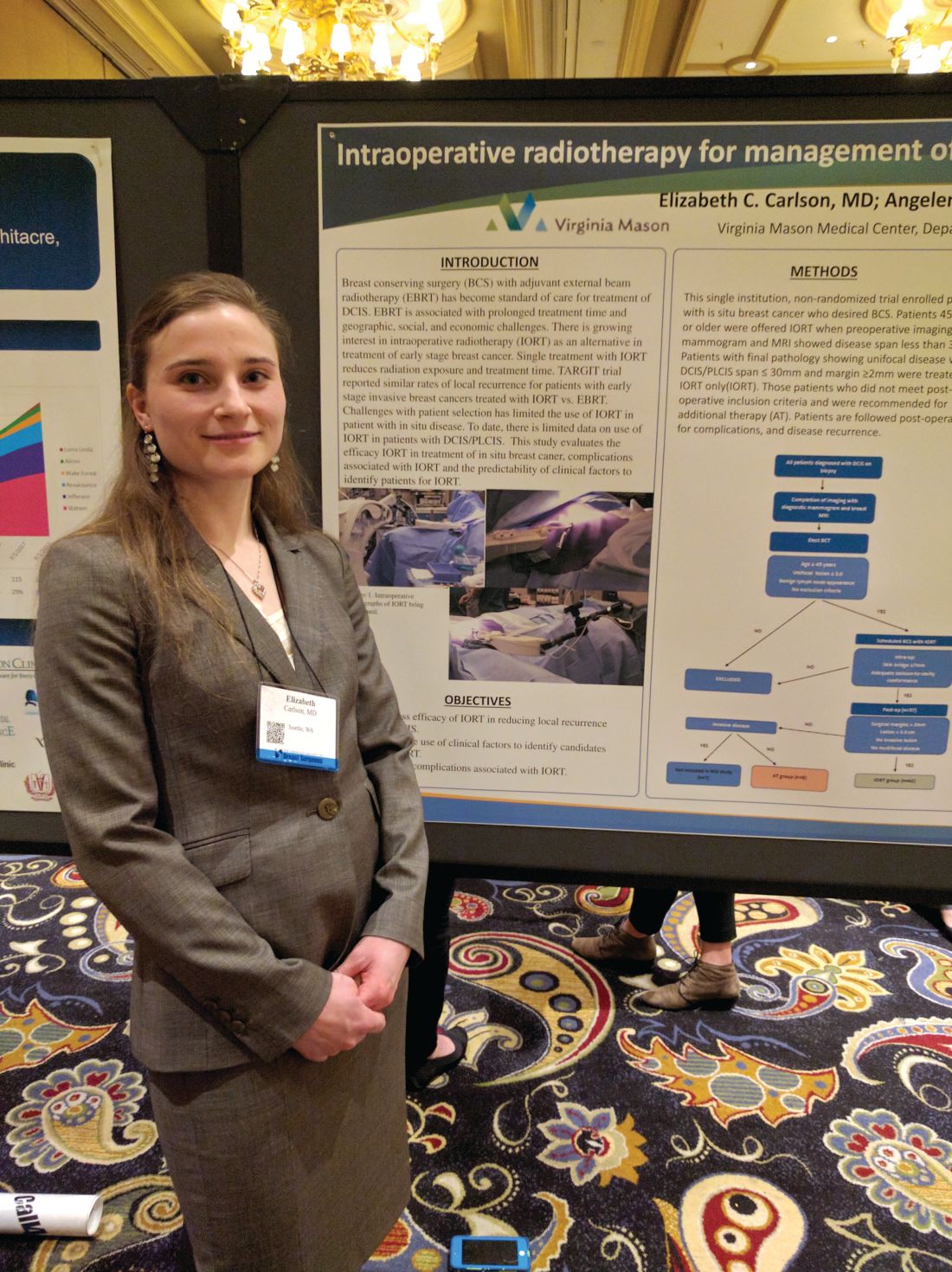
The researchers conducted a prospective, nonrandomized study of DCIS in women who underwent core biopsy between February 2012 and July 2016 at Virginia Mason Medical Center. Those who elected breast conserving therapy were assessed for IORT using bilateral digital mammography and contrast enhanced MRI.
For criteria, the researchers selected patients over age 44 years with unifocal DCIS and a lesion size of up to 3.0 cm. Physicians recommended additional therapy when the pathology report revealed DCIS larger than 3 cm and/or margins of up to 2 mm.
Of the 57 patients initially enrolled, 7 were found to have invasive disease and were excluded. Another eight patients were excluded because of margins, tumor size, or multifocal disease, and were recommended for additional treatment.
The remaining 42 patients with DCIS were treated with IORT. At a mean follow-up time of 32 months, the researchers observed no local recurrences, and no Radiation Therapy Oncology Group grade 3 or 4 complications were reported.
Hyperpigmentation occurred more often in the IORT group, at 40% (17 of 42 patients), than in the additional treatment group, at 13% (1 of 8), a nonsignificant difference. The hyperpigmentation tended to be minimal and focused on the surgical scar. There were no differences between the groups with respect to superficial wound separation, infection, seroma, fat necrosis, transient radiation dermatitis, or rib fracture.
“Our conclusions are that are selection criteria with mammography and MRI are effective in identifying patients who are appropriate for IORT, and importantly, at 32 months follow-up, no patients had recurrences, so we’re not negatively impacting oncological outcome,” said Dr. Carlson.
The fact that eight patients were recommended for additional treatment based on margins, a disease span greater than 3 cm, or multifocal disease “is actually quite heartening because it implies that the mammogram and the MRI are quite effective at identifying patients that met our criteria,” said Dr. Carlson.
The study is small and had no control group, but Dr. Carlson is confident of the results. “We didn’t do a randomized trial because we believed strongly that IORT will be effective in these patients with good cosmesis. When you compare it to local recurrence rates in the TARGIT-A trial, we’re still doing quite well, so to some extent that can be used as a baseline benchmark,” she said.
AT ASBS 2017
Key clinical point: Intraoperative radiation appears safe and effective, and is more convenient than postoperative radiation.
Major finding: There were no ductal carcinoma in situ recurrences at a median 32 months’ follow-up.
Data source: Uncontrolled, prospective study of 42 patients.
Disclosures: The study was funded by the National Institutes of Health. Dr. Carlson reported having no financial disclosures.
Positive node risk defined for elderly breast cancer patients
LAS VEGAS – In women aged 70 years or older with hormone receptor–positive invasive breast cancer, their tumor size, grade, and histology – but not human epidermal growth factor receptor–2 status – predicted nodal positivity, according to a retrospective analysis.
Investigators at the Mayo Clinic, Rochester, Minn., reviewed 52,532 women in the National Cancer Database from 2010 to 2013 who were at least 70 years old with hormone receptor–positive invasive breast cancer and clinically node negative disease, who had axillary surgery performed. Two-thirds of the cohort was used to identify risk factors, and the remaining third to validate them. About 16% in both groups had cancer in their axillary lymph nodes.
On multivariate analysis, higher clinical T stage, higher grade, and invasive lobular and invasive mammary histology were all associated with positive nodes. Although significant on univariate analysis, age (P = .57) and HER2 status (P = .32) fell out on multivariate analysis.
Nodal positivity was more than five times as likely with clinical T2 tumors, compared with T1a tumors, and far less likely for patients with invasive mucinous carcinoma than for those with invasive ductal carcinoma.
The team expects to release a nomogram for general use in clinical practice to predict the risk of positive nodes for various combinations of tumor size, grade, and histology in older women. When the model predicted a less than 10% chance of node positive disease, the actual rate in the validation set was around 5.5%. When it predicted a 30%-39% chance, the actual rate was 32.6%. The area under the receiver operating characteristic curve was 0.7 in both the development and validation sets, indicating good discrimination.
The work grew out of an effort to implement the Society of Surgical Oncologists’ recommendation not to do routine sentinel node biopsies in clinically node negative, hormone receptor–positive invasive breast cancer in women over the age of 70 years, a recommendation the group made as part of its contribution to the Choosing Wisely campaign.
“After the guideline was released, we were sitting in the clinic thinking how to apply it to our patients,” lead investigator Jessemae Welsh, MD, a surgeon at the Mayo Clinic, said at the American Society of Breast Surgeons annual meeting.
The problem is that nodal positivity is important to know for other aspects of care, including regional nodal irradiation and duration of systemic hormone therapy, and axillary lymph node staging might still be indicated if older women are truly at high risk. “We [wanted] to develop a multivariate model that gives a precise estimate of nodal risk,” to help “patients and surgeons to decide together based on an individual risk” how best to proceed. Also, a prediction of low risk “can help reassure patients that they will do well without axillary surgery,” she said.
Mayo’s nomogram is unique in that it focuses specifically on women 70 years and older. Development of previous nomograms incorporated older women, but did not focus on them specifically, Dr. Welsh said.
Dr. Welsh said she had no relevant disclosures.
aotto@frontlinemedcom.com
AT ASBS 2017
Key clinical point:
Major finding: On multivariate analysis, higher clinical T stage, higher grade, and invasive lobular and invasive mammary histology were all associated with positive nodes.
Data source: An analysis of data from 52,532 women in the National Cancer Database during 2010-2013.
Disclosures: The lead investigator had no disclosures.
Forgo axillary dissection for single suspicious node on ultrasound
LAS VEGAS – About half of breast cancer patients without palpable lymphadenopathy but with preoperative ultrasound-guided, biopsy-proven axillary lymph node metastases have N1 disease, according to a review of 129 women.
Among the 30 women with a primary tumor 2 cm or smaller and only one abnormal lymph node on axillary ultrasound, 22 (73%) had metastases limited to the one lymph node, suggesting that such patients “may undergo ... sentinel lymph node biopsy” instead of a complete axillary lymph node dissection (ALND), the investigators concluded.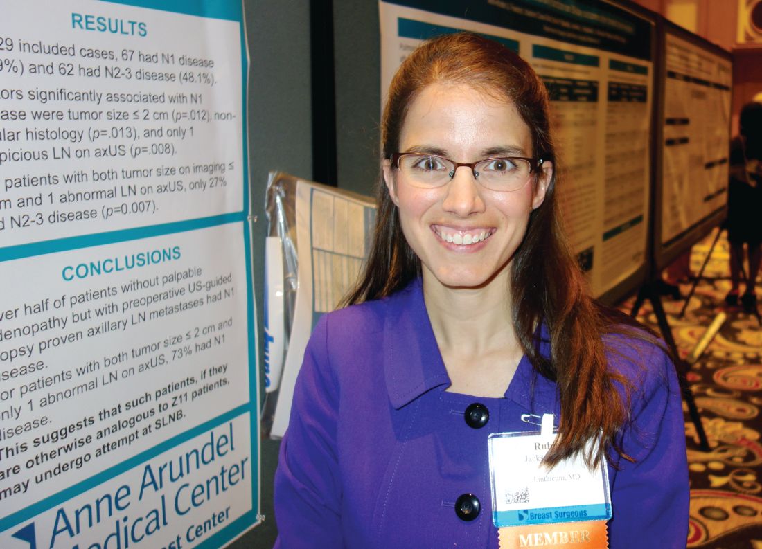
The problem that has come up in clinical practice is that axillary ultrasound is now a routine part of breast cancer workup, but the original trial didn’t address ultrasound, said senior investigator Rubie Sue Jackson, MD, at the annual meeting of the American Society of Breast Surgeons. As a result, “surgeons don’t know what to do with an ultrasound-detected suspicious node. I think a lot of surgeons, if they detect a positive lymph node by ultrasound, even if it’s nonpalpable, would not consider the patient a candidate for sentinel lymph node biopsy. Our data suggest that many of these patients are being overtreated if they have an upfront axillary lymph node dissection,” she said.
The 129 women had 1-3 suspicious, nonpalpable nodes on ultrasound that turned out to have metastatic disease on needle biopsy. They all had subsequent ALNDs.
On final pathology, 67 women (52%) had only one metastatic node. For those women, a sentinel lymph node biopsy was likely all that they required. “They probably did not benefit from having an ALND,” said Dr. Jackson, a breast surgeon at the Anne Arundel Medical Center in Annapolis, Md. The other 62 women (48%) had N2-3 disease.
A primary tumor sized 2 cm or smaller (P = .012); nonlobular histology (P = .013), and having only one suspicious nonpalpable node on ultrasound (P = .008) were all associated with NI disease. Of the women who met the criteria, only eight (27%) had N 2-3 disease (P = .007).
“Patients meeting the three criteria are particularly unlikely to have three or more positive sentinel lymph nodes” and require subsequent ALND. “You don’t need to do a complete axillary lymph node dissection upfront, as long as they are getting a lumpectomy and whole breast radiation,” Dr. Jackson said.
These days at Anne Arundel, “we do the axillary ultrasound, we biopsy the lymph node if it looks suspicious, but we don’t feel forced to do an ALND. If a patient has tumor biology that’s likely to be highly responsive, we do upfront chemotherapy. If they have luminal tumor biology that’s not going to be very responsive to neoadjuvant therapy,” with one or two suspicious nodes, “and they are planning to get breast conserving therapy, I would do a sentinel lymph node biopsy and x-ray the specimen to make sure that I’ve retrieved the clip,” she said.
Dr. Jackson didn’t have any disclosures, and there was no industry funding for the work.
aotto@frontlinemedcom.com
AT ASBS 2017
Key clinical point:
Major finding: Among the 30 women with tumors 2 cm or smaller, and only one abnormal lymph node on axillary ultrasound, 22 (73%) had metastasis limited to the one lymph node.
Data source: A review of 129 women with breast cancer.
Disclosures: There was no industry funding, and the senior investigator had no disclosures.
Decision to remove breast cancer metastases depends on location of lesions
LAS VEGAS – Determination of whether excision of persistent breast cancer metastases can benefit the patient and even prolong survival depends on the location of the metastatic lesions, an investigator said at the annual meeting of the American Society of Breast Surgeons.
Before making the decision, it’s important to restage the patient’s disease and to recheck the receptor status if a biopsy is accessible, said Roshni Rao, MD, of the University of Texas Southwestern Medical Center.
Brain

Survival is worse when there are concurrent extracranial metastases or when brain metastases are greater than 5 cm, in patients with triple negative tumors, and in patients with a Karnofsky score of 70 or less, Dr. Rao said.
Surgery has the greatest benefit in patients with a single metastasis, with no extracranial disease, and who are able to undergo adjuvant whole brain radiation. In these cases, surgery improves survival, lowers recurrence rates, and reduces the risk of death neurological causes, she said.
Long-term survival is most common with continuous adjuvant therapy, either trastuzumab or hormonal, and in patients with a longer time interval to development of metastases. One series showed a 20-month survival increase.
Liver
Approximately 15% of patients with synchronous metastases will have liver metastases, and about half of stage IV patients will experience liver metastases at some point during treatment.
There is evidence from colorectal cancer that removing liver metastases is beneficial, and that has prompted interest in a similar approach in breast cancer. Liver resection has also become safer with new advances.
Dr. Rao discussed a single-institution study which took an aggressive approach to liver resection in 85 patients (Ann Surg. 2006;244:897-907). The researchers found that increased survival was associated with a good response to adjuvant chemotherapy, an r0 or r1 resection, and in patients who had a previous liver resection and were healthy enough to undergo another resection.
Overall, existing studies support liver resection if there are one to three lesions, if negative margins can be achieved, if the tumors are hormone receptor positive, and if the cancer is hormone positive and has good response to chemotherapy.
Dr. Rao emphasized that liver resections should be performed with a multidisciplinary team and should only be attempted at centers with low morbidity and where the doctors are experienced with liver resection.
When it’s possible, liver resection is beneficial. “There have been multiple reports of long-term survivors with no evidence of disease. There is likely a survival benefit with careful selection of these patients,” Dr. Rao said.
Lung
Lung surgeries are becoming safer, especially with the availability of video-assisted techniques, and pulmonary metastases are increasingly being spotted using more sensitive techniques such as higher resolution computed tomography.
A lung metastasis registry analysis showed three factors improved survival: prolonged disease-free survival, especially longer than 36 months; a complete resection; and a small number of metastases and success in resecting them all (Eur J Cardiothorac Surg. 2002;22:335-44). A more recent meta-analysis showed the same results (J Thorac Dis. 2015;7:1441-51).
Bone
Bone metastases remain rare choices for surgical treatment. Most of the time, morbidity will be too high, and there are good options for systemic treatment. That leaves surgery reserved mostly for stabilization or the treatment of fractures.
However, there are a couple of exceptions, according to Dr. Rao. One multi-institutional randomized trial looked at metastatic epidural spinal cord compression. Subjects either underwent decompressive surgery with stabilization and radiation or radiation alone. Patients in the surgical group had a longer ambulatory period and had a lower usage rate of steroids and opioids. Morbidity outcomes were similar in both groups. Patients whose primary tumor was in the breast seemed to benefit the most with respect to ambulatory time (Lancet. 2005;9486:643-8; J Neurosurg Spine. 2008;8:271-8).
Sternal metastases represent another special case. 70% of the time, patients with sternal metastases have it as their only metastatic site. A French series of 33 patients who underwent aggressive chest wall and rib resection reported a 36% complication rate, while another study of 28 patients showed a 21% complication rate. Those complications are a problem, “but if you’re able to perform this in a resected tumor, there are long term survivors. As usual, triple negative breast cancers predicted a worse prognosis,” said Dr. Rao.
Dr. Rao concluded that resection of metastatic sites has a role. “I think it’s our responsibility as breast surgeons who are many times continuously following these patients to consider appropriate operations,” said Dr. Rao.
However, she did sound one note of caution. Surgery can interrupt therapy that is helping a patient. “Let’s say you have someone get a big liver resection, and then they have a tough time with recovery. There could be a long period of time they can’t get the therapy that was keeping them alive. That’s the real concern,” she said.
LAS VEGAS – Determination of whether excision of persistent breast cancer metastases can benefit the patient and even prolong survival depends on the location of the metastatic lesions, an investigator said at the annual meeting of the American Society of Breast Surgeons.
Before making the decision, it’s important to restage the patient’s disease and to recheck the receptor status if a biopsy is accessible, said Roshni Rao, MD, of the University of Texas Southwestern Medical Center.
Brain
Survival is worse when there are concurrent extracranial metastases or when brain metastases are greater than 5 cm, in patients with triple negative tumors, and in patients with a Karnofsky score of 70 or less, Dr. Rao said.
Surgery has the greatest benefit in patients with a single metastasis, with no extracranial disease, and who are able to undergo adjuvant whole brain radiation. In these cases, surgery improves survival, lowers recurrence rates, and reduces the risk of death neurological causes, she said.
Long-term survival is most common with continuous adjuvant therapy, either trastuzumab or hormonal, and in patients with a longer time interval to development of metastases. One series showed a 20-month survival increase.
Liver
Approximately 15% of patients with synchronous metastases will have liver metastases, and about half of stage IV patients will experience liver metastases at some point during treatment.
There is evidence from colorectal cancer that removing liver metastases is beneficial, and that has prompted interest in a similar approach in breast cancer. Liver resection has also become safer with new advances.
Dr. Rao discussed a single-institution study which took an aggressive approach to liver resection in 85 patients (Ann Surg. 2006;244:897-907). The researchers found that increased survival was associated with a good response to adjuvant chemotherapy, an r0 or r1 resection, and in patients who had a previous liver resection and were healthy enough to undergo another resection.
Overall, existing studies support liver resection if there are one to three lesions, if negative margins can be achieved, if the tumors are hormone receptor positive, and if the cancer is hormone positive and has good response to chemotherapy.
Dr. Rao emphasized that liver resections should be performed with a multidisciplinary team and should only be attempted at centers with low morbidity and where the doctors are experienced with liver resection.
When it’s possible, liver resection is beneficial. “There have been multiple reports of long-term survivors with no evidence of disease. There is likely a survival benefit with careful selection of these patients,” Dr. Rao said.
Lung
Lung surgeries are becoming safer, especially with the availability of video-assisted techniques, and pulmonary metastases are increasingly being spotted using more sensitive techniques such as higher resolution computed tomography.
A lung metastasis registry analysis showed three factors improved survival: prolonged disease-free survival, especially longer than 36 months; a complete resection; and a small number of metastases and success in resecting them all (Eur J Cardiothorac Surg. 2002;22:335-44). A more recent meta-analysis showed the same results (J Thorac Dis. 2015;7:1441-51).
Bone
Bone metastases remain rare choices for surgical treatment. Most of the time, morbidity will be too high, and there are good options for systemic treatment. That leaves surgery reserved mostly for stabilization or the treatment of fractures.
However, there are a couple of exceptions, according to Dr. Rao. One multi-institutional randomized trial looked at metastatic epidural spinal cord compression. Subjects either underwent decompressive surgery with stabilization and radiation or radiation alone. Patients in the surgical group had a longer ambulatory period and had a lower usage rate of steroids and opioids. Morbidity outcomes were similar in both groups. Patients whose primary tumor was in the breast seemed to benefit the most with respect to ambulatory time (Lancet. 2005;9486:643-8; J Neurosurg Spine. 2008;8:271-8).
Sternal metastases represent another special case. 70% of the time, patients with sternal metastases have it as their only metastatic site. A French series of 33 patients who underwent aggressive chest wall and rib resection reported a 36% complication rate, while another study of 28 patients showed a 21% complication rate. Those complications are a problem, “but if you’re able to perform this in a resected tumor, there are long term survivors. As usual, triple negative breast cancers predicted a worse prognosis,” said Dr. Rao.
Dr. Rao concluded that resection of metastatic sites has a role. “I think it’s our responsibility as breast surgeons who are many times continuously following these patients to consider appropriate operations,” said Dr. Rao.
However, she did sound one note of caution. Surgery can interrupt therapy that is helping a patient. “Let’s say you have someone get a big liver resection, and then they have a tough time with recovery. There could be a long period of time they can’t get the therapy that was keeping them alive. That’s the real concern,” she said.
EXPERT ANALYSIS FROM ASBS 2017
Androgen receptor screening not ready for triple-negative breast cancer
LAS VEGAS – Despite the early promise of antiandrogen therapy, it’s not time yet to routinely screen women with triple-negative breast cancer for androgen receptors, according to Tiffany A. Traina, MD, the head of research into the disease at Memorial Sloan Kettering Cancer Center, New York.

There’s no standardized test for androgen receptors in breast cancer, so people “are doing different kinds of testing.” In the literature, “the range of AR positivity is anywhere from 12% to 79%, which reflects how we are all over the map in methodology; you might just as well throw a dart at the board. I would encourage screening in the context of the ongoing trials,” Dr. Traina said.
More than a decade ago, Memorial Sloan Kettering found a subset of TNBC that had ARs, which was peculiar because the tumors weren’t otherwise responsive to hormones. Androgen exposure increased growth, but the AR antagonist flutamide (Eulexin)blocked it. “It was thought provoking. There are a lot of drugs in the prostate cancer world” such as flutamide that shut down androgens, she said (Oncogene. 2006 Jun 29;25[28]:3994-4008).
Several have been tried, and investigations are ongoing. The work matters because TNBC is a particularly bad diagnosis. Blocking androgens seems to give some women a few more months of life.
Dr. Traina was the senior author in an early proof-of-concept study for AR blockade that involved 26 women with metastatic TNBC who had been through up to eight prior chemotherapy regimens. The women received 150 mg daily of the prostate cancer AR antagonist bicalutamide (Casodex). Disease remained stable in five (19%) for more than 6 months. Median progression-free survival was 12 weeks, which was “not that far off from what you get with [standard] chemotherapies. This was encouraging, and it led to multiple other trials looking at targeted therapies,” she said (Clin Cancer Res. 2013 Oct 1; 19[19]: 5505-12).
Dr. Traina led a phase II investigation of the prostate cancer AR antagonist enzalutamide (Xtandi) in 118 women with advanced AR-positive TNBC. Her team created an androgen-driven gene signature as a potential biomarker of response. Median progression-free survival was 32 weeks in the 56 women (47%) who were positive for the gene signature, but 9 weeks in those who were not. There were two complete responses and five partial responses with enzalutamide. Currently, “we are looking at using enzalutamide for patients with AR-positive TNBC in the early stage after failure of standard therapies,” she said.
French investigators recently reported a 6-month clinical benefit – including one complete response – in 7 (21%) of 34 women with locally advanced or metastatic TNBC who were treated with 1,000 mg daily of abiraterone acetate (Zytiga), an androgen biosynthesis inhibitor approved for prostate cancer (Ann Oncol. 2016 May;27[5]:812-8).
“We still have a ways to go” before AR treatment reaches the clinic for routine breast cancer treatment, “but there’s reason for hope,” Dr. Traina said.
Dr. Traina reported funding, honoraria, and steering committing payments from a number of companies working on or marketing TNBC AR drugs, including Pfizer, Astellas, Innocrin, AstraZeneca, Eisai, and Merck.
LAS VEGAS – Despite the early promise of antiandrogen therapy, it’s not time yet to routinely screen women with triple-negative breast cancer for androgen receptors, according to Tiffany A. Traina, MD, the head of research into the disease at Memorial Sloan Kettering Cancer Center, New York.
There’s no standardized test for androgen receptors in breast cancer, so people “are doing different kinds of testing.” In the literature, “the range of AR positivity is anywhere from 12% to 79%, which reflects how we are all over the map in methodology; you might just as well throw a dart at the board. I would encourage screening in the context of the ongoing trials,” Dr. Traina said.
More than a decade ago, Memorial Sloan Kettering found a subset of TNBC that had ARs, which was peculiar because the tumors weren’t otherwise responsive to hormones. Androgen exposure increased growth, but the AR antagonist flutamide (Eulexin)blocked it. “It was thought provoking. There are a lot of drugs in the prostate cancer world” such as flutamide that shut down androgens, she said (Oncogene. 2006 Jun 29;25[28]:3994-4008).
Several have been tried, and investigations are ongoing. The work matters because TNBC is a particularly bad diagnosis. Blocking androgens seems to give some women a few more months of life.
Dr. Traina was the senior author in an early proof-of-concept study for AR blockade that involved 26 women with metastatic TNBC who had been through up to eight prior chemotherapy regimens. The women received 150 mg daily of the prostate cancer AR antagonist bicalutamide (Casodex). Disease remained stable in five (19%) for more than 6 months. Median progression-free survival was 12 weeks, which was “not that far off from what you get with [standard] chemotherapies. This was encouraging, and it led to multiple other trials looking at targeted therapies,” she said (Clin Cancer Res. 2013 Oct 1; 19[19]: 5505-12).
Dr. Traina led a phase II investigation of the prostate cancer AR antagonist enzalutamide (Xtandi) in 118 women with advanced AR-positive TNBC. Her team created an androgen-driven gene signature as a potential biomarker of response. Median progression-free survival was 32 weeks in the 56 women (47%) who were positive for the gene signature, but 9 weeks in those who were not. There were two complete responses and five partial responses with enzalutamide. Currently, “we are looking at using enzalutamide for patients with AR-positive TNBC in the early stage after failure of standard therapies,” she said.
French investigators recently reported a 6-month clinical benefit – including one complete response – in 7 (21%) of 34 women with locally advanced or metastatic TNBC who were treated with 1,000 mg daily of abiraterone acetate (Zytiga), an androgen biosynthesis inhibitor approved for prostate cancer (Ann Oncol. 2016 May;27[5]:812-8).
“We still have a ways to go” before AR treatment reaches the clinic for routine breast cancer treatment, “but there’s reason for hope,” Dr. Traina said.
Dr. Traina reported funding, honoraria, and steering committing payments from a number of companies working on or marketing TNBC AR drugs, including Pfizer, Astellas, Innocrin, AstraZeneca, Eisai, and Merck.
EXPERT ANALYSIS FROM ASBS 2017
Breast density is no reason to perform MRI
LAS VEGAS – In women with higher density (HD) breasts, preoperative MRI revealed more abnormalities than were seen in women with lower density (LD) breasts, but there was no difference in the number of secondary cancers detected or long-term recurrence rates.
Breast density is often cited by radiologists as a reason to conduct a preoperative MRI, but the study suggests that it should not be a driving factor. “It’s a real challenge when our radiologists provide us reports that say, ‘Due to increased density, we recommend MRI,’ because it’s really hard to then disregard that. I think this is very important data,” said Judy Boughey, MD, professor of surgery at the Mayo Clinic, Rochester, MN, who moderated the session at the annual meeting of the American Society of Breast Surgeons where the research was presented.
“MRI is a valuable tool, and we’re still trying to figure out who it should be performed in,” said lead author Sarah McLaughlin, MD, of the Mayo Clinic, Jacksonville, FL, in an interview.
The researchers retrospectively analyzed data from 683 women at their institution who underwent preoperative MRI between 2007 and 2011. They grouped them by mammography results into LD (33%; Breast Imaging–Reporting and Data System density, 1 and 2) and HD (67%; BI-RADS density, 3 and 4).
Patients in the HD group more often had ipsilateral MRI findings (42% vs. 31%; P = .005), but ,of those with MRI findings, a similar number of patients in each group needed a second site biopsy (HD 65% vs. LD 67%; P = .78).
In all patients who had an additional MRI finding, the odds of detecting an additional ipsilateral cancer were not statistically significant between HD (32%) and LD (23%; P = .15) patients.
HD patients were also more likely to have abnormalities in the contralateral breast (25% vs. 14%; P = .009), but there were no statistically significant differences in rates of second-site biopsy recommendations or in the percentages of abnormalities that turned out to be cancerous (HD 6% vs. LD 3%; P = 1.0).
Following MRI, 70% of LD patients expressed a preference for breast-conserving surgery, compared with 53% of HD patients (P = .0001).
Over a median 7 years of follow-up, there was no difference in freedom from recurrence rates between the two groups (91% in LD vs. 90% in HD; P = .57).
“To me, it says that you don’t have to order an MRI just because they have cancer in a high density breast. You can feel reassured by your surgical plan and treatment recommendations based on conventional imaging,” said Dr. McLaughlin.
The researchers can’t determine if having an MRI done increased patient worry and potentially led to the higher rate of mastectomies chosen by women in the HD group. “Is that a result of the MRI? I don’t think we can say that, but there’s this whole other discussion piece that goes into it. You definitely see patients who say, ‘But it found these other things, and I’m going to have a mastectomy.’ So, there’s that patient preference and worry piece,” said Dr. McLaughlin.
The study results should offer some reassurance to patients. “There were no differences in local recurrence rates according to density. Maybe the next angle is allaying some of that fear, because the outcomes were the same. It’s really driven more by tumor biology and multimodality therapy,” said Dr. McLaughlin.
The study doesn’t provide the final word on breast density and MRI, according to Dr. Boughey. “I think this is an area that needs to be studied more with a clinical trial. There are several going on in different countries, and this is an area where we need level 1 data. This study does fit with what many other studies have shown, which is that MRI probably doesn’t have as much benefit as patients believe it does, so our role really is to try to help educate patients.”
AT ASBS 2017
Key clinical point: High breast density is probably not cause enough to order preoperative MRI.
Major finding: Freedom from recurrence rates were 90% in high density and 91% in low.
Data source: Retrospective analysis of 683 women at a single institution.
Disclosures: The study was funded internally. Dr. McLaughlin and Dr. Boughey reported having no relevant financial relationships.
Radiation bests mastectomy for occult breast cancer
LAS VEGAS – Overall survival was better when women with occult breast cancer had axillary lymph node dissection and radiation, instead of mastectomy, in a database review of 934 cases by the University of Maryland Medical Center, Baltimore, the largest review to date of how best to handle the problem.
Five- and 10-year overall survival was 90.8% and 84.8%, respectively, among the 342 women treated with axillary lymph node dissection (ALND) plus adjuvant radiation, versus 80.0% and 69.8% among the 592 who had ALND and mastectomies, plus or minus radiation, according to an analysis of the National Cancer Database from 2004-2013. The results were presented at the annual meeting of the American Society of Breast Surgeons.
ALND plus radiation was independently associated with overall survival on multivariate analysis (HR 0.51, 95% CI 0.32-0.81, P = .004), and was associated with fewer comorbidities, use of chemotherapy, number of positive nodes, and number of nodes examined, compared with mastectomy.
Women treated with ALND plus radiation “had significantly better overall survival than those treated with mastectomy, even after adjusting for other covariates. We believe the study supports overall use of this treatment approach in patients with occult breast cancer,” said lead investigator, Lindsay Hessler, MD, of University of Maryland, Baltimore.
Occult breast cancer – axillary lymph node metastases without clinical or radiologic evidence of a primary breast tumor – is rare and accounted for less than 0.1% of the 2.03 million breast cancer cases in the database. It’s been unclear how best to treat it; most of the previous investigations were small single-center series and case reports.
The only other significant review was smaller, with 750 women in the Surveillance, Epidemiology, and End Results database treated from 1983 to 2006, the “vast majority” before routine use of breast MRI. It showed that “definitive locoregional treatment with either mastectomy or [radiation therapy] improves [overall survival] in patients with occult breast cancer and axillary metastasis who undergo ALND,” but it didn’t suggest which option is best. The National Comprehensive Cancer Network recommends either approach (Cancer. 2010 Sep 1;116[17]:4000-6).
The new University of Maryland findings “confirm that women do not need to have a mastectomy if you can’t find the cancer in their breast. Women do better if you radiate the breast instead of removing it. A lot of academic centers are doing this now, but some people don’t know about it. This needs to be implemented in a more widespread fashion,” said Shelley Hwang, MD, a surgical oncologist at Duke University, Durham, N.C., who moderated Dr. Hessler’s presentation.
Indeed, patients were most likely to be treated with radiation and ALND at an academic center (OR 2.03, 95% CI 1.5-2.74, P less than .001), the only factor on multivariate analysis related to treatment choice.
The review excluded women with only internal mammary lymph node involvement, those with lumpectomies, and women who had less than four nodes recovered on ALND. Mastectomy and radiation patients were similar in nodal stage, race, income, insurance, estrogen receptor status, comorbidities, and year of diagnosis. On pathology, a tumor was found in about a third of the patients who had mastectomies. MRI use and recurrence rates were unavailable in the National Cancer Database.
The findings are subject to all the limits of database reviews, including the possible confounder that women treated at university hospitals might also have had more optimal systemic therapy, as an audience member noted.
The investigators said they had no financial disclosures.
AT ASBS 2017
Key clinical point:
Major finding: Five- and 10-year overall survival was 90.8% and 84.8%, respectively, among the 342 women treated with axillary lymph node dissection (ALND) plus adjuvant radiation, versus 80.0% and 69.8% among the 592 who had ALND and mastectomies, plus or minus radiation.
Data source: Review of 934 cases in the National Cancer Database.
Disclosures: The investigators said they had no financial disclosures.
Mastectomy unnecessary for some breast cancer recurrences
LAS VEGAS – Although mastectomy is the standard of care for tumor recurrence following lumpectomy and whole breast irradiation, a second lumpectomy with partial breast irradiation is a sound alternative under certain circumstances, according to Manjeet Chadha, MD, professor of radiation oncology and director of the department of radiation oncology at Icahn School of Medicine at Mount Sinai, New York.
It depends on whether the new lesion is a true recurrence, or simply another primary tumor. In the absence of a genetic footprint to compare the two, Dr. Chadha and her colleagues use several of what she called “soft criteria” to make the call and counsel women.
True ipsilateral recurrence of an aggressive tumor tends to happen early, and in the same quadrant. However, if breast cancer recurs more than 3 years after treatment of the primary tumor and in a different quadrant, and if the patient is negative for BRCA mutation, and if the new growth is small, localized on MRI, histologically different from the primary tumor, and likely to be resected with clean margins, Dr. Chadha said she is comfortable offering a second lumpectomy and partial breast radiation – usually multicatheter brachytherapy – to women who do not want a mastectomy.
“Second cancer in a previously irradiated breast is not an uncommon clinical entity. Based on patient preference, the option of repeat breast conservation and reirradiation may be offered selectively as an alternative to mastectomy,” followed by systemic therapy, she said at the annual meeting of the American Society of Breast Surgeons.
“I think all of us across the country are discussing presentations like this in tumor boards,” but it’s not always offered as an option. Sometimes, “the mindset of the treating surgeon is ‘oh, this breast has had radiation; I can’t give radiation again.’ Clearly, whole breast reirradiation is not recommended,” but it seems possible based on a growing body of literature to differentiate new primaries with new biology from true recurrences, and to treat them safely with breast conserving surgery and partial irradiation, she said.
The largest series to date of salvage lumpectomy with multicatheter brachytherapy followed 217 women for a median of 3.9 years. Median tumor size was 1.2 cm. The 5-year local control rate – effectively, the mastectomy-free survival – was 94.4%, and the overall survival was 88.7%, which mirrors the success of first-time lumpectomy with whole breast irradiation, and lends support to the notion that some recurrences are, in fact, entirely new disease. The European team reported excellent or good cosmetic results in 85% of women (Radiother Oncol. 2013 Aug;108[2]:226-31).
The series used high-dose radiation. Dr. Chadha said she and her colleagues have had similar success with low-dose multicatheter brachytherapy, with similarly good aesthetic results. To avoid cosmetic impact, however, she noted it’s important to work with radiation oncologists “mindful of the nuances of what’s needed,” including how far to separate the skin from the radiation.
Brachytherapy has the most support in the literature, but external beam therapy is also an option. “Whatever technique you use, the delineation of the target and the geometric coverage of the lumpectomy cavity [must be] complete in all cases,” she said.
Dr. Chadha had no financial conflicts of interest.
EXPERT ANALYSIS FROM ASBS 2017