User login
American Society for Dermatologic Surgery (ASDS): Annual Meeting
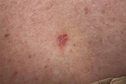
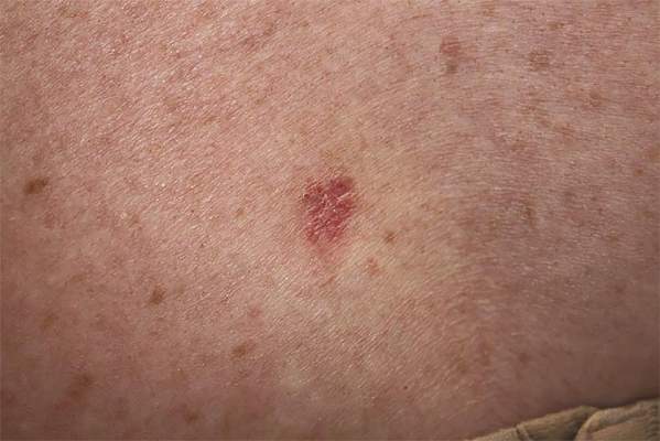
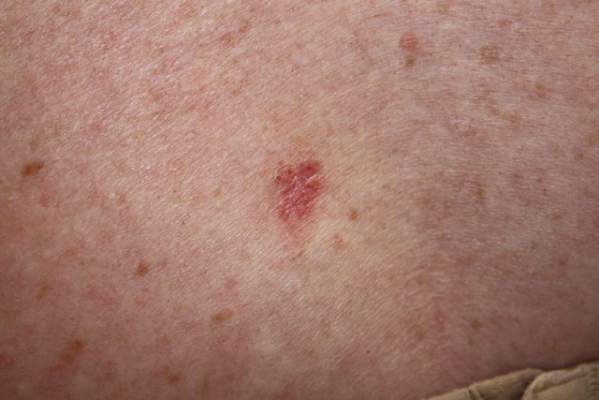
RNA sequencing characterized high-risk squamous cell carcinomas
SAN DIEGO – Cutaneous squamous cell carcinomas from organ transplant recipients had a more aggressive molecular profile than did tumor samples from immunocompetent patients, according to an RNA sequencing study presented at the annual meeting of the American Society for Dermatologic Surgery.
Specimens from organ transplant recipients showed greater induction of biologic pathways related to cancer signaling, fibrosis, and extracellular matrix remodeling, said Dr. Cameron Chesnut, a dermatologist in private practice in Spokane, Wash., who carried out the research while he was a dermatologic surgery resident at the University of California, Los Angeles.
Furthermore, the TP53 tumor suppressor gene was inhibited at least five times more in samples from organ transplant recipients, compared with those from immunocompetent patients, Dr. Chesnut said in an interview.
Squamous cell carcinoma (SCC) is the most common cancer to occur after organ transplantation, Dr. Chesnut and his associates noted. The malignancy is 65-250 times more common, is more than 4 times more likely to metastasize, and has a mortality rate of 5% compared with a rate of less than 1% in immunocompetent patients, based on data published online in the journal F1000 Prime Reports, they said.
To characterize these high-risk SCCs and compare them with lower-risk SCCs, the researchers performed RNA sequencing of three normal skin samples and SCC specimens from 15 patients – 7 organ transplant recipients and 8 otherwise healthy individuals. The researchers used an Illumina GAIIx RNA Seq instrument to generate RNA sequencing libraries of the specimens. They also used the web-based Ingenuity Pathway Analysis technique to identify the major biological pathways regulated within the tumors.
In all, 690 highly expressed genes were induced at least fivefold in SCCs from organ transplant recipients compared with those from otherwise healthy patients. These genes encoded pathways related to fibrosis, extracellular remodeling, the cell cycle, and tumor signaling, the investigators said. The COX-2 pathway for prostaglandin synthesis also was induced fivefold or more in the high-risk SCCs compared with those from immunocompetent patients, Dr. Chesnut added.
The researchers also identified 1,290 highly expressed genes that were inhibited at least fivefold in SCCs from organ transplant recipients compared with specimens from immunocompetent patients. The most strongly inhibited pathways were related to sterol biosynthesis and epithelial differentiation, followed by nucleotide excision repair, interleukin-6 and IL-17, and apoptosis, they said.
Based on these findings, novel therapeutics might someday be able to target specific biologic pathways that are highly induced in SCCs from organ transplant recipients, Dr. Chesnut said. “It’s hard to say what the most likely candidates are,” but based on the study findings, “regulating inflammation may be a target,” he added. Dr. Chesnut and his associates reported no external funding sources or conflicts of interest.
Key clinical point: Squamous cell carcinomas from organ transplant recipients showed a more aggressive molecular profile than did those from immunocompetent individuals.
Major finding: The high-risk tumors showed greater induction of biologic pathways related to cancer signaling, fibrosis, and extracellular matrix remodeling, and inhibition of the tp53 tumor suppressor gene.
Data source: RNA sequencing of 15 squamous cell carcinomas, including seven from organ transplant recipients.
Disclosures: The investigators reported no external funding sources or conflicts of interest.
Ionizing radiation linked to BCC
SAN DIEGO – Patients treated with ionizing radiation were 2.65 to 3.78 times more likely to develop basal cell carcinoma than were controls, and exposure at younger ages or relatively high doses further increased that risk, according to a pooled analysis presented at the annual meeting of the American Society for Dermatologic Surgery.
“Concomitant exposure to ultraviolet radiation may potentiate this effect,” said Dr. Min Deng, a dermatology resident at the University of Chicago. “With newer treatment protocols and improved shielding, it would be interesting to see if the effect of ionizing radiation has changed, or whether we as dermatologists should more actively screen this at-risk population.”
Basal cell carcinoma (BCC) is the most common skin cancer worldwide, but relatively few dermatology papers have assessed the effects of ionizing radiation on the incidence of BCC, said Dr. Deng and coauthor Dr. Diana Bolotin, also of the University of Chicago.
To better understand the link, the researchers searched PubMed for controlled studies on the topic by using the terms “radiation,” “risk,” and “basal cell carcinoma.” They excluded case reports, animal studies, studies published in languages other than English, and trials of radiation as a treatment of BCC, they said. They also excluded studies of atomic bomb survivors, because exposure was uncontrolled and methods to estimate exposure in this group have changed over time, they noted.
In all, six studies published between 1991 and 2012 met the inclusion criteria, and all six showed a statistically significant relative risk or odds of BCC with ionizing radiation exposure, the investigators reported. Three analyses calculated the relative risk of BCC in patients who received radiation for tinea capitis or who underwent total-body irradiation before hematopoietic cell transplantation, they said. Two studies evaluated the odds of BCC in patients with a past history of radiation exposure, and one study assessed time to subsequent BCCs in patients with a history of BCC.
For the three studies that calculated relative risk, the researchers calculated a pooled RR of BCC after ionizing radiation treatment of 2.65 (95% confidence interval, 1.22-5.72) compared with controls. The study of total-body irradiation (TBI) did not report or control for the primary disease for which patients were treated, “which may have confounded the results,” they said. When they excluded that study from their calculation, the overall RR rose to 3.78 (95% CI, 2.62 to 5.44).
Notably, in the two studies of patients with tinea capitis, the relative risk of BCC fell by 12% to 16% for every additional year of age at which patients received radiation treatment, the researchers said. Similarly, risk of BCC dropped by 10.9% for every 1-year increase in age at total-body irradiation prior to cell transplantation.
The combined odds ratio for the next two studies was 4.28 (1.45-12.63). “Likewise, both studies found an elevated odds ratio with younger age at radiation exposure,” the researchers added. One study that stratified patients by type of medical condition detected a “markedly elevated” 8.7 odds of BCC after radiation treatment for acne (2.0 to 38.0), they noted.
The sixth study was a nested case-control analysis that found a statistically significant increase in the odds of BCC starting at a 1-Gy dose of ionizing radiation, and rising linearly up to doses of 35-63.3 Gy. “The risk for developing multiple BCCs also appears to be elevated in patients with a history of radiation therapy,” the researchers said. Patients who had been exposed to ionizing ratio were 2.3 times more likely to develop new BCCs compared with unexposed patients (1.7 to 3.1), they said.
The investigators reported no funding sources or conflicts of interest.
Key clinical point: Ionizing radiation therapy significantly increased risk of basal cell carcinoma, especially when patients were younger or were treated at relatively high doses.
Major finding: The pooled relative risk of BCC after ionizing radiation treatment was 2.65 (95% confidence interval, 1.22-5.72) compared with controls.
Data source: Pooled analysis of six studies of ionizing radiation exposure and risk or odds of basal cell carcinoma.
Disclosures: The researchers reported no funding sources or conflicts of interest.
Ultrasound offers noninvasive option for body contouring, skin tightening
SAN DIEGO – Ultrasound treatments are an alternative for patients looking to avoid the pain and prolonged recovery associated with surgical body contouring or skin tightening procedures, according to Dr. Elizabeth L. Tanzi. Currently, several types of ultrasound devices are available for such procedures, she said at the annual meeting of the American Society for Dermatologic Surgery.
High-intensity, focused ultrasound and pulsed focused ultrasound devices are used for body contouring, and the micro-focused high-intensity ultrasound device is used for skin tightening, Dr. Tanzi explained. Body contouring has rapidly gained popularity in the United States and elsewhere, but surgical contouring procedures can be painful and can require prolonged recovery times, she noted.

Ultrasound for body contouring works via two noninvasive mechanisms, both of which cause selective damage of adipose tissue, said Dr. Tanzi, codirector of the Washington (D.C.) Institute of Dermatologic Laser Surgery.
First, adipose tissue directly absorbs ultrasonic energy during treatment, leading to immediate thermal coagulation and necrosis of fat cells, she said. Mechanical processes also break down tissue, such as when shear forces are created at high levels of pressure during treatment. Circulating macrophages then absorb the damaged adipocytes and the triglycerides they release, leading to a gradual reduction of the fat layer at the treatment site, she added.
When using ultrasound for body contouring, “patient selection is paramount,” Dr. Tanzi emphasized. Although the procedure carries no risk of infection or scarring, ultrasound yields subtler cosmetic results than would liposuction, and patients should understand that difference up front, she said. “This is body contouring, not weight loss,” she added. Patients can typically expect to see a slow reduction in adipose tissue at the treatment area about 1-4 months after treatment, and they also should understand that they might need multiple treatments to achieve their cosmetic goals, Dr. Tanzi said.
“Discuss side effects with patients,” Dr. Tanzi added. When used for body contouring, ultrasound can cause temporary erythema, bruising at the treatment site, soreness, and contour defects, although the latter are rare, she noted. Dr. Tanzi said she now treats patients with lower levels of ultrasonic energy to improve their comfort. “We are much more aggressive now with the overall number of lines we place on the body,” she added. “Go over the areas again and again.”
For example, in one clinical study, high-intensity focused ultrasound yielded statistically significant decreases in waist circumference at 12 weeks with treatment of 150-180 Joules per square centimeter, she noted. However, six passes at just 30 J/cm2 was more comfortable and gave similar results, she said (Dermatol. Surg. 2014;40:641-51).
Micro-focused high-intensity ultrasound can be used for skin tightening, particularly on the face. The procedure targets precise areas of adipose tissue within the skin to create small foci of thermal necrosis, and also stimulates localized collagen production for about 3-6 months afterward, Dr. Tanzi said. As with body contouring, patients should expect subtler results than would be expected from a surgical procedure, but the absence of scarring and the opportunity to immediately return to regular activities can bolster their satisfaction, she said.
Dr. Tanzi is a consultant for Ulthera, Solta, Cynosure/Palomar, Syneron & Candela, and Cutera, and has received financial support from Zeltiq, miraDry, and Clarisonic.
SAN DIEGO – Ultrasound treatments are an alternative for patients looking to avoid the pain and prolonged recovery associated with surgical body contouring or skin tightening procedures, according to Dr. Elizabeth L. Tanzi. Currently, several types of ultrasound devices are available for such procedures, she said at the annual meeting of the American Society for Dermatologic Surgery.
High-intensity, focused ultrasound and pulsed focused ultrasound devices are used for body contouring, and the micro-focused high-intensity ultrasound device is used for skin tightening, Dr. Tanzi explained. Body contouring has rapidly gained popularity in the United States and elsewhere, but surgical contouring procedures can be painful and can require prolonged recovery times, she noted.
Ultrasound for body contouring works via two noninvasive mechanisms, both of which cause selective damage of adipose tissue, said Dr. Tanzi, codirector of the Washington (D.C.) Institute of Dermatologic Laser Surgery.
First, adipose tissue directly absorbs ultrasonic energy during treatment, leading to immediate thermal coagulation and necrosis of fat cells, she said. Mechanical processes also break down tissue, such as when shear forces are created at high levels of pressure during treatment. Circulating macrophages then absorb the damaged adipocytes and the triglycerides they release, leading to a gradual reduction of the fat layer at the treatment site, she added.
When using ultrasound for body contouring, “patient selection is paramount,” Dr. Tanzi emphasized. Although the procedure carries no risk of infection or scarring, ultrasound yields subtler cosmetic results than would liposuction, and patients should understand that difference up front, she said. “This is body contouring, not weight loss,” she added. Patients can typically expect to see a slow reduction in adipose tissue at the treatment area about 1-4 months after treatment, and they also should understand that they might need multiple treatments to achieve their cosmetic goals, Dr. Tanzi said.
“Discuss side effects with patients,” Dr. Tanzi added. When used for body contouring, ultrasound can cause temporary erythema, bruising at the treatment site, soreness, and contour defects, although the latter are rare, she noted. Dr. Tanzi said she now treats patients with lower levels of ultrasonic energy to improve their comfort. “We are much more aggressive now with the overall number of lines we place on the body,” she added. “Go over the areas again and again.”
For example, in one clinical study, high-intensity focused ultrasound yielded statistically significant decreases in waist circumference at 12 weeks with treatment of 150-180 Joules per square centimeter, she noted. However, six passes at just 30 J/cm2 was more comfortable and gave similar results, she said (Dermatol. Surg. 2014;40:641-51).
Micro-focused high-intensity ultrasound can be used for skin tightening, particularly on the face. The procedure targets precise areas of adipose tissue within the skin to create small foci of thermal necrosis, and also stimulates localized collagen production for about 3-6 months afterward, Dr. Tanzi said. As with body contouring, patients should expect subtler results than would be expected from a surgical procedure, but the absence of scarring and the opportunity to immediately return to regular activities can bolster their satisfaction, she said.
Dr. Tanzi is a consultant for Ulthera, Solta, Cynosure/Palomar, Syneron & Candela, and Cutera, and has received financial support from Zeltiq, miraDry, and Clarisonic.

Biopsy can underestimate diversity, aggressiveness of basal cell carcinomas
SAN DIEGO – Histology of basal cell carcinomas removed by Mohs micrographic surgery showed that presurgical biopsies had not revealed all tumor subtypes in 64% of cases, and had underestimated the aggressiveness of the tumors 24% of the time, according to data from a large, multicenter, retrospective study.
“Unfortunately, while cheap and cost-effective, biopsies are a subsample of the full malignancy,” said Dr. Murad Alam, professor of dermatology, otolaryngology, and surgery at Northwestern University in Chicago. “Skin biopsy of basal cell carcinoma [BCC] may fail to detect all BCC subtypes, and as such may underestimate the aggressiveness of an individual BCC tumor.”

Basal cell carcinoma is the most common skin cancer worldwide, and can broadly be grouped into aggressive and indolent types, Dr. Alam said at the annual meeting of the American Society for Dermatologic Surgery. But tumors often show mixed histology, and cancer treatment needs to target the most aggressive subtype present in the tumor, he added. Results of past studies suggested that biopsies of BCCs could miss tumor subtypes, but the current research is the first large, multicenter study to confirm these findings, he and his associates said.
For the study, the investigators compared biopsy reports and microscopic slides of Mohs micrographic surgery (MMS) specimens from 871 consecutive cases of BCC treated at three hospitals in Illinois from 2013 to 2014. Patients first underwent biopsies, followed by complete excision of their tumors during MMS. Almost 59% of patients were male, and tumors were most commonly removed from the nose or cheek. In all, 78% of biopsies were obtained by the shave technique, but punch and excisional biopsies also were performed, the researchers noted.
Using standard definitions of BCC subtypes, the investigators compared levels of concordance between biopsy and MMS histology findings, Dr. Alam said. They also grouped tumor specimens as high risk (that is, infiltrative, morpheic, micronodular, basosquamous) or low risk (superficial or nodular), and determined whether tumor biopsy and MMS histology yielded the same or discordant risk assessments, he added.
Biopsies identified only 18% of tumors as being of mixed histology, compared with 57% of MMS specimens, said Dr. Alam. Biopsy results matched MMS histologies in only 31% of cases, while in 64% of cases, the MMS specimen yielded more tumor subtypes than the biopsy specimen. The researchers noted that biopsy yielded more subtypes than did MMS in 4% of cases, and that MMS and biopsy subtypes were fully discordant in only four cases.
Dr. Alam and his associates declared no external funding sources or conflicts of interest.
SAN DIEGO – Histology of basal cell carcinomas removed by Mohs micrographic surgery showed that presurgical biopsies had not revealed all tumor subtypes in 64% of cases, and had underestimated the aggressiveness of the tumors 24% of the time, according to data from a large, multicenter, retrospective study.
“Unfortunately, while cheap and cost-effective, biopsies are a subsample of the full malignancy,” said Dr. Murad Alam, professor of dermatology, otolaryngology, and surgery at Northwestern University in Chicago. “Skin biopsy of basal cell carcinoma [BCC] may fail to detect all BCC subtypes, and as such may underestimate the aggressiveness of an individual BCC tumor.”
Basal cell carcinoma is the most common skin cancer worldwide, and can broadly be grouped into aggressive and indolent types, Dr. Alam said at the annual meeting of the American Society for Dermatologic Surgery. But tumors often show mixed histology, and cancer treatment needs to target the most aggressive subtype present in the tumor, he added. Results of past studies suggested that biopsies of BCCs could miss tumor subtypes, but the current research is the first large, multicenter study to confirm these findings, he and his associates said.
For the study, the investigators compared biopsy reports and microscopic slides of Mohs micrographic surgery (MMS) specimens from 871 consecutive cases of BCC treated at three hospitals in Illinois from 2013 to 2014. Patients first underwent biopsies, followed by complete excision of their tumors during MMS. Almost 59% of patients were male, and tumors were most commonly removed from the nose or cheek. In all, 78% of biopsies were obtained by the shave technique, but punch and excisional biopsies also were performed, the researchers noted.
Using standard definitions of BCC subtypes, the investigators compared levels of concordance between biopsy and MMS histology findings, Dr. Alam said. They also grouped tumor specimens as high risk (that is, infiltrative, morpheic, micronodular, basosquamous) or low risk (superficial or nodular), and determined whether tumor biopsy and MMS histology yielded the same or discordant risk assessments, he added.
Biopsies identified only 18% of tumors as being of mixed histology, compared with 57% of MMS specimens, said Dr. Alam. Biopsy results matched MMS histologies in only 31% of cases, while in 64% of cases, the MMS specimen yielded more tumor subtypes than the biopsy specimen. The researchers noted that biopsy yielded more subtypes than did MMS in 4% of cases, and that MMS and biopsy subtypes were fully discordant in only four cases.
Dr. Alam and his associates declared no external funding sources or conflicts of interest.
Key clinical point: Definitive excision by Mohs micrographic surgery reveals more information about basal cell carcinoma subtypes and tumor behavior than does biopsy.
Major finding: Compared with Mohs specimens, biopsy underestimated the diversity of tumor subtypes in 64% of cases, and underestimated tumor aggressiveness in 24% of cases.
Data source: Multicenter retrospective study of 871 basal cell carcinomas that were biopsied and then removed by Mohs micrographic surgery.
Disclosures: The investigators declared no external funding sources or conflicts of interest.
Collagen filler succeeds against acne scars
SAN DIEGO – An injectable collagen-based filler significantly outperformed saline placebo for treating acne scars, with durable effects at 12 months, according to a randomized, double-blind crossover trial of 147 adults.
The study “successfully demonstrates the effectiveness and safety of polymethylmethacrylate in atrophic acne scars,” said Dr. James Spencer, who conducted the research while at Mount Sinai School of Medicine in New York. Dr. Spencer is now in private practice in St. Petersburg, Fla.
Six months after treatment, 64% of patients who received the polymethylmethacrylate-collagen filler (or PMMA) had at least half their scars improve by at least 2 points on an acne rating scale, Dr. Spencer and his associates said at the annual meeting of the American Society for Dermatologic Surgery. Only 32% of the control group achieved that result (P = .0005). Response rates for the filler were 61% at 9 months and 70% at 12 months, and “crossover subjects subsequently treated with PMMA collagen showed similar response levels,” they said.
The PMMA-collagen filler is easy to administer; “works well on deep, severe scars; and should also work very well on shallow scars,” the researchers noted. The treatment “may enable practitioners to effectively treat acne scarring with no need for capital equipment expenditure or the risks associated with resurfacing procedures,” they said.
About 61% of patients in the study were female, participants averaged 44 years of age, and 20% were Fitzpatrick skin type V or VI. To enter the study, participants had to have at least four facial acne scars that were soft contoured, rolling, distensible, and rated moderate to severe (3-4) on a 4-point acne rating scale.
The patients were treated every 2 weeks for a month, and again at months 3 and 6. At 6 months, patients in the placebo group crossed over and received the filler, and all patients were followed for another 6 months.
Adverse effects were uncommon and included mild transient pain at the injection site, swelling, bruising, and acne, the researchers said. “There was no evidence of granulomas, changes in pigmentation, or hypertrophic scarring,” they added.
No funding source was reported for the study. Dr. Spencer and his coauthors reported financial relationships with Photomedex, Genentech, and Leo Pharma.
Key clinical point: A collagen-based filler significantly improved the appearance of acne scars in adults, with persistent improvement at 12 months.
Major finding: At 6-month evaluation, 64% of the intervention group were considered responders, compared with 32% of the control group (P = .0005).
Data source: A prospective, randomized, double-blind, controlled, multicenter crossover study of 147 adults with acne scars.
Disclosures: The researchers did not report funding sources. Dr. Spencer and his coauthors reported financial relationships with Photomedex, Genentech, and Leo Pharma.
Mixed results seen for imaging skin tumors before Mohs surgery
SAN DIEGO – Dermoscopy was no better than visual clinical inspection for delineating skin tumor margins before Mohs micrographic surgery, based on data from a pooled analysis, according to Dr. Syril Que.
But her review showed that confocal microscopy and optical coherence tomography devices “may be of potential diagnostic value – both for defining BCC [basal cell carcinoma] tumor margins prior to excision and for minimizing the number of stages needed subsequently,” said Dr. Que, a dermatology resident at the University of Connecticut Health Center in Farmington. “Nevertheless, there are limitations to both imaging modalities, and adjustments that need to be made before they are incorporated as a standard of practice in Mohs surgery,” she emphasized.
In a search of PubMed, Dr. Que found 27 original research studies that compared noninvasive imaging devices for delineating skin tumor margins with Mohs histology results. The studies were published between 2004 and 2014; 26 studies included BCC specimens, 3 included squamous cell carcinomas, and only 1 looked at lentigo maligna and melanoma. When broken down by type of imaging device, 17 of the studies focused on confocal microscopy, 5 were of dermoscopy, and 5 were of optical coherence tomography, she added. The number of specimens per study ranged from 2 to 115.
Only one study found an added diagnostic benefit from dermoscopy of skin tumor margins, compared with visual inspection, and that was a report of only two cases, said Dr. Que. The larger studies of 40-60 patients showed no significant difference between clinical inspection and dermoscopy for delineating margins. “Currently available evidence shows that dermoscopy has no advantage over clinical inspection when used prior to Mohs surgery,” she concluded.
Studies of confocal microscopy of BCCs were more promising, Dr. Que noted. These papers reported sensitivities of 73%-100%, and specificities of 89%-99%. “Sensitivity and specificity tended to be lower for micronodular or infiltrative BCCs,” she added. The papers did not report sensitivity or specificity values for squamous cell carcinomas, but did state that it was difficult to use confocal microscopy alone to distinguish between these tumors and actinic keratoses, she said at the annual meeting of the American Society for Dermatologic Surgery. Few studies addressed the use of confocal microscopy for melanoma or lentigo maligna, she added.
Finally, the papers on optical coherence tomography reported that the device “showed excellent correlation with histopathology, and was 84% accurate in predicting surgical margins,” said Dr. Que. “Optical coherence tomography appropriately assessed the subclinical spread of tumor, based on the close proximity of optical coherence tomography margins to the final Mohs defect,” she added.
Dr. Que said she had no relevant financial disclosures.
Vismodegib offers promise for basal cell carcinoma, with caveats
SAN DIEGO – Patients treated with vismodegib for locally advanced or metastatic basal cell carcinoma went a median of 15 months before their disease progressed or they stopped treatment because of side effects, according to a 30-month update of the pivotal ERIVANCE basal cell carcinoma study.
Median progression-free survival on the first-in-class oral hedgehog-pathway inhibitor was 9 months, reported Dr. Seaver Soon at the annual meeting of the American Society for Dermatologic Surgery.
Data from two other trials of vismodegib resemble results from ERIVANCE, added Dr. Soon, a dermatologist in private practice in La Jolla, Calif. An expanded access study (J. Am. Acad. Dermatol 2014;70:60-9) of 119 patients with advanced basal cell carcinoma (BCC) reported comparable objective response rates (46.4% for patients with locally advanced BCC and 30.8% for patients with metastatic disease), and an interim analysis of data from the STEVIE trial had findings that were “very similar” to ERIVANCE, he said.

Thus far, vismodegib “offers a hope in treating otherwise difficult to manage, unresectable basal cell carcinoma tumors,” said Dr. Iren Kossintseva, a dermatologist in Vancouver, B.C. But the drug “may not be as tissue sparing as promised, she added. In a patient with chronic lymphocytic leukemia who had a large BCC on his lower eyelid and cheek, 7.5 months of vismodegib reduced the exophyticity and erosiveness of the tumor, but “likely did not substantially reduce the overall extent of necessary reconstruction,” she reported.
Vismodegib can cause potentially severe side effects. All seven patients who Dr. Kossintseva treated with 150 mg vismodegib per day during 2013-2014 developed “notable” adverse effects – including polycyclic rash, sensory and motor problems within the tumor area, bilateral edema of the lower limbs, congestive heart failure, and renal failure that has been slow to improve after stopping vismodegib, she said. “These are unique patients, and it’s often an uphill battle with these patients,” she added.
Tumors also can exhibit primary and secondary resistance to vismodegib, Dr. Soon noted. Studies have shown primary resistance characterized by tumor progression after as little as 2 months of treatment (Mol. Oncol. 2014; S1574-7891:00216-6) while secondary (or acquired) resistance occurs after an initial response to treatment and is linked to a mutation that interferes with drug binding, he said. Acquired resistance typically occurs when patients have been on vismodegib for about a year, Dr. Soon added. “Concurrent treatment with an alternative smoothened inhibitor, such as itraconazole, and downstream target inhibitors may overcome resistance,” he said. Dr. Kossintseva declared no conflicts of interest. Dr. Soon reported receiving honoraria and research grants from Genentech, the maker of vismodegib.
SAN DIEGO – Patients treated with vismodegib for locally advanced or metastatic basal cell carcinoma went a median of 15 months before their disease progressed or they stopped treatment because of side effects, according to a 30-month update of the pivotal ERIVANCE basal cell carcinoma study.
Median progression-free survival on the first-in-class oral hedgehog-pathway inhibitor was 9 months, reported Dr. Seaver Soon at the annual meeting of the American Society for Dermatologic Surgery.
Data from two other trials of vismodegib resemble results from ERIVANCE, added Dr. Soon, a dermatologist in private practice in La Jolla, Calif. An expanded access study (J. Am. Acad. Dermatol 2014;70:60-9) of 119 patients with advanced basal cell carcinoma (BCC) reported comparable objective response rates (46.4% for patients with locally advanced BCC and 30.8% for patients with metastatic disease), and an interim analysis of data from the STEVIE trial had findings that were “very similar” to ERIVANCE, he said.
Thus far, vismodegib “offers a hope in treating otherwise difficult to manage, unresectable basal cell carcinoma tumors,” said Dr. Iren Kossintseva, a dermatologist in Vancouver, B.C. But the drug “may not be as tissue sparing as promised, she added. In a patient with chronic lymphocytic leukemia who had a large BCC on his lower eyelid and cheek, 7.5 months of vismodegib reduced the exophyticity and erosiveness of the tumor, but “likely did not substantially reduce the overall extent of necessary reconstruction,” she reported.
Vismodegib can cause potentially severe side effects. All seven patients who Dr. Kossintseva treated with 150 mg vismodegib per day during 2013-2014 developed “notable” adverse effects – including polycyclic rash, sensory and motor problems within the tumor area, bilateral edema of the lower limbs, congestive heart failure, and renal failure that has been slow to improve after stopping vismodegib, she said. “These are unique patients, and it’s often an uphill battle with these patients,” she added.
Tumors also can exhibit primary and secondary resistance to vismodegib, Dr. Soon noted. Studies have shown primary resistance characterized by tumor progression after as little as 2 months of treatment (Mol. Oncol. 2014; S1574-7891:00216-6) while secondary (or acquired) resistance occurs after an initial response to treatment and is linked to a mutation that interferes with drug binding, he said. Acquired resistance typically occurs when patients have been on vismodegib for about a year, Dr. Soon added. “Concurrent treatment with an alternative smoothened inhibitor, such as itraconazole, and downstream target inhibitors may overcome resistance,” he said. Dr. Kossintseva declared no conflicts of interest. Dr. Soon reported receiving honoraria and research grants from Genentech, the maker of vismodegib.

Counsel cryolipolysis patients about paradoxical adipose hyperplasia
SAN DIEGO – Clinicians who perform cryolipolysis should inform patients about the risk of paradoxical adipose hyperplasia, “a newly described, rare side effect” consisting of a “usually permanent” increase in fatty tissue at the treatment site, said Dr. Andrew Nelson, a dermatologist in private practice in St. Petersburg, Fla.
Based on case reports to date, the estimated incidence of paradoxical adipose hyperplasia (PAH) is only about 0.0051%, or 1 case per 20,000 treatment cycles, Dr. Nelson added. But use of cryolipolysis is growing rapidly, including use among physician extenders, he noted. “We should be counseling all our patients on the possibility of PAH, and we need to generate more data to determine its cause and the best treatment options.”

Even a single cycle of cryolipolysis can trigger PAH, which seems to occur at least 2-3 months later and requires invasive treatment to resolve, Dr. Nelson said at the annual meeting of the American Society for Dermatologic Surgery (ASDS). “Most patients report an initial reduction of fat before PAH develops,” he added.
Histology at the site of PAH typically shows disorganized adipocytes that vary more in size and shape than does normal panniculus, but the overlying dermis and epidermis appear normal. About half of cases reported to date have involved men, “but women are treated at a higher rate than men, so there may be a preponderance of men,” he said.
No one knows why PAH occurs, Dr. Nelson noted. Possibilities include recruitment of local or circulating preadipocytes or stem cells; changes in the expression of receptors or soluble factors that play a role in adipocyte metabolism; reduced sympathetic innervation; and hypoxic injury, he said. Hypoxic injury is known to spur capillary growth, which can cause fat hypertrophy, he added.
In a case reported at the ASDS meeting by Dr. Brian Raphael, a 75-year-old woman developed PAH approximately 4 months after undergoing two sessions of cryolipolysis that had been spaced 1 month apart. The adipose tissue had accumulated in the center of the treatment site “in a distribution identical to the device applicator,” Dr. Raphael and his associates reported. They performed a wedge biopsy to confirm that the fat accumulation was benign, and then carried out a pannectomy to remove the unwanted fat, they said.
Cryolipolysis works by slowly cooling adipocytes, which are more sensitive to temperature decreases than epidermal and dermal cells, Dr. Nelson said. The adipocytes crystallize, which is thought to trigger their apoptosis and gradual elimination from the body in the months after treatment. Adverse effects besides PAH are usually mild and include edema, pain, and temporary redness at the treatment site, he noted. About two-thirds of patients also have a temporary, localized loss of sensation that can last up to 8 weeks, he added.
Dr. Nelson and Dr. Raphael reported no conflicts of interest.
SAN DIEGO – Clinicians who perform cryolipolysis should inform patients about the risk of paradoxical adipose hyperplasia, “a newly described, rare side effect” consisting of a “usually permanent” increase in fatty tissue at the treatment site, said Dr. Andrew Nelson, a dermatologist in private practice in St. Petersburg, Fla.
Based on case reports to date, the estimated incidence of paradoxical adipose hyperplasia (PAH) is only about 0.0051%, or 1 case per 20,000 treatment cycles, Dr. Nelson added. But use of cryolipolysis is growing rapidly, including use among physician extenders, he noted. “We should be counseling all our patients on the possibility of PAH, and we need to generate more data to determine its cause and the best treatment options.”
Even a single cycle of cryolipolysis can trigger PAH, which seems to occur at least 2-3 months later and requires invasive treatment to resolve, Dr. Nelson said at the annual meeting of the American Society for Dermatologic Surgery (ASDS). “Most patients report an initial reduction of fat before PAH develops,” he added.
Histology at the site of PAH typically shows disorganized adipocytes that vary more in size and shape than does normal panniculus, but the overlying dermis and epidermis appear normal. About half of cases reported to date have involved men, “but women are treated at a higher rate than men, so there may be a preponderance of men,” he said.
No one knows why PAH occurs, Dr. Nelson noted. Possibilities include recruitment of local or circulating preadipocytes or stem cells; changes in the expression of receptors or soluble factors that play a role in adipocyte metabolism; reduced sympathetic innervation; and hypoxic injury, he said. Hypoxic injury is known to spur capillary growth, which can cause fat hypertrophy, he added.
In a case reported at the ASDS meeting by Dr. Brian Raphael, a 75-year-old woman developed PAH approximately 4 months after undergoing two sessions of cryolipolysis that had been spaced 1 month apart. The adipose tissue had accumulated in the center of the treatment site “in a distribution identical to the device applicator,” Dr. Raphael and his associates reported. They performed a wedge biopsy to confirm that the fat accumulation was benign, and then carried out a pannectomy to remove the unwanted fat, they said.
Cryolipolysis works by slowly cooling adipocytes, which are more sensitive to temperature decreases than epidermal and dermal cells, Dr. Nelson said. The adipocytes crystallize, which is thought to trigger their apoptosis and gradual elimination from the body in the months after treatment. Adverse effects besides PAH are usually mild and include edema, pain, and temporary redness at the treatment site, he noted. About two-thirds of patients also have a temporary, localized loss of sensation that can last up to 8 weeks, he added.
Dr. Nelson and Dr. Raphael reported no conflicts of interest.
AT THE ASDS ANNUAL MEETING