User login
Caribbean Dermatology Symposium, Inc.: Annual Coastal Dermatology Symposium
The Art and Science of Detecting Allergic Contact Dermatitis
WOODINVILLE, WASH. – Identifying the culprit in allergic contact dermatitis often requires careful sleuthing and tailored patch testing, according to Dr. James G. Marks Jr.
Dr. Marks reviewed the current options for patch testing trays, described several cases of allergic contact dermatitis, and shared pointers for diagnosis and management at the annual Coastal Dermatology Symposium.
Allergen-screening series
Dermatologists can now choose from a variety of standard screening trays for patch testing, said Dr. Marks, professor and chair of the department of dermatology, Pennsylvania State University, Hershey. The TRUE test (the only one approved by the Food and Drug Administration) contains 35 antigens.
However, dermatologists can supplement and customize these screening trays to create a system specific to their practice and geographic area. Dr. Marks said he uses a customized tray with 100 antigens.
"The important point is there is no universal standard screening tray, so pick what works for you," he said.
"Those of you who use the TRUE test, great; as you know in the last year you have had another panel [added], so you get more screening antigens," he said. "It makes sense intuitively, and it’s proven by publications of the North American [Contact Dermatitis] Group and others that the more antigens you test with, the more positives you get and the more relevant reactions you can get," he explained.
"Then create your own," Dr. Marks advised. "So if you use the TRUE test, maybe supplement ... with a few more allergens."
Alpha-methylene-gamma-butyrolactone
Florists may come in contact with alpha-methylene-gamma-butyrolactone through handling Alstroemeria (also known as Peruvian lily), a flower popular because of its long-lasting blooms.
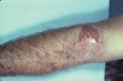
The compound is found in the sap that leaks out from cut stems; thus, the presentation is typically finger dermatitis, Dr. Marks said at the meeting, which was presented by the Caribbean Dermatology Symposium.
"It is the most common cause of allergic contact dermatitis in florists. So if you see florists, this is the allergen until proven otherwise," he said.
The compound is also found in the white epidermis of tulips, in which case it is known as tuliposide A. About half of tulip bulb sorters are allergic to it.
"You either patch test with parts of the Alstroemeria plant or get the allergen alpha-methylene-gamma-butyrolactone" commercially, Dr. Marks said. "I test everyone to alpha-methylene-gamma-butyrolactone, even though it’s a small subset. That’s one of my 100 [antigens]."
Methylisothiazolinone
"Methylisothiazolinone has become a very important and hot allergen," Dr. Marks commented. This allergen is increasingly used in personal care products and requires a special approach to patch testing. It is found in many wet wipes, use of which can produce, for example, perioral dermatitis.
The standard combination test antigen, applied at 100 parts per million, contains 3 parts methylchloroisothiazolinone (MCI) and 1 part methylisothiazolinone (MI), he noted. Thus, "you are really only patch testing to 25 parts per million of MI."
"The recommended concentration of patch test to MI is a bit in flux," said Dr. Marks; the North American group currently uses 2,000 parts per million but is considering halving that number, he noted.
"You can see how you can miss patients who are allergic to MI if you only patch test to MCI/MI," he commented. "So those of you who are using the TRUE test, you are going to miss patients who are allergic to MI."
"The important point is you’ve got to test both – MCI/MI and MI alone. ... You should supplement what you are patch testing with MI, certainly at least at 1,000 parts per million, if not at 2,000," Dr. Marks advised.
"The Cosmetic Ingredient Review, which sets limits for [MI] in the U.S., is going to be reevaluating, and I’m sure will be having lower limits in leave-on and rinse-off products," he noted.
Rubber accelerators
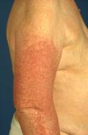
Surgeons may develop particularly problematic allergic contact dermatitis as a reaction to the rubber accelerators used in the manufacturing of many surgical gloves, Dr. Marks noted.
He described the case of a surgeon who developed severe hand dermatitis and eventually a generalized dermatitis. "In this case, if you used the TRUE test, you would make the diagnosis; he was positive to thiuram and carba mix."
Allergen avoidance entailed finding an alternative, rubber-free surgical glove, the Derma Prene Ultra glove (manufactured by Ansell), which is made of neoprene. Also, the surgeon switched to vinyl exam gloves for outpatient care.
"There may be other surgical gloves ...," Dr. Marks acknowledged. "But be sure and keep this some place because some time in the future when you have your surgeon friend with hand dermatitis, you can recommend that glove after you patch test them and prove that they are rubber-accelerator positive."
Cocamidopropyl betaine contaminants
Patients may develop allergic contact dermatitis after using shampoos and bath gels containing cocamidopropyl betaine, a surfactant.
In fact, they are actually reacting to a contaminant or impurity generated in the manufacturing process, either 3-dimethylaminopropylamine or amidoamine, according to Dr. Marks.
"So if you have pure cocamidopropyl betaine, there will be no allergy," he noted. But if you test for "cocamidopropyl betaine, and what you are patch testing with is from, say, Chemotechnique or Allergeaze, it’s going to have presumably the contaminants or the impurities in it, 3-dimethylaminopropylamine and amidoamine."
Treatment entails careful reading of labels on personal care products and avoidance of those containing cocamidopropyl betaine.
Acrylates
Don’t rule out acrylates – either acrylic or methacrylic acid – monomers that are polymerized with heat or light to form solid plastics that can cause reactions.
"The monomers are both irritants and allergens, so you need the right concentration to patch test to," Dr. Marks noted. "They are found in all sorts of things – adhesives, inks, artificial nails, dental resins, bone cement, and plastics."
Presentations may vary widely, including, for example, finger dermatitis in patients who have sculptured nails, and gum stomatitis in patients who have undergone procedures involving dental resin, said Dr. Marks.
"If you have workers or patients who have exposure to acrylates, you need more extensive screening," Dr. Marks advised, noting that his acrylate patch test series contains six compounds.
"No one is a screen for all of them," he commented. "Some [experts] feel that ethyl acrylate is the best screen; certainly, for sculptured nails it’s good." Others in his series include methyl methacrylate (found in bone cement) and ethyl cyanoacrylate (found in Super Glue adhesive).
Glyceryl thioglycolate
Allergy to glyceryl thioglycolate, found in acid permanent waves, can manifest as hand dermatitis in hairdressers and as dermatitis of the face, neck, and ears in their clients.
"If you see hairdressers [in your practice], you should consider strongly having this antigen as part of your [patch test] armamentarium," Dr. Marks recommended.
Alkaline perms, by contrast, do not contain glyceryl thioglycolate and thus provide a simple solution. "You can cure that hairdresser, and she or he can continue to do perms just by switching from an acid to an alkaline perm," he explained.
Dr. Marks said he had no relevant financial disclosures.
WOODINVILLE, WASH. – Identifying the culprit in allergic contact dermatitis often requires careful sleuthing and tailored patch testing, according to Dr. James G. Marks Jr.
Dr. Marks reviewed the current options for patch testing trays, described several cases of allergic contact dermatitis, and shared pointers for diagnosis and management at the annual Coastal Dermatology Symposium.
Allergen-screening series
Dermatologists can now choose from a variety of standard screening trays for patch testing, said Dr. Marks, professor and chair of the department of dermatology, Pennsylvania State University, Hershey. The TRUE test (the only one approved by the Food and Drug Administration) contains 35 antigens.
However, dermatologists can supplement and customize these screening trays to create a system specific to their practice and geographic area. Dr. Marks said he uses a customized tray with 100 antigens.
"The important point is there is no universal standard screening tray, so pick what works for you," he said.
"Those of you who use the TRUE test, great; as you know in the last year you have had another panel [added], so you get more screening antigens," he said. "It makes sense intuitively, and it’s proven by publications of the North American [Contact Dermatitis] Group and others that the more antigens you test with, the more positives you get and the more relevant reactions you can get," he explained.
"Then create your own," Dr. Marks advised. "So if you use the TRUE test, maybe supplement ... with a few more allergens."
Alpha-methylene-gamma-butyrolactone
Florists may come in contact with alpha-methylene-gamma-butyrolactone through handling Alstroemeria (also known as Peruvian lily), a flower popular because of its long-lasting blooms.
The compound is found in the sap that leaks out from cut stems; thus, the presentation is typically finger dermatitis, Dr. Marks said at the meeting, which was presented by the Caribbean Dermatology Symposium.
"It is the most common cause of allergic contact dermatitis in florists. So if you see florists, this is the allergen until proven otherwise," he said.
The compound is also found in the white epidermis of tulips, in which case it is known as tuliposide A. About half of tulip bulb sorters are allergic to it.
"You either patch test with parts of the Alstroemeria plant or get the allergen alpha-methylene-gamma-butyrolactone" commercially, Dr. Marks said. "I test everyone to alpha-methylene-gamma-butyrolactone, even though it’s a small subset. That’s one of my 100 [antigens]."
Methylisothiazolinone
"Methylisothiazolinone has become a very important and hot allergen," Dr. Marks commented. This allergen is increasingly used in personal care products and requires a special approach to patch testing. It is found in many wet wipes, use of which can produce, for example, perioral dermatitis.
The standard combination test antigen, applied at 100 parts per million, contains 3 parts methylchloroisothiazolinone (MCI) and 1 part methylisothiazolinone (MI), he noted. Thus, "you are really only patch testing to 25 parts per million of MI."
"The recommended concentration of patch test to MI is a bit in flux," said Dr. Marks; the North American group currently uses 2,000 parts per million but is considering halving that number, he noted.
"You can see how you can miss patients who are allergic to MI if you only patch test to MCI/MI," he commented. "So those of you who are using the TRUE test, you are going to miss patients who are allergic to MI."
"The important point is you’ve got to test both – MCI/MI and MI alone. ... You should supplement what you are patch testing with MI, certainly at least at 1,000 parts per million, if not at 2,000," Dr. Marks advised.
"The Cosmetic Ingredient Review, which sets limits for [MI] in the U.S., is going to be reevaluating, and I’m sure will be having lower limits in leave-on and rinse-off products," he noted.
Rubber accelerators
Surgeons may develop particularly problematic allergic contact dermatitis as a reaction to the rubber accelerators used in the manufacturing of many surgical gloves, Dr. Marks noted.
He described the case of a surgeon who developed severe hand dermatitis and eventually a generalized dermatitis. "In this case, if you used the TRUE test, you would make the diagnosis; he was positive to thiuram and carba mix."
Allergen avoidance entailed finding an alternative, rubber-free surgical glove, the Derma Prene Ultra glove (manufactured by Ansell), which is made of neoprene. Also, the surgeon switched to vinyl exam gloves for outpatient care.
"There may be other surgical gloves ...," Dr. Marks acknowledged. "But be sure and keep this some place because some time in the future when you have your surgeon friend with hand dermatitis, you can recommend that glove after you patch test them and prove that they are rubber-accelerator positive."
Cocamidopropyl betaine contaminants
Patients may develop allergic contact dermatitis after using shampoos and bath gels containing cocamidopropyl betaine, a surfactant.
In fact, they are actually reacting to a contaminant or impurity generated in the manufacturing process, either 3-dimethylaminopropylamine or amidoamine, according to Dr. Marks.
"So if you have pure cocamidopropyl betaine, there will be no allergy," he noted. But if you test for "cocamidopropyl betaine, and what you are patch testing with is from, say, Chemotechnique or Allergeaze, it’s going to have presumably the contaminants or the impurities in it, 3-dimethylaminopropylamine and amidoamine."
Treatment entails careful reading of labels on personal care products and avoidance of those containing cocamidopropyl betaine.
Acrylates
Don’t rule out acrylates – either acrylic or methacrylic acid – monomers that are polymerized with heat or light to form solid plastics that can cause reactions.
"The monomers are both irritants and allergens, so you need the right concentration to patch test to," Dr. Marks noted. "They are found in all sorts of things – adhesives, inks, artificial nails, dental resins, bone cement, and plastics."
Presentations may vary widely, including, for example, finger dermatitis in patients who have sculptured nails, and gum stomatitis in patients who have undergone procedures involving dental resin, said Dr. Marks.
"If you have workers or patients who have exposure to acrylates, you need more extensive screening," Dr. Marks advised, noting that his acrylate patch test series contains six compounds.
"No one is a screen for all of them," he commented. "Some [experts] feel that ethyl acrylate is the best screen; certainly, for sculptured nails it’s good." Others in his series include methyl methacrylate (found in bone cement) and ethyl cyanoacrylate (found in Super Glue adhesive).
Glyceryl thioglycolate
Allergy to glyceryl thioglycolate, found in acid permanent waves, can manifest as hand dermatitis in hairdressers and as dermatitis of the face, neck, and ears in their clients.
"If you see hairdressers [in your practice], you should consider strongly having this antigen as part of your [patch test] armamentarium," Dr. Marks recommended.
Alkaline perms, by contrast, do not contain glyceryl thioglycolate and thus provide a simple solution. "You can cure that hairdresser, and she or he can continue to do perms just by switching from an acid to an alkaline perm," he explained.
Dr. Marks said he had no relevant financial disclosures.
AT THE COASTAL DERMATOLOGY SYMPOSIUM

The art and science of detecting allergic contact dermatitis
WOODINVILLE, WASH. – Identifying the culprit in allergic contact dermatitis often requires careful sleuthing and tailored patch testing, according to Dr. James G. Marks Jr.
Dr. Marks reviewed the current options for patch testing trays, described several cases of allergic contact dermatitis, and shared pointers for diagnosis and management at the annual Coastal Dermatology Symposium.

Allergen-screening series
Dermatologists can now choose from a variety of standard screening trays for patch testing, said Dr. Marks, professor and chair of the department of dermatology, Pennsylvania State University, Hershey. The TRUE test (the only one approved by the Food and Drug Administration) contains 35 antigens.
However, dermatologists can supplement and customize these screening trays to create a system specific to their practice and geographic area. Dr. Marks said he uses a customized tray with 100 antigens.
"The important point is there is no universal standard screening tray, so pick what works for you," he said.
"Those of you who use the TRUE test, great; as you know in the last year you have had another panel [added], so you get more screening antigens," he said. "It makes sense intuitively, and it’s proven by publications of the North American [Contact Dermatitis] Group and others that the more antigens you test with, the more positives you get and the more relevant reactions you can get," he explained.
"Then create your own," Dr. Marks advised. "So if you use the TRUE test, maybe supplement ... with a few more allergens."
Alpha-methylene-gamma-butyrolactone
Florists may come in contact with alpha-methylene-gamma-butyrolactone through handling Alstroemeria (also known as Peruvian lily), a flower popular because of its long-lasting blooms.
The compound is found in the sap that leaks out from cut stems; thus, the presentation is typically finger dermatitis, Dr. Marks said at the meeting, which was presented by the Caribbean Dermatology Symposium.
"It is the most common cause of allergic contact dermatitis in florists. So if you see florists, this is the allergen until proven otherwise," he said.
The compound is also found in the white epidermis of tulips, in which case it is known as tuliposide A. About half of tulip bulb sorters are allergic to it.
"You either patch test with parts of the Alstroemeria plant or get the allergen alpha-methylene-gamma-butyrolactone" commercially, Dr. Marks said. "I test everyone to alpha-methylene-gamma-butyrolactone, even though it’s a small subset. That’s one of my 100 [antigens]."
Methylisothiazolinone
"Methylisothiazolinone has become a very important and hot allergen," Dr. Marks commented. This allergen is increasingly used in personal care products and requires a special approach to patch testing. It is found in many wet wipes, use of which can produce, for example, perioral dermatitis.
The standard combination test antigen, applied at 100 parts per million, contains 3 parts methylchloroisothiazolinone (MCI) and 1 part methylisothiazolinone (MI), he noted. Thus, "you are really only patch testing to 25 parts per million of MI."
"The recommended concentration of patch test to MI is a bit in flux," said Dr. Marks; the North American group currently uses 2,000 parts per million but is considering halving that number, he noted.
"You can see how you can miss patients who are allergic to MI if you only patch test to MCI/MI," he commented. "So those of you who are using the TRUE test, you are going to miss patients who are allergic to MI."
"The important point is you’ve got to test both – MCI/MI and MI alone. ... You should supplement what you are patch testing with MI, certainly at least at 1,000 parts per million, if not at 2,000," Dr. Marks advised.
"The Cosmetic Ingredient Review, which sets limits for [MI] in the U.S., is going to be reevaluating, and I’m sure will be having lower limits in leave-on and rinse-off products," he noted.
Rubber accelerators
Surgeons may develop particularly problematic allergic contact dermatitis as a reaction to the rubber accelerators used in the manufacturing of many surgical gloves, Dr. Marks noted.
He described the case of a surgeon who developed severe hand dermatitis and eventually a generalized dermatitis. "In this case, if you used the TRUE test, you would make the diagnosis; he was positive to thiuram and carba mix."
Allergen avoidance entailed finding an alternative, rubber-free surgical glove, the Derma Prene Ultra glove (manufactured by Ansell), which is made of neoprene. Also, the surgeon switched to vinyl exam gloves for outpatient care.
"There may be other surgical gloves ...," Dr. Marks acknowledged. "But be sure and keep this some place because some time in the future when you have your surgeon friend with hand dermatitis, you can recommend that glove after you patch test them and prove that they are rubber-accelerator positive."
Cocamidopropyl betaine contaminants
Patients may develop allergic contact dermatitis after using shampoos and bath gels containing cocamidopropyl betaine, a surfactant.
In fact, they are actually reacting to a contaminant or impurity generated in the manufacturing process, either 3-dimethylaminopropylamine or amidoamine, according to Dr. Marks.
"So if you have pure cocamidopropyl betaine, there will be no allergy," he noted. But if you test for "cocamidopropyl betaine, and what you are patch testing with is from, say, Chemotechnique or Allergeaze, it’s going to have presumably the contaminants or the impurities in it, 3-dimethylaminopropylamine and amidoamine."
Treatment entails careful reading of labels on personal care products and avoidance of those containing cocamidopropyl betaine.
Acrylates
Don’t rule out acrylates – either acrylic or methacrylic acid – monomers that are polymerized with heat or light to form solid plastics that can cause reactions.
"The monomers are both irritants and allergens, so you need the right concentration to patch test to," Dr. Marks noted. "They are found in all sorts of things – adhesives, inks, artificial nails, dental resins, bone cement, and plastics."
Presentations may vary widely, including, for example, finger dermatitis in patients who have sculptured nails, and gum stomatitis in patients who have undergone procedures involving dental resin, said Dr. Marks.
"If you have workers or patients who have exposure to acrylates, you need more extensive screening," Dr. Marks advised, noting that his acrylate patch test series contains six compounds.
"No one is a screen for all of them," he commented. "Some [experts] feel that ethyl acrylate is the best screen; certainly, for sculptured nails it’s good." Others in his series include methyl methacrylate (found in bone cement) and ethyl cyanoacrylate (found in Super Glue adhesive).
Glyceryl thioglycolate
Allergy to glyceryl thioglycolate, found in acid permanent waves, can manifest as hand dermatitis in hairdressers and as dermatitis of the face, neck, and ears in their clients.
"If you see hairdressers [in your practice], you should consider strongly having this antigen as part of your [patch test] armamentarium," Dr. Marks recommended.
Alkaline perms, by contrast, do not contain glyceryl thioglycolate and thus provide a simple solution. "You can cure that hairdresser, and she or he can continue to do perms just by switching from an acid to an alkaline perm," he explained.
Dr. Marks said he had no relevant financial disclosures.
WOODINVILLE, WASH. – Identifying the culprit in allergic contact dermatitis often requires careful sleuthing and tailored patch testing, according to Dr. James G. Marks Jr.
Dr. Marks reviewed the current options for patch testing trays, described several cases of allergic contact dermatitis, and shared pointers for diagnosis and management at the annual Coastal Dermatology Symposium.
Allergen-screening series
Dermatologists can now choose from a variety of standard screening trays for patch testing, said Dr. Marks, professor and chair of the department of dermatology, Pennsylvania State University, Hershey. The TRUE test (the only one approved by the Food and Drug Administration) contains 35 antigens.
However, dermatologists can supplement and customize these screening trays to create a system specific to their practice and geographic area. Dr. Marks said he uses a customized tray with 100 antigens.
"The important point is there is no universal standard screening tray, so pick what works for you," he said.
"Those of you who use the TRUE test, great; as you know in the last year you have had another panel [added], so you get more screening antigens," he said. "It makes sense intuitively, and it’s proven by publications of the North American [Contact Dermatitis] Group and others that the more antigens you test with, the more positives you get and the more relevant reactions you can get," he explained.
"Then create your own," Dr. Marks advised. "So if you use the TRUE test, maybe supplement ... with a few more allergens."
Alpha-methylene-gamma-butyrolactone
Florists may come in contact with alpha-methylene-gamma-butyrolactone through handling Alstroemeria (also known as Peruvian lily), a flower popular because of its long-lasting blooms.
The compound is found in the sap that leaks out from cut stems; thus, the presentation is typically finger dermatitis, Dr. Marks said at the meeting, which was presented by the Caribbean Dermatology Symposium.
"It is the most common cause of allergic contact dermatitis in florists. So if you see florists, this is the allergen until proven otherwise," he said.
The compound is also found in the white epidermis of tulips, in which case it is known as tuliposide A. About half of tulip bulb sorters are allergic to it.
"You either patch test with parts of the Alstroemeria plant or get the allergen alpha-methylene-gamma-butyrolactone" commercially, Dr. Marks said. "I test everyone to alpha-methylene-gamma-butyrolactone, even though it’s a small subset. That’s one of my 100 [antigens]."
Methylisothiazolinone
"Methylisothiazolinone has become a very important and hot allergen," Dr. Marks commented. This allergen is increasingly used in personal care products and requires a special approach to patch testing. It is found in many wet wipes, use of which can produce, for example, perioral dermatitis.
The standard combination test antigen, applied at 100 parts per million, contains 3 parts methylchloroisothiazolinone (MCI) and 1 part methylisothiazolinone (MI), he noted. Thus, "you are really only patch testing to 25 parts per million of MI."
"The recommended concentration of patch test to MI is a bit in flux," said Dr. Marks; the North American group currently uses 2,000 parts per million but is considering halving that number, he noted.
"You can see how you can miss patients who are allergic to MI if you only patch test to MCI/MI," he commented. "So those of you who are using the TRUE test, you are going to miss patients who are allergic to MI."
"The important point is you’ve got to test both – MCI/MI and MI alone. ... You should supplement what you are patch testing with MI, certainly at least at 1,000 parts per million, if not at 2,000," Dr. Marks advised.
"The Cosmetic Ingredient Review, which sets limits for [MI] in the U.S., is going to be reevaluating, and I’m sure will be having lower limits in leave-on and rinse-off products," he noted.
Rubber accelerators
Surgeons may develop particularly problematic allergic contact dermatitis as a reaction to the rubber accelerators used in the manufacturing of many surgical gloves, Dr. Marks noted.
He described the case of a surgeon who developed severe hand dermatitis and eventually a generalized dermatitis. "In this case, if you used the TRUE test, you would make the diagnosis; he was positive to thiuram and carba mix."
Allergen avoidance entailed finding an alternative, rubber-free surgical glove, the Derma Prene Ultra glove (manufactured by Ansell), which is made of neoprene. Also, the surgeon switched to vinyl exam gloves for outpatient care.
"There may be other surgical gloves ...," Dr. Marks acknowledged. "But be sure and keep this some place because some time in the future when you have your surgeon friend with hand dermatitis, you can recommend that glove after you patch test them and prove that they are rubber-accelerator positive."
Cocamidopropyl betaine contaminants
Patients may develop allergic contact dermatitis after using shampoos and bath gels containing cocamidopropyl betaine, a surfactant.
In fact, they are actually reacting to a contaminant or impurity generated in the manufacturing process, either 3-dimethylaminopropylamine or amidoamine, according to Dr. Marks.
"So if you have pure cocamidopropyl betaine, there will be no allergy," he noted. But if you test for "cocamidopropyl betaine, and what you are patch testing with is from, say, Chemotechnique or Allergeaze, it’s going to have presumably the contaminants or the impurities in it, 3-dimethylaminopropylamine and amidoamine."
Treatment entails careful reading of labels on personal care products and avoidance of those containing cocamidopropyl betaine.
Acrylates
Don’t rule out acrylates – either acrylic or methacrylic acid – monomers that are polymerized with heat or light to form solid plastics that can cause reactions.
"The monomers are both irritants and allergens, so you need the right concentration to patch test to," Dr. Marks noted. "They are found in all sorts of things – adhesives, inks, artificial nails, dental resins, bone cement, and plastics."
Presentations may vary widely, including, for example, finger dermatitis in patients who have sculptured nails, and gum stomatitis in patients who have undergone procedures involving dental resin, said Dr. Marks.
"If you have workers or patients who have exposure to acrylates, you need more extensive screening," Dr. Marks advised, noting that his acrylate patch test series contains six compounds.
"No one is a screen for all of them," he commented. "Some [experts] feel that ethyl acrylate is the best screen; certainly, for sculptured nails it’s good." Others in his series include methyl methacrylate (found in bone cement) and ethyl cyanoacrylate (found in Super Glue adhesive).
Glyceryl thioglycolate
Allergy to glyceryl thioglycolate, found in acid permanent waves, can manifest as hand dermatitis in hairdressers and as dermatitis of the face, neck, and ears in their clients.
"If you see hairdressers [in your practice], you should consider strongly having this antigen as part of your [patch test] armamentarium," Dr. Marks recommended.
Alkaline perms, by contrast, do not contain glyceryl thioglycolate and thus provide a simple solution. "You can cure that hairdresser, and she or he can continue to do perms just by switching from an acid to an alkaline perm," he explained.
Dr. Marks said he had no relevant financial disclosures.
AT THE COASTAL DERMATOLOGY SYMPOSIUM

Expert offers insight on photocontact dermatitis
WOODINVILLE, WASH. – The diagnosis of photocontact dermatitis can be challenging and often requires careful photopatch testing, according to Dr. Vincent A. DeLeo.
He reviewed the finer points of diagnosing the two main types of photocontact dermatitis – photoirritant and photoallergic – and differentiating them from other conditions with a similar clinical presentation at the annual Coastal Dermatology Symposium.
Photoirritant contact dermatitis
In photoirritant contact dermatitis, a chemical deposited on the skin absorbs radiation and then causes damage directly, without any immune reaction. The diagnosis is a clinical one, said Dr. DeLeo, chairman of the department of dermatology at St. Luke’s–Roosevelt Hospital Center and Beth Israel Medical Center in New York.
"You as a clinician know it is a photodistributed disease," he said. "You take a good history, you get a history of the patient being exposed topically to the chemical, and that’s how you make the diagnosis," he commented. "This is universal; it will happen to any of us if we get exposed to these photosensitizing chemicals in enough concentration and we get enough light to develop this kind of reaction."
Photoirritant contact dermatitis is almost always induced by UVA radiation; therefore, it can occur even if a patient is wearing a sunscreen that protects against UVB light.
The reaction has the appearance of a sunburn, with erythema, edema, and possibly blistering. It usually resolves with hyperpigmentation. "The interesting thing is that a lot of these reactions are subclinical, at least the erythema component that you don’t see. All you do see is the resulting hyperpigmentation," he said at the meeting, presented by the Caribbean Dermatology Symposium.
A common culprit is tar, in which case the condition is known as tar smarts. These cases usually result from occupational exposure in roofers, but can be seen as well in people who use tar shampoos and then go out in the sun.
The furocoumarins (psoralens) also can produce photoirritant contact dermatitis. The most common form is phytophotodermatitis, which involves plant compounds, especially those in the exocorp (green peel) of limes. The hallmark is tiny blisters on involved skin.
"It’s a delayed reaction. So people are squeezing limes into their gin and tonics on the weekend, and then Monday or Tuesday, when they are back at work, all of a sudden they develop these blisters, and they don’t associate it with what they did on weekend," Dr. DeLeo noted.
Phytophotodermatitis also can result from contact with celery that has been bred to have a high psoralen content so that it stays fresh longer. "You see this in grocery workers primarily, people who are exposed to a lot of celery, handling it and then going into the sun and getting the reaction," Dr. DeLeo said.
Photoallergic contact dermatitis
In photoallergic contact dermatitis, the chemical deposited on the skin – most commonly a sunscreen, fragrance, or medication – absorbs radiation and becomes an allergen. Only sensitized people have a reaction.
This dermatitis is exematous and pruritic, and shows a sun-exposed distribution, although it is usually patchy. The reaction is delayed, typically starting 1-2 days after sun exposure.
"The photodistributed eruption will usually be in all photoexposed areas. And this is important in making the distinction between this and other photosensitivities," Dr. DeLeo said. "Usually it will occur on all areas including the face ... and usually it spares upper lids and beneath the chin, although not always," he noted.
"The diagnosis of photoallergic contact dermatitis is confirmed by photopatch testing," he said. "Almost all photosensitive patients with eczematous morphology should be photopatch tested."
However, survey data suggest that only about two-thirds of dermatologists do this testing (Am. J. Contact Dermat. 2003;14:5-11), largely because this dermatitis is uncommon today as most sensitizing agents are identified before products come to market.
Photopatch testing can be done in the office; it requires at least a UVA source, photoantigens, and general patch test supplies. A UVB source is helpful for distinguishing photoallergic contact dermatitis from conditions such as chronic actinic dermatitis.
"If I were to buy four photoantigens, I would probably buy oxybenzone, musk ambrette, padimate O, and avobenzone," Dr. DeLeo said. "You can also test directly to the patient’s products, and usually it’s going to be sunscreens, so you can just have them bring in whatever they are using."
In a study by the North American Contact Dermatitis Group in which 363 consecutive patients with a history of photosensitivity were given a final diagnosis after photopatch testing, the leading diagnosis was allergic contact dermatitis (122 patients), followed by photoallergic contact dermatitis (75 patients) and polymorphous light eruption (72 patients).
The most common photoallergen was oxybenzone. Others included ketoprofen, avobenzone, sulisobenzone, and musk ambrette.
"In the Northwest, mainly in Washington and Oregon, people are compounding ketoprofen, which is a nonsteroidal anti-inflammatory agent, into a topical agent ... that can induce a photoallergic contact dermatitis," Dr. DeLeo explained.
Newer photoallergens include the sunscreen methylene bis-benzotriazolyl tetramethylbutylphenol (bisoctrizole, brand name Tinosorb M) (Dermatitis 2011;22:106-11).
"This [product] is not available in the U.S., but is available in Europe," Dr. DeLeo noted. However, reactions have been seen among patients using Ahava, an Israeli product sold in the United States. The manufacturers "are apparently saying they are not using it as a sunscreen but as a sort of preservative. So you need to watch out for this one," he said.
In fact, the Food and Drug Administration is considering approval of several new sunscreens, and bisoctrizole is among them, so more cases may be seen, according to Dr. DeLeo.
Another emerging photoallergen is the sunscreen octocrylene. "We haven’t seen any positives to this as yet, but it is being reported in Europe as a very important new photoallergic agent. It may be that they are using a lot more of that as a photostabilizer for avobenzone in Europe than we do in our own cosmetics," he said.
Importantly, there is cross-reactivity among oxybenzone, octocrylene, and ketoprofen, said Dr. DeLeo. Therefore, "those of you who live in the Northwest and see this reaction to ketoprofen may want your patients to avoid oxybenzone and octocrylene as well," he added.
Dr. DeLeo disclosed that he is a consultant for Pfizer, Prous Science, L’Oréal, Limited Brands, Goodyear, Estée Lauder, Mary Kay, and La Roche-Posay.
AT THE COASTAL DERMATOLOGY SYMPOSIUM

Management of keloids draws on clinical wisdom
WOODINVILLE, WASH. – Make decisions about whether to surgically or medically manage keloids according to features of the lesion and patient-related factors, especially likely treatment compliance, Dr. Hilary E. Baldwin advised.
"Step one when we are dealing with our patients with keloids is trying to decide what the patients actually hope for," Dr. Baldwin said at the annual Coastal Dermatology Symposium. That might be eradication of the keloid; palliation of symptoms such as itching or pain; flattening, lightening, or softening of the keloid; ability to wear clip-on earrings; or restoration of mobility.

"Most patients, in my opinion, are actually not surgical candidates, and most need to be convinced to pursue other options," said Dr. Baldwin of the dermatology department at the State University of New York, Brooklyn.
Surgical management
Size and shape should factor into decisions about surgical removal of keloids, said Dr. Baldwin. Larger keloids are not much more difficult to remove than smaller ones, and pedunculated keloids are generally the most amenable to surgery, she noted.
Keloid age also comes into play, as the older, mature, brown lesions are less likely to recur after surgery than the younger, pink, symptomatic ones. Location affects outcome as well, Dr. Baldwin said. Keloids on the jaw, the deltoid, mid-back, mid-chest, and upper back are most likely to recur if treated surgically, and those in low-tension areas are less likely to do so.
Surgery also may be considered for keloids that are truly unresponsive to intralesional corticosteroids, but those are uncommon as previous lack of response is usually because of an inadequate dose, Dr. Baldwin explained.
"Most important is patient commitment. They often believe that cutting is a quick fix and they are not going to have to do anything afterward, and they are poor flight risks because of that," she said. Statistics suggest that the nearly 100% of keloids recur with surgery alone, but the value falls to 50% with adjunctive corticosteroids, and 20% with adjunctive radiation therapy, she added.
Adjunctive therapies
When adjunctive corticosteroids are used after surgery, the steroid is injected into the base and walls of the excision site, according to Dr. Baldwin; her preference is to use 40 mg/cc triamcinolone, with injections given for at least 6 months.
"Radiation therapy ... works really well postop; in fact, it’s our best adjunctive therapy, if you’re not afraid of it and the patient’s not afraid of it," she said. At her institution, this therapy is completed within a week after surgery, "so even if patients are a flight risk, they always return for that first visit, and then they are done with at least one adjunctive therapy."
The main barrier to use has been a fear of secondary malignancy. But reports of cancer after radiation therapy are rare and questionably associated with the treatment, Dr. Baldwin said.
Pressure dressings also are effective, and these are most practical in the form of compression earrings worn after excision of earlobe keloids, usually for at least 6 months. The earrings also come in a form called "sleepers" that may be more acceptable to patients.
Interferon injections also can help prevent recurrence. "I treat the very first day ... into the wall and into the base, just as I would with steroids, and then repeat 1 week later," Dr. Baldwin explained. "So if you use this and radiation therapy, 1 week later, you have finished two adjunctive therapies."
Especially challenging are keloids nearly obliterating normal tissue, such as extensive earlobe keloids. "I call them last-chance keloids," Dr. Baldwin commented. "If I don’t do a good job and don’t have good adjunctive therapy, it’s going to regrow and that’s the last time I am going to be able to cut. The earlobe will be eaten away with keloid, and we are never going to be able to give [the patient] an earlobe on which to hang an earring."
After excision of last-chance keloids, "I recommend that you do everything ... Use everything that is available to you," Dr. Baldwin advised. In addition to excision, that includes "intralesional steroids, intralesional interferon, radiation therapy, and earrings. Why pick one? Use them all if appropriate, and you are much less likely to have a recurrence," she said.
Medical management
Medical therapy is indicated for large, sessile keloids and for those located in areas where surgery could lead to complications, according to Dr. Baldwin.
In these cases, intralesional corticosteroids are the mainstay, and her preference is to use a high dose of triamcinolone. "I only use 40 [mg/cc]—that’s it. I never use anything less," she said. "I don’t care where the keloid is: A keloid on the face is the same as a keloid on the back. Most of the failures that I see are due to inadequate concentration. It’s almost always on the face where people are a little bit shy about using the extraordinarily high doses," she added.
Dr. Baldwin dismissed the dogma of not exceeding 40 mg per month, given that this translates to a dose of just 50 mg of prednisone. "I will sometimes use 80 [mg] in one sitting. I don’t really have that kind of a restriction because it doesn’t make a ton of sense," she said.
For pre-injection anesthesia, Dr. Baldwin recommended applying topical anesthetic followed by a lidocaine ring block. When treating hard keloids, to prevent backwash of the corticosteroid along the needle track, she said she preapplies Krazy Glue and, immediately after removing the needle, applies a strip of precut tape.
Patients should be advised of the possible complications of hypopigmentation, telangiectasia, and atrophy. "You also have to warn your patients about what I call the Play-Doh effect," whereby the keloid spreads as it softens, Dr. Baldwin said. "So the footprint is ultimately much bigger than it was originally, although it’s now flatter."
Intralesional injection of 5-fluorouracil can be used alone or with admixed steroids. However, Dr. Baldwin said that in her hands, the combination has been disappointing, perhaps because the triamcinolone is diluted in the process. "So I’ve reconsidered recently the possibility of doing the steroids the way I like it and following [with 5-fluoruracil] in addition to it, rather than diluting my triamcinolone," she said.
The newest nonsurgical therapy is intralesional cryotherapy, whereby the keloid is frozen from the inside out with a probe attached to a cryotherapy device, so it eventually undergoes necrosis while the skin is preserved. The reduction in volume achieved has ranged from 51% to 93% across studies, with minimal to no hypopigmentation.
"The probe itself unfortunately costs $450 and is not reusable, which is a problem. At the present time this is not covered by insurance," said Dr. Baldwin, who disclosed no relevant conflicts of interest. "But for keloids that really can’t be treated any other way, it has been quite helpful."
The meeting was presented by the Caribbean Dermatology Symposium.
WOODINVILLE, WASH. – Make decisions about whether to surgically or medically manage keloids according to features of the lesion and patient-related factors, especially likely treatment compliance, Dr. Hilary E. Baldwin advised.
"Step one when we are dealing with our patients with keloids is trying to decide what the patients actually hope for," Dr. Baldwin said at the annual Coastal Dermatology Symposium. That might be eradication of the keloid; palliation of symptoms such as itching or pain; flattening, lightening, or softening of the keloid; ability to wear clip-on earrings; or restoration of mobility.
"Most patients, in my opinion, are actually not surgical candidates, and most need to be convinced to pursue other options," said Dr. Baldwin of the dermatology department at the State University of New York, Brooklyn.
Surgical management
Size and shape should factor into decisions about surgical removal of keloids, said Dr. Baldwin. Larger keloids are not much more difficult to remove than smaller ones, and pedunculated keloids are generally the most amenable to surgery, she noted.
Keloid age also comes into play, as the older, mature, brown lesions are less likely to recur after surgery than the younger, pink, symptomatic ones. Location affects outcome as well, Dr. Baldwin said. Keloids on the jaw, the deltoid, mid-back, mid-chest, and upper back are most likely to recur if treated surgically, and those in low-tension areas are less likely to do so.
Surgery also may be considered for keloids that are truly unresponsive to intralesional corticosteroids, but those are uncommon as previous lack of response is usually because of an inadequate dose, Dr. Baldwin explained.
"Most important is patient commitment. They often believe that cutting is a quick fix and they are not going to have to do anything afterward, and they are poor flight risks because of that," she said. Statistics suggest that the nearly 100% of keloids recur with surgery alone, but the value falls to 50% with adjunctive corticosteroids, and 20% with adjunctive radiation therapy, she added.
Adjunctive therapies
When adjunctive corticosteroids are used after surgery, the steroid is injected into the base and walls of the excision site, according to Dr. Baldwin; her preference is to use 40 mg/cc triamcinolone, with injections given for at least 6 months.
"Radiation therapy ... works really well postop; in fact, it’s our best adjunctive therapy, if you’re not afraid of it and the patient’s not afraid of it," she said. At her institution, this therapy is completed within a week after surgery, "so even if patients are a flight risk, they always return for that first visit, and then they are done with at least one adjunctive therapy."
The main barrier to use has been a fear of secondary malignancy. But reports of cancer after radiation therapy are rare and questionably associated with the treatment, Dr. Baldwin said.
Pressure dressings also are effective, and these are most practical in the form of compression earrings worn after excision of earlobe keloids, usually for at least 6 months. The earrings also come in a form called "sleepers" that may be more acceptable to patients.
Interferon injections also can help prevent recurrence. "I treat the very first day ... into the wall and into the base, just as I would with steroids, and then repeat 1 week later," Dr. Baldwin explained. "So if you use this and radiation therapy, 1 week later, you have finished two adjunctive therapies."
Especially challenging are keloids nearly obliterating normal tissue, such as extensive earlobe keloids. "I call them last-chance keloids," Dr. Baldwin commented. "If I don’t do a good job and don’t have good adjunctive therapy, it’s going to regrow and that’s the last time I am going to be able to cut. The earlobe will be eaten away with keloid, and we are never going to be able to give [the patient] an earlobe on which to hang an earring."
After excision of last-chance keloids, "I recommend that you do everything ... Use everything that is available to you," Dr. Baldwin advised. In addition to excision, that includes "intralesional steroids, intralesional interferon, radiation therapy, and earrings. Why pick one? Use them all if appropriate, and you are much less likely to have a recurrence," she said.
Medical management
Medical therapy is indicated for large, sessile keloids and for those located in areas where surgery could lead to complications, according to Dr. Baldwin.
In these cases, intralesional corticosteroids are the mainstay, and her preference is to use a high dose of triamcinolone. "I only use 40 [mg/cc]—that’s it. I never use anything less," she said. "I don’t care where the keloid is: A keloid on the face is the same as a keloid on the back. Most of the failures that I see are due to inadequate concentration. It’s almost always on the face where people are a little bit shy about using the extraordinarily high doses," she added.
Dr. Baldwin dismissed the dogma of not exceeding 40 mg per month, given that this translates to a dose of just 50 mg of prednisone. "I will sometimes use 80 [mg] in one sitting. I don’t really have that kind of a restriction because it doesn’t make a ton of sense," she said.
For pre-injection anesthesia, Dr. Baldwin recommended applying topical anesthetic followed by a lidocaine ring block. When treating hard keloids, to prevent backwash of the corticosteroid along the needle track, she said she preapplies Krazy Glue and, immediately after removing the needle, applies a strip of precut tape.
Patients should be advised of the possible complications of hypopigmentation, telangiectasia, and atrophy. "You also have to warn your patients about what I call the Play-Doh effect," whereby the keloid spreads as it softens, Dr. Baldwin said. "So the footprint is ultimately much bigger than it was originally, although it’s now flatter."
Intralesional injection of 5-fluorouracil can be used alone or with admixed steroids. However, Dr. Baldwin said that in her hands, the combination has been disappointing, perhaps because the triamcinolone is diluted in the process. "So I’ve reconsidered recently the possibility of doing the steroids the way I like it and following [with 5-fluoruracil] in addition to it, rather than diluting my triamcinolone," she said.
The newest nonsurgical therapy is intralesional cryotherapy, whereby the keloid is frozen from the inside out with a probe attached to a cryotherapy device, so it eventually undergoes necrosis while the skin is preserved. The reduction in volume achieved has ranged from 51% to 93% across studies, with minimal to no hypopigmentation.
"The probe itself unfortunately costs $450 and is not reusable, which is a problem. At the present time this is not covered by insurance," said Dr. Baldwin, who disclosed no relevant conflicts of interest. "But for keloids that really can’t be treated any other way, it has been quite helpful."
The meeting was presented by the Caribbean Dermatology Symposium.
EXPERT ANALYSIS FROM THE COASTAL DERMATOLOGY SYMPOSIUM