User login
The T-cell repertoire in NSCLC: Therapeutic implications
SAN FRANCISCO – An analysis of the T-cell repertoire in nearly 400 patients with stage I-III non–small cell lung cancer (NSCLC) suggests that patients with a more tumor-focused repertoire have better outcomes.
The findings, which suggest that patients with fewer T cells and with lower clonality in tumor-adjacent normal lung tissue fare better, could have implications for the use of tumor-infiltrating lymphocyte (TIL) therapy and checkpoint blockade – and possibly other therapies – in these patients, according to Alexandre Reuben, PhD, of the University of Texas MD Anderson Cancer Center, Houston.
Studying the T-cell repertoire in the lung can be rather “messy,” because many T cells may be responding to the outside environment, but comparing findings in the normal lung with those in tumor tissue helps to clarify things, Dr. Reuben said at the ASCO-SITC Clinical Immuno-Oncology Symposium.
Why study the T-cell repertoire?
The successes seen with immune checkpoint blockade in recent years are largely a result of the ability of these therapies to enhance the antitumor T-cell response. Interestingly, checkpoint blockade works better in tumor types like lung cancer that have a high mutational load, Dr. Reuben said, explaining that this is largely attributable to the ability of the mutations to increase tumor immunogenicity through generation of tumor-specific antigens, which can then be targeted by the T-cell response.
This relationship between the mutational and neoantigen burden and patient outcomes has been described, but the role of the T-cell repertoire and how it relates to patient outcomes is less clear.
Hypothesizing that T-cell repertoire would be associated with survival in patients with NSCLC, he and his colleagues collected peripheral blood, normal lung, and tumor tissue, and performed T cell–receptor sequencing, among other analyses, in 398 patients.
“T cells recognize antigens through their T-cell receptor, as a result of which they undergo clonal expansion. Therefore, by sequencing the variable region of the T-cell receptor, one can gain insight into the T cells that are responding within the sample, as well as the overall T-cell repertoire,” he explained.
This provides information on T-cell density and richness, and thus on the diversity of the T-cell repertoire, he said, adding that it is possible to go beyond that and plot T cells based on their frequency in a sample to study clonality.
Since these T cells tend to expand clonally as a result of activation, uneven distribution would be associated with a reactive T-cell repertoire, as described by a high T-cell clonality.
An assessment to determine how the T-cell repertoire relates to clinical characteristics and response revealed a few interesting correlations. For example, adenocarcinomas tended to be more densely infiltrated than squamous cell carcinomas, smaller tumors were also more densely infiltrated by T cells than were their larger counterparts, and in smokers the T-cell repertoire appeared much more reactive than in nonsmokers.
“But ultimately, we didn’t see the [direct correlations between the T-cell repertoire and outcomes] we were hoping to see,” Dr. Reuben said, noting that this could be because of environmental influences.
“Obviously the lung is exposed to the outside environment, which could be masking some of the antitumor T-cell responses we were hoping to study,” he explained. “So we used a more holistic approach, integrating the peripheral blood and normal lung with tumor repertoire going forward.”
T cells in normal lung vs. tumor
Measuring the proportion of the T-cell repertoire that is shared in peripheral blood vs. normal lung and vs. tumor tissue showed that there is very little in common between them.
“However, when you compare the normal lung to the tumor, there’s much more homology in the T-cell repertoire,” he said, noting that, given the T-cell expansion resulting from antigenic stimulation, focusing on the most dominant cells in a sample highlights those most likely to be responding to antigens. “When we did that ... we saw even more of an enrichment in the homology between the normal lung and tumor T-cell repertoire, suggesting certain parallels in the ongoing immune responses across both these compartments.”
Further, T-cell density and diversity were actually higher in the tumor than in the normal lung in about two-thirds of patients, he said.
“However, surprisingly ... clonality appears to be much higher in the normal lung than in the tumor,” he added, noting that this was the case in about 75% of patients.
These findings raise three key questions:
Why is clonality higher in the normal lung?
T cells are not confined to a specific part of the host and are free to circulate, Dr. Reuben said.
“However, the closer you get to a site of inflammation, the higher the enrichment for T cells that are relevant to that specific site of inflammation, so you can use these statistical methods to enrich for T cells that are more relevant and try to subtract out T cells that are simply circulating through the organ,” he noted.
He and his colleagues used these methods and compared both normal lung and tumor to the peripheral blood, focusing only on clones that were statistically enriched in these two compartments “to really eliminate a lot of the background that may have been caused by the low-frequency T cells in these samples.”
When you look at the lung enriched T-cell repertoire between normal lung vs. tumor, the homology increases quite significantly, suggesting that by subtracting these T cells that are circulating through the host and not likely relevant to the antigenic response, you’re increasing the homology and further highlighting some of the aforementioned parallels in the ongoing immune responses between both sites, he said.
“Now if you look at clonality, there’s really no clear trend ... in the total T-cell repertoire or the enriched repertoire focusing on the normal lung, but if you look at the tumor, there’s a trend toward increased clonality in all patients – to the extent where you no longer see a difference in clonality between the normal lung and tumor, suggesting that this enrichment is allowing us to focus increasingly on T cells relevant to the antitumor response,” he added.
T-cell clonality is highly reliant on the ability of T cells to expand as a result of antigenic stimulation, and immune profiling showed that programmed cell death–1 (PD-1) and programmed death–ligand 1 (PD-L1) were higher within the tumor, suggesting that there is some dysfunction on both sides of this interaction, which could also explain the lower clonality originally seen within the tumor, he said.
Why is the T-cell repertoire so similar across normal lung and tumor (and what are these T cells really recognizing)?
“Well, we performed whole-exome sequencing and it’s really no surprise that mutational load is substantial in the tumor, but what was surprising was the amount of mutations we detected in the normal lung,” Dr. Reuben said.
The number was lower than in the tumor, though still considerable, and included a large proportion that were shared mutations between the normal lung and the tumor, he noted.
A closer look at the shared mutations showed that they correlated positively with the proportion of shared dominant T cells between the normal lung and the tumor, suggesting that some of the shared T cells may be targeting shared mutations between the normal lung and the tumor. The correlation was weak, but statistically significant, so while it doesn’t account for all of the overlap, it likely accounts for some of the homology, he said.
In a paper published last year, Mark M Davis, PhD, of Stanford (Calif.) University and his colleagues went beyond standard analysis of the T-cell repertoire and identified residues specific to certain antigens in order to classify T cells based on their likely reactivity. Dr. Reuben and his colleagues collaborated with that group to determine whether T cells were predominantly viral or nonviral.
“If you focus on the normal lung and tumor, you don’t see much of a trend. In some patients there are more viral motifs, and in others are more nonviral motifs, but what was striking was the enrichment for viral motifs that we saw when we focused on the T cells that were shared between the normal lung and tumor,” Dr Reuben said.
In fact, 88% of patients had more viral motifs within their shared T cells vs. only 33% in the normal lung and 30% in tumor.
“So T cells that are shared may be recognizing a combination of shared mutations and/or viruses,” he explained.
How does the T-cell repertoire relate to outcomes?
A focus on the normal lung showed that patients with fewer T cells and lower clonality had better outcomes.
“What does this mean? It suggests that potentially, in these patients, the immune response in the lung is less distracted by outside pathogens and agents unrelated to the tumor, potentially providing the opportunity for a more focused antitumor T-cell response,” Dr. Reuben said, concluding that “T-cell density is higher, but clonality is lower in tumor vs. normal lung, there’s a substantial overlap in the T-cell repertoire between the normal lung and the tumor (including many T cells which may be reactive to shared mutations and/or viruses), and it seems like a more tumor-focused T-cell repertoire in the lung may be associated with improved outcomes.”
In an interview, Dr. Reuben said the findings have certain therapeutic implications, because most current therapies target the T-cell response whether by design or consequence.
“Considering the large proportion of T cells found in lung tumors which are unrelated to tumor responses, expansion of the wrong T cells – whether these target viruses or shared mutations between the normal lung and tumor – could potentially offer no benefit to the patient, because it would likely not contribute to eradicating their tumor,” he explained. “Furthermore, targeting T cells (through checkpoint blockade or TIL therapy) that are reactive to shared mutations could increase the potential for toxicity within these patients. Therefore, a better understanding of the T-cell repertoire in the lung is necessary to increase the specificity and success rates of current immunotherapies.”
Invited discussant, Antoni Ribas, MD, of the University of California, Los Angeles, suggested that the finding of a substantial number of shared T-cells is likely a baseline phenomenon, and that on-therapy biopsies in patients who respond to treatment would better separate and expand the T cells that responded from those that did not.
In fact, Dr. Reuben and his colleagues have expanded their research in this manner.
“We are now studying this phenomenon longitudinally in patients receiving checkpoint blockade to see how these factors evolve over the course of therapy,” he said.
Dr. Reuben reported having no disclosures. Dr. Ribas owns stock in Advaxis, Arcus Ventures, Compugen, CytomX Therapeutics, Five Prime Therapeutics, FLX Bio, and Kite Pharma, and has served as a consultant or advisor for Amgen, Genentech/Roche, Merck, Novartis, and Pierre Fabre.
sworcester@frontlinemedcom.com
SOURCE: Reuben A et al. Clinical Immuno-Oncology Symposium Abstract 140.
REPORTING FROM THE CLINICAL IMMUNO-ONCOLOGY SYMPOSIUM
Key clinical point:
Major finding: Patients with fewer T cells and lower clonality in the normal lung had better outcomes.
Study details: An analysis of the T-cell repertoire in 398 patients with stage I-III NSCLC.
Disclosures: Dr. Reuben reported having no disclosures. Dr. Ribas owns stock in Advaxis, Arcus Ventures, Compugen, CytomX Therapeutics, Five Prime Therapeutics, FLX Bio, and Kite Pharma, and has served as a consultant or advisor for Amgen, Genentech/Roche, Merck, Novartis, and Pierre Fabre.
Source: Reuben A et al. Clinical Immuno-Oncology Symposium Abstract 140.
Study explores biological implications of MHC-II expression in tumor cells
SAN FRANCISCO – , and a recent analysis of MHC-II–positive tumor features provided some insight into the evolution of that response.
The analysis, which involved RNA sequencing on 58 patients with anti–programmed cell death-1 (PD-1)–treated melanoma and lung tumors and on a subset of matched pretreatment specimens at acquired resistance, also highlighted the Fc-receptor–like 6 (FCRL6) molecule as a potential novel immunotherapy target, Justin M. Balko, PharmD, PhD, of Vanderbilt University Medical Center, Nashville, Tenn., reported at the ASCO-SITC Clinical Immuno-Oncology Symposium.
MHC-II
“MHC-II functions to present class-II restricted antigens to CD4+ T cells, especially T helper cells,” he said, explaining that the expression is typically confined to the professional antigen presenting cell (pAPC) population, but has also previously been shown to be both constitutively and dynamically expressed on tumor cells.
He and his colleagues showed in a 2016 study that MHC-II expression on tumor cells had potential as a biomarker for anti-PD-1 response.
The current study was undertaken to further explore the biological implications of MHC-II expression in tumor cells.
“Importantly here, instead of using mRNA for MHC-II, which could be confounded by other cells in the stroma or microenvironment, we performed immunohistochemistry (IHC) for MHC-II specifically on the tumor compartment within these samples,” Dr. Balko said, noting that he and his colleagues were specifically looking for what was different in gene expression patterns in the MHC-II+ tumor cells.
They compared the gene sets that were enriched in HLA-DR+, or MHC-II+, tumor cells within human tumors with those from melanoma cell lines grown ex vivo in culture (which eliminated any confounding factors of RNA data from contaminating stroma or immune cells), and found substantial gene set overlap.
“These were signatures of innate autoimmunity or inflammation, including those describing allograft rejection gene sets, viral myocarditis, and asthma, suggesting there’s a tumor-intrinsic inflammation signal associated with class II expression on tumor cells,” he said. “We also previously showed in the melanoma data set that HLA-DR expression specifically on tumor cells had a strong association with CD4 infiltrate, and a slightly weaker association with the degree of CD8 infiltration within the tumors.”
Similarly, quantitative immunofluorescence of MHC-II expression in 100 triple negative breast cancer tumors showed that those tumors with HLA-DR or MHC-II expression on tumor cells had a greater degree of CD4 infiltrate than did the negative tumors. CD8 infiltrate was also increased, but enrichment was greater toward the CD4 compartment – an interesting finding given that MHC-II presents antigen to T helper cells, Dr. Balko noted.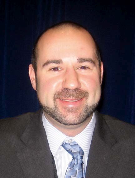
A closer look at individual genes that were different between class II–negative and positive tumors showed that LAG-3 mRNA was more enriched in the HLA-DR+ tumors, and also in patients who experienced a significant response to anti-PD-1 therapy.
“We also had a small population of samples that were derived from relapsed specimens,” he said. “We performed IHC within a small subset where we had paired tumors from pre-PD-1 response and relapse [to look at] LAG-3+ lymphocytes in the tumor ... and saw significant enrichment of LAG-3 infiltrate in the relapsed specimens. Importantly, all of these tumors were MHC-II+.”
Findings in a mouse model
The functional significance of this was explored using an MHC-II–negative orthotopic model cell line unlikely to induce expression of MHC-II when treated with interferon-gamma in culture; MHC-class-II transactivator (CIITA), the master regulator of MHC-II, was used to transduce the cells, resulting in cells that were “constitutively 100% class II+.”
Immunocompetent mice injected with these cells rejected tumors at a much higher rate, but IHC showed more nonregulatory CD4 cells in mice that did not reject the MHC-II+ tumors, Dr. Balko noted.
Gene expression analysis of the rejection-escaped tumors showed more mRNA for PD-1 and LAG-3, similar to what was seen in the study subjects.
“To see if the effect was truly an increase in PD-1 and LAG-3 on lymphocytes within the tumor microenvironment or in lymphoid tissues, we injected immunocompetent mice with either control or CIITA-positive tumors, and then at 7 days harvested either the contralateral lymph node, the spleen, or the proximal or tumor-draining lymph node,” he said.
This showed increased amounts of LAG-3 and PD-1-positive CD4 and CD8 cells in the tumor-draining lymph node, and more LAG-3 PD-1-positive CD8 cells within the tumor itself.
“To perform a therapeutic study, but also to eliminate any confounding factors of the rejecting mice, we waited 14 days after injection of the tumor cells and only enrolled mice with actively growing tumors. We randomized the mice to treatment with either IgG vehicle control, or anti-PD-1, or the combination of anti-PD1 plus LAG-3, and we had a very substantial [75%] complete response rate in the mice with class II–positive tumors treated with the combined PD-1 and LAG-3,” he said. “Importantly, all of the mice in this study were reinoculated with the [MHC-II–negative] cell line and had complete rejection of any subsequent injection of tumor cells.”
To assess whether any other MHC-II receptors could be expressed in the tumor microenvironment, Dr. Balko and his colleagues turned their attention to the FCRL6 molecule, which has previously been shown to be an MHC-II receptor that is expressed on cytolytic cells.
FCRL6
“[FCRL6] actually has an [immunoreceptor tyrosine-based inhibitory motif] domain in the intracellular portion of the human ortholog, which suggests that it could have some inhibitory function,” Dr. Balko said, adding that it has been shown to be expressed in a substantial proportion of natural killer cells and CD8 cells, and in a minor fraction of CD4+ T cells, which have been described as “cytotoxic CD4 cells.”
An immortalized FCRL6-negative natural killer cell line know as NK-92 was used to test for inhibitory function.
“We co-cultured it with K562 cells, which are a leukemia cell line that is both class I and class II negative; because they have a missing-self signal, the natural killer cells will naturally lyse the K562 cells, which can be measured by chromium release,” he explained.
When MHC-II was reconstituted on K562 cells, the natural killer cells still had effective lysis of the K562 cells, but when FCRL6 was also transduced on the natural killer cells, this interaction was stopped, and there was suppression of cytotoxic activity, or chromium release, in the co-cultures, suggesting that FCRL6 may have a checkpoint-like functionality, he said.
In the melanoma dataset, a look at FLCR6 mRNA in the tumor microenvironment showed that it was also much more highly expressed in HLA-DR–positive tumors and in the relapsed specimens.
In the tumors with paired specimens (three of which were MHC-II positive and three of which were MHC-II negative), IHC for FCRL6 identified greater enrichment of lymphocytes in the MHC-II–positive tumors, but the difference was not statistically significant.
In the breast cancer samples, where more LAG-3 and FCRL6 was seen in the triple-negative breast tumors, quantitative immunofluorescence showed that FCRL6-postive lymphocytes and LAG-3-positive lymphocytes had a substantial suppression of CD8-sel-positive granzyme B-positive cells within the microenvironment that was more substantial than that observed with PD-L1 expression, he noted.
“So our conclusions are that MHC-II tumors demonstrate enhanced T cell-mediated inflammation and immunity and anti-tumor immunity is circumvented through adaptive resistance by PD-1 and potentially LAG-3/MHC-II engagement in some tumors, and that ... FCRL6 may be a novel MHC-II receptor with inhibitory functionality, and could be a new immunotherapy target,” he said.
MHC-II expression could be useful for stratifying patients to combined anti-PD-1/anti-LAG-3 therapy, and eventually to combined anti-PD-1/anti-FCRL6 therapy, he added.
Combined anti-PD-1 and anti-LAG-3 therapy
The findings are of particular interest given recent findings regarding LAG-3 antibodies in development, said invited discussant Antoni Ribas, MD.
In a study reported by Ascierto et al. at ASCO 2017, for example, combined anti-PD-1 and anti-LAG-3 therapy had a 13% overall response rate in metastatic melanoma patients who progressed on anti-PD-1 therapy alone (20% and 7.1% in those with and without LAG-3 expression, respectively), said Dr. Ribas of the University of California, Los Angeles.
The 20% response rate seen in those with LAG-3 expression suggests “there could be a biomarker for this combined therapy,” he said, noting that while the overall response rate of 13% is low, “it is relevant because it is rescuing some patients who progressed on therapy, and it follows Dr. Balko’s science of why that would be the case.”
Dr. Balko has received research funding from Incyte, and holds a patent on use of HLA-DR/MHC expression to predict response to immunotherapies. Dr. Ribas owns stock in Advaxis, Arcus Ventures, Compugen, CytomX Therapeutics, Five Prime Therapeutics, FLX Bio, and Kite Pharma, and has served as a consultant or adviser for Amgen, Genentech/Roche, Merck, Novartis, and Pierre Fabre.
sworcester@frontlinemedcom.com
SOURCE: Balko J et al., ASCO-SITC abstract 180
REPORTING FROM THE CLINICAL IMMUNO-ONCOLOGY SYMPOSIUM
Key clinical point: MHC-II expression could be useful for stratifying patients to anti-PD-1/anti-LAG-3 and other therapies.
Major finding: The ORR was 75% for class II–positive tumors treated with combined anti-PD-1/anti-LAG-3
Study details: RNA sequencing on 58 patients with anti-PD-1-treated tumors and on matched pretreatment specimens.
Disclosures: Dr. Balko has received research funding from Incyte, and holds a patent on use of HLA-DR/MHC expression to predict response to immunotherapies. Dr. Ribas owns stock in Advaxis, Arcus Ventures, Compugen, CytomX Therapeutics, Five Prime Therapeutics, FLX Bio, and Kite Pharma, and has served as a consultant or adviser for Amgen, Genentech/Roche, Merck, Novartis, and Pierre Fabre.
Source: Balko J et al. ASCO-SITC abstract 180.
ERV expression may predict response to immune checkpoint blockade in ccRCC, other solid tumors
SAN FRANCISCO – The (ccRCC), according to findings from an analysis of nearly 5,000 tumor samples.
Similar associations may exist in other solid tumors, Shridar Ganesan, MD, reported at the ASCO-SITC Clinical Immuno-Oncology Symposium.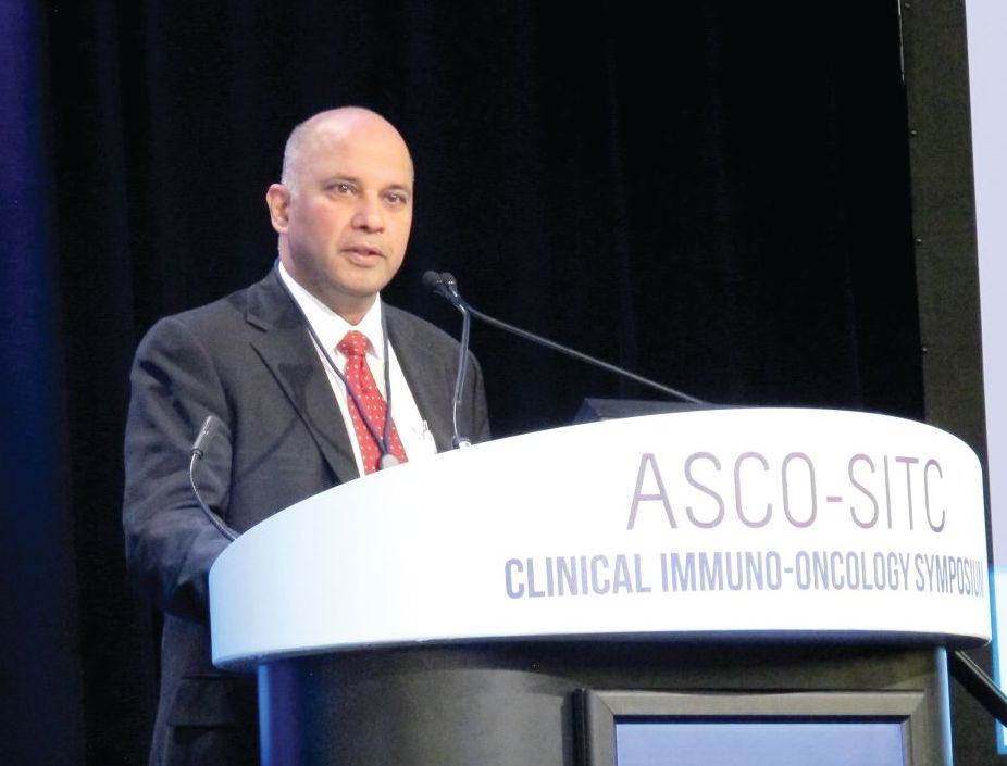
Merkel cell carcinoma, for example, has a high response rate despite its relatively low mutation burden. This is explained by the presence of infection with the Merkel cell polyomavirus in a low mutation burden subset, he said, noting that expression of the virus likely leads to expression of antigens that makes the disease both highly immunogenic and responsive to immune checkpoint therapy.
Expression of exogenous viruses is associated with response to immune checkpoint therapy in other cancers with low mutation burden, such as Epstein-Barr virus-positive gastric cancer, natural killer lymphoma, and Hodgkin disease.
“Intriguingly, renal cell carcinoma also has a relatively higher response rate to immune checkpoint therapy than would be anticipated by its relatively low mutation burden. However, this has no evidence of exogenous virus infection. Therefore, we turned our attention to endogenous retroviruses, which are an abundant potential stimulant of innate immunity in cancer,” he said.
Endogenous retroviruses (ERVs) now comprise about 5-8% of the human genome, and almost all of them have mutations that disable key coding genes of the retrovirus, meaning most cannot produce all the proteins necessary for viral replication.
“They’re essentially genomic fossils,” Dr. Ganesan said, adding that they all are silenced epigenetically in most normal adult tissues, both by DNA methylation and histone methylation. “However, inappropriate expression of endogenous retroviruses has been reported by multiple groups in some cancers and has been associated with evidence of immune activation. One can imagine that endogenous retroviral expression can be immunogenic both by expression of some [open reading frames] that are not disabled ... or just the viral RNA itself, which can be detected by cytoplasmic sensors ... and activate innate immunity,” he said.
To assess whether ERV expression corresponds with evidence of immune checkpoint activation in cancer, he and his colleagues conducted a pan-cancer analysis of more than 4,900 tumors across 21 cancer types using data from the Cancer Genome Atlas (TCGA).
“When we did this for about 66 annotated endogenous retroviruses in the TCGA database, we could see that different cancers had different amounts of retrovirus associated with immune activation ... in fact the strongest signal seen in this analysis was in clear cell renal cell carcinoma,” he said.
Signals were also seen in estrogen receptor–positive/human epidermal growth factor receptor 2-negative breast cancer, head and neck squamous cell carcinoma, and colon cancer.
A variety of ERVs were upregulated in RCC; different panels were associated with different cancer types. But two – ERVK.2 and ERV3.2 – were consistently upregulated across all the tumor types.
A closer look at ERV expression showed three clear clusters: extremely-high, intermediate, and low expression of ERV, and the very-high expression cluster had increased expression of numerous checkpoint genes, including CD8A, PD1, CTLA4, and LAG3 among others, compared with the low expression cluster.
This leads to the question of why a subset of RCCs have ERV expression.
In an attempt to answer that question, a transcript analysis was conducted to determine which transcripts are differentially regulated between the high and low ERV-expressing groups, and gene ontology analysis was performed.
“The results were quite striking. If you look at the modules that are differentially expressed between the high-ERV and the low-ERV groups, what pops up is really a lot of histone methyltransferase modules and chromatin regulation modules, ” he said, noting that this makes sense because of the silencing of ERV expression by histone methylation and DNA methylation, and suggests that “there is some deep abnormality in chromatin modulation in this subset of RCCs.
“This is intriguing, because RCCs are known to have mutations in chromatin modifying genes,” he said.
A look into whether the high-ERV–expressing group was enriched in any of these mutations showed some enrichment of BAP1 in the high- vs. low-expressing group, and that is currently being looked at further, he noted.
The next question was whether ERV expression correlated with response to immune checkpoint blockade in ccRCC, and this was looked at in a nonrandomized group of 15 patients with metastatic ccRCC who were treated with single-agent immune checkpoint therapy who had either clearly documented partial response or progressive disease. In 13 patient samples for which RNA expression of ERV3.2 was successfully measured by quantitative real-time polymerase chain reaction using two primer sets, ERV3.2 expression was significantly higher in responders vs. nonresponders in both primer sets (P less than .05 and .005).
“In summary, we have shown that expression of ERVs correlates with immune activation and increased expression of immune checkpoint genes in a subset of ccRCC, and perhaps several other solid tumor classes, and the expression of ERV3.2 is perhaps associated with response to PD1 blockade in this small preliminary cohort of ccRCC patients,” he said, noting that abnormal expression of ERV may be a biomarker of immune checkpoint therapy response in some cancers with a low mutation burden. “Mechanisms underlying ERV expression need to be investigated and may reflect underlying chromatin alterations or epigenetic abnormalities.”
Dr. Ganesan reported that his spouse is employed by Merck and that he is a consultant and/or advisory board member for Novartis, Roche, and Inspirata. He also holds patents with Inspirata.
sworcester@frontlinemedcom.com
SOURCE: Panda A et al., ASCO-SITC, Abstract #104.
REPORTING FROM THE CLINICAL IMMUNO-ONCOLOGY SYMPOSIUM
Key clinical point: ERV3.2 expression is associated with immune checkpoint blockade response in ccRCC.
Major finding: ERV3.2 expression was significantly higher in responders vs. nonresponders in two primer sets (P less than .05 and.005).
Study details: A pan-cancer analysis of more 4,900 tumors.
Disclosures: Dr. Ganesan reported that his spouse is employed by Merck and that he is a consultant and/or advisory board member for Novartis, Roche, and Inspirata. He also holds patents with Inspirata
Source: Panda A et al. ASCO-SITC, Abstract #104.
Study: Test for PD-L1 amplification in solid tumors
SAN FRANCISCO – Amplification of programmed death-ligand 1 (PD-L1), also known as cluster of differentiation 274 (CD274), is rare in most solid tumors, but findings from an analysis in which a majority of patients with the alteration experienced durable responses to PD-1/PD-L1 blockade suggest that testing for it may be warranted.
Of 117,344 deidentified cancer patient samples from a large database, only 0.7% had PD-L1 amplification, which was defined as 6 or more copy number alterations (CNAs). The CNAs were found across more than 100 tumor histologies, Aaron Goodman, MD, reported at the ASCO-SITC Clinical Immuno-Oncology Symposium.
Of a subset of 2,039 clinically annotated patients from the database, who were seen at the University of California, San Diego (UCSD) Center for Personalized Cancer Therapy, 13 (0.6%) had PD-L1 CNAs, and 9 were treated with immune checkpoint blockade, either alone or in combination with another immunotherapeutic or targeted therapy, after a median of four prior systemic therapies.
The PD-1/PD-L1 blockade response rate in those nine patients was 67%, and median progression-free survival was 15.2 months; three objective responses were ongoing for at least 15 months, said Dr. Goodman of UCSD.
The findings are notable, because in unselected patients, the rates of response to immune checkpoint blockade range from 10% to 20%.
Lessons from cHL and solid tumors
“Over the past few years, investigators have identified numerous biomarkers that can select subgroups of patients with increased likelihoods of responding to PD-1 blockade,” he said, adding that biomarkers include PD-L1 expression by immunohistochemistry, microsatellite instability – with microsatellite instability–high tumors responding extremely well to immunotherapy, tumor mutational burden measured by whole exome sequencing and next generation sequencing, and possibly PD-L1 amplification.
Of note, response rates are high in patients with classical Hodgkin lymphoma (cHL). In general, cHL patients respond well to treatment, with the majority being cured by way of multiagent chemotherapy and radiation.
“But for the subpopulation that fails to respond to chemotherapy or relapses, outcomes still remain suboptimal. Remarkably, in the relapsed/refractory population of Hodgkin lymphoma ... response rates to single agent nivolumab and pembrolizumab were 65% to 87% [in recent studies],” he said. “Long-term follow-up demonstrates that the majority of these responses were durable and lasted over a year.”
The question is why relapsed/refractory cHL patients treated with immune checkpoint blockade have such a higher response rate than is typically seen in patients with solid tumors.
One answer might lie in the recent finding that nearly 100% of cHL tumors harbor amplification of 9p24.1; the 9p24.1 amplicon encodes the genes PD-L1, PD-L2, and JAK2, (and thus is also known as the PDJ amplicon), he explained, adding that “through gene dose-dependent increased expression of PD-L1 ligand on the Hodgkin lymphoma Reed-Sternberg cells, there is also JAK-STAT mediation of further expression of PD-L1 on the Reed-Sternberg cells.
An encounter with a patient with metastatic basal cell carcinoma – a “relatively unusual situation, as the majority of patients are cured with local therapy”– led to interest in looking at 9p24.1 alterations in solid tumors.
The patient had extensive metastatic disease, and had progressed through multiple therapies. Given his limited treatment options, next generation sequencing was performed on a biopsy from his tumor, and it revealed the PTCH1 alteration typical in basal cell carcinoma, as well as amplification of 9p24.1 with PD-L1, PD-L2, and JAK2 amplification. Nivolumab monotherapy was initiated.
“Within 2 months, he had an excellent partial response to therapy, and I’m pleased to say that he’s in an ongoing complete response 2 years later,” Dr. Goodman said.
It was that case that sparked the idea for the current study.
9p24.1 alterations and checkpoint blockade
“With my interest in hematologic malignancies, I was unaware that [9p24.1] amplification could occur in solid tumors, so the first aim was to determine the prevalence of chromosome 9p24.1 alterations in solid tumors. The next was to determine if patients with solid tumors and chromosome 9p24.1 alterations respond to PD-1/PD-L1 checkpoint blockade.
“What is astounding is [that PD-L1 amplification] was found in over 100 unique tumor histologies, although rare in most histologies,” Dr. Goodman said, noting that histologies with a statistically increased prevalence of PD-L1 amplification included breast cancer, head and neck squamous cell carcinoma, lung squamous cell carcinoma, and soft tissue sarcoma.
There also were some rare histologies with increased prevalence of PD-L1 amplification, including nasopharyngeal carcinoma, renal sarcomatoid carcinoma, bladder squamous cell carcinoma, and liver mixed hepatocellular cholangiocarcinoma, he said.
Tumors with a paucity of PD-L1 amplification included colorectal cancer, pancreatic cancer, and cutaneous melanoma, although even these still harbored a few patients with amplification, he said.
A closer look at the mutational burden in amplified vs. unamplified tumors showed a median of 7.4 vs. 3.6 mut/mb, but in the PD-L1 amplified group, 85% still had a low-to intermediate mutational burden of 1-20 mut/mb.
“Microsatellite instability and PD-L1 amplification were not mutually exclusive, but a rare event. Five of the 821 cases with PD-L1 amplification were microsatellite high; these included three carcinomas of unknown origin and two cases of gastrointestinal cancer,” he noted.
Treatment outcomes
In the 13 UCSD patients with PD-L1 amplification, nine different malignancies were identified, and all patients had advanced or metastatic disease and were heavily pretreated. Of the nine treated patients, five received anti-PD-1 monotherapy, one received anti-CTLA4/anti-PD-1 combination therapy, and three received a PD-1/PD-L1 inhibitor plus an investigational agent, which was immunotherapeutic, Dr. Goodman said.
The 67% overall response rate was similar to that seen in Hodgkin lymphoma, and many of the responses were durable; median overall survival was not reached.
Of note, genomic analysis in the 13 UCSD patients found to have PD-L1 amplification showed there were 143 total alterations in 70 different genes. All but one patient had amplification of PD-L1, PD-L2, and JAK2, and that one had amplification of PD-L1 and PD-L2.
Of six tumors with tissue available to test for PD-L1 expression by immunohistochemistry, four (67%) tested positive. None were microsatellite high, and tumor-infiltrating lymphocytes were present in five cases.
The tumors that tested negative for PD-L1 expression were from the patient with the rare basal cell cancer, and another with glioblastoma. Both responded to anti-PD1/PD-L1 therapy.
The glioblastoma patient was a 40-year-old man with progressive disease, who underwent standard surgical debulking followed by concurrent radiation therapy plus temozolomide. He progressed soon after completing the concurrent chemoradiation therapy, and genomic profiling revealed 12 alterations, including 9p24.1 amplification, Dr. Goodman said, adding that nivolumab therapy was initiated.
“By week 12, much of the tumor mass had started to resolve, and by week 26 it continued to decrease further. He continues to be in an ongoing partial response at 5.2 months,” he said.
Recommendations
The findings of this study demonstrate that PD-Ll amplification is rare in solid tumors.
“However, PD-L1 amplification appears to be tissue agnostic, as we have seen in over 100 tumor histologies. We also noted that PD-L1 amplification was enriched in many rare tumors with limited treatment options, including anaplastic thyroid cancer, sarcomatoid carcinoma, and some sarcomas. We believe testing for PD-L1 amplification may be warranted given the frequent responses that were durable and seemed to be independent of mutational burden,” he concluded.
Ravindra Uppaluri, MD, session chair and discussant for Dr. Goodman’s presentation, said that Dr. Goodman’s findings should be considered in the context of “the complex biology [of PD-L1/PD-L2] that has evolved over the last few years.”
He specifically mentioned the two patients without PD-L1 expression despite amplification, but with response to immune checkpoint blockade, and noted that “there are several things going on here ... and we really want to look at all these things.”
The PDJ amplicon, especially given “the ability to look at this with the targeted gene panels that many patients are getting,” is clearly contributing to biomarker stratification, said Dr. Uppaluri of Dana-Farber Cancer Institute and Brigham and Women’s Hospital, Boston.
However, it should be assessed as part of a “global biomarker” that includes tumor-infiltrating lymphocytes and tumor mutational burden, he said.
Dr. Goodman reported having no disclosures. Dr. Uppaluri has received grant/research support from NIH/NIDCR, Merck, and V Foundation, and has received honoraria from Merck.
sworcester@frontlinemedcom.com
SOURCE: Goodman A et al. ASCO-SITC, Abstract 47
REPORTING FROM THE CLINICAL IMMUNO-ONCOLOGY SYMPOSIUM
Key clinical point: Solid tumor patients with PD-L1 amplification had durable responses to PD-1/PD-L1 blockade.
Major finding: The overall response rate was 67% in nine patients treated with PD-1/PD-L1 blockade.
Study details: An analysis of more than 117,000 patient samples.
Disclosures: Dr. Goodman reported having no disclosures. Dr. Uppaluri has received grant/research support from NIH/NIDCR, Merck, and V Foundation, and has received honoraria from Merck.
Source: Goodman A et al. ASCO-SITC, Abstract 47.
TRANSCEND NHL trial identifies window for CAR T expansion
for CAR T expansion
SAN FRANCISCO – The CD19-directed 4-1BB chimeric antigen receptor (CAR) T-cell product JCAR017 demonstrated increased CAR T-cell expansion and persistence, and higher durability of response at higher dose levels – with manageable toxicities – in a pivotal phase 1 trial of relapsed/refractory B-cell non-Hodgkin lymphoma.
However, preliminary modeling data suggest that a therapeutic window exists for the CAR T-cell expansion, which means that development of strategies for pushing patients into that window could enhance efficacy and limit toxicity associated with JCAR017, Tanya Siddiqi, MD, of City of Hope Comprehensive Cancer Center, Duarte, Calif., reported at the ASCO-SITC Clinical Immuno-Oncology Symposium.
TRANSCEND NHL 001 is a multicenter, seamless design, pivotal trial, which started as a phase 1 first-in-human study of JCAR017, a defined composition CAR T-cell product also known as lisocabtagene maraleucel and administered at precise doses of CD4+ and CD8+ CAR T cells. Dose-finding and dose-expansion cohorts have been investigated, and currently the pivotal diffuse large B-cell lymphoma (DLBCL) cohort is enrolling, Dr. Saddiqi said, noting that dose level 2 (1 x 10⁸ cells given as a single dose) was selected for that cohort.
The current findings are based on the TRANSCEND core population – a set of patients selected from the dose-finding and dose-expansion cohorts. This population includes patients with DLBCL not otherwise specified, transformed follicular lymphoma, or high-grade double- or triple-hit lymphomas, she said, explaining that she and her colleagues looked at prelymphodepletion baseline patient characteristics and biomarkers to assess how they related to outcomes and toxicities. This precise dosing of JCAR017 reduces variability, enabling the identification of potential patient factors associated with clinical outcomes, she said.
Response rates among 27 patients who received dose level 2 were high, with an 81% overall response rate and a 63% complete response rate.
“Patients with complete remission seemed to have more of a durable response,” she said. “In the core population at dose level 2, 50% with 6 months of follow-up seemed to remain in complete response, so there’s a dose-response effect in these patients.”
It doesn’t appear that the dose affects development of cytokine release syndrome (CRS) or neurotoxicity (NT), as the rates of these (30% and 20%, respectively) did not differ by dose level or schedule, but certain baseline features, such as a lactate dehydrogenase (LDH) level above 500 U/L and tumor burden measured as the sum of the product of diameters of 50 cm or greater, do appear to affect the development of these toxicities.
For example, 10 of 13 (77%) of patients with both of those baseline characteristics developed CRS, and 7 of 13 (54%) developed NT; for those without these characteristics, the odds of developing CRS and NT were significantly lower, she said.
Of note, higher Cmax, or peak expansion of CAR T cells in vivo, was seen at dose level 2, and exposure (area under the curve) was also higher, as was expected at that dose level, Dr. Siddiqi said.
“It also appears that there is a trend of patients with higher tumor burden to have higher expansion of their CAR T cells in vivo,” she said, noting that some of those patients were “superexpanders” with very high expansion of CAR T cells. “Similarly there were certain cytokines at the prelymphodepletion time point that seem to be higher … in patients who also go on to have higher expansion of their CAR T cells.”
These included interleukin-7, IL-15, MIP (macrophage inflammatory protein)–1alpha, and tumor necrosis factor (TNF)–alpha.
“Patients with certain higher inflammatory cytokines and biomarkers of inflammation also were noted to have higher events of CRS and neurotoxicity, so not just higher tumor burden, but also higher inflammatory state at baseline seems to affect patients in terms of getting any grade CRS or any grade neurotoxicity,” she said.
Those associated with CRS included ferritin, C-reactive protein (CRP), IL-10, IL-15, IL-16, TNF-alpha, and MIP-1beta levels, and those associated with NT included ferritin, CRP, d-dimer, IL-6, IL-15, TNF-alpha, and MIP-1alpha levels.
“Interestingly, there’s an inverse relationship between the expression of some of these biomarkers and inflammatory markers and the durability of response at 3 months,” she said.
Patients with higher tumor burden, LDH, and other markers either had no response at 3 months, or they had a very rapid response at the 1-month mark and then lost their response by 3 months, she noted.
“One of the theories could be that these patients with higher tumor burden, higher inflammatory state – they have such a high peak expansion rapidly after receiving their CAR T cells that potentially those CAR T cells may be getting exhausted or dying off very quickly, and therefore patients can lose their response,” she said.
When these data are considered together and modeled, there seems to be a therapeutic window where patients who have optimal expansion of CAR T cells in vivo may have lesser toxicity, higher overall response rates, and better durability of response, she said.
That is, on one end of the spectrum, there are patients with lower CAR T-cell expansion who have lower toxicities, but who also have lower overall response rates and lower durability of response, and on the other end, there are patients with superhigh expansion of CAR T cells, who have higher response rates, but also higher rates of toxicity, and who lose their response quickly.
“So if we can identify patients who would be in this window of optimal target expansion of CAR T cells, and if we could find strategies or mechanisms to move patients who are on either end of that spectrum into this window, we may be able to get patients with better efficacy and lower toxicities, and there could be combination strategies that could help get us there,” she said.
TRANSCEND NHL 001 is sponsored by Juno Therapeutics. Dr. Siddiqi reported serving as a consultant or adviser for Juno Therapeutics, and as a member of the speakers bureau for Pharmacyclics/Janssen and Seattle Genetics. Research funding was provided to her institution by Juno Therapeutics and several other companies.
sworcester@frontlinemedcom.com
SOURCE: Siddiqi T et al. ASCO-SITC, abstract 122.
for CAR T expansion
for CAR T expansion
REPORTING FROM THE CLINICAL IMMUNO-ONCOLOGY SYNDROME
Key clinical point:
Major finding: In all, 77% of patients with a lactate dehydrogenase level greater than 500 U/L and a sum of the product of diameters of 50 cm or greater developed cytokine release syndrome, and 54% of patients with those characteristics developed neurotoxicity.
Study details: A cohort of 27 patients from the pivotal phase 1 TRANSCEND NHL 001 trial.
Disclosures: TRANSCEND NHL 001 is sponsored by Juno Therapeutics. Dr. Siddiqi reported serving as a consultant or adviser for Juno Therapeutics, and as a member of the speakers bureau for Pharmacyclics/Janssen and Seattle Genetics. Research funding was provided to her institution by Juno Therapeutics and several other companies.
Source: Siddiqi T et al. ASCO-SITC, abstract 122.
AML immune profiles correlate with relapse-free survival
SAN FRANCISCO – Immune-enriched acute myeloid leukemias might be amenable to immunotherapy that is tailored to the bone marrow tumor microenvironment, according to findings from a pan-cancer analysis of bone marrow samples.
The analysis, performed with 3D biology technology and an RNA pan-cancer immune profiling panel to characterize bone marrow specimens from 46 children and 28 adults with acute myeloid leukemia (AML), identified heterogeneous immune profiles that correlated with relapse-free survival (RFS) and overall survival (OS), Jayakumar Vadakekolathu, PhD, reported at the ASCO-SITC Clinical Immuno-Oncology Symposium.
The specimens, including 63 from nonpromyelocytic de novo AML, 7 from AML in children with complete remission, 3 from adults with secondary AML, and 1 from an adult with treatment-related AML, were analyzed with the nCounter system from NanoString Technologies, and were visualized via digital spatial profiling, said Dr. Vadakekolathu of Nottingham Trent University in England.
The investigators identified two distinct immune gene expression profiles (GEPs) that were largely age-differentiated: Cluster A (myeloid-enriched specimens) included 26 children and 8 adults, and cluster B (myeloid-depleted specimens) included 9 children and 18 adults. These GEPs predicted clinical outcome; relapse free survival was 2.2 months in cluster A versus 18.3 months in cluster B (hazard ratio, 2.58) and overall survival was 6.3 months in cluster A, compared with 22.4 months in cluster B (HR, 2.39), Dr. Vadakekolathu reported.
The findings could have implications for the development of new treatment strategies, he said, noting that AML is a highly heterogeneous disease in terms of genetics, clinical manifestations, and outcome.
“Prognosis is determined by cytogenetic and molecular abnormalities, as well as by response to chemotherapy. De novo AML is cured in roughly 70% of children, 35%-40% of adults, and 5%-15% of elderly patients,” he said. Some patients with AML fail to respond to induction chemotherapy, and others eventually relapse despite the lack of adverse risk factors, he added.
The general therapeutic strategy in patients with AML has not changed substantially in more than 30 years, he said.
“High degrees of molecular complexity in AML present a considerable challenge in clinical implementation, and there is an urgent need to discover better biomarkers to identify high-risk patients before starting chemotherapy, which would enable testing of investigational therapeutic strategies in clinical trials,” he said.
In an effort to identify immune gene signatures across the spectrum of AML genotypes and to correlate transcriptomic and proteomic profiles with patient outcomes, he and his colleagues used a pediatric cohort from Children’s Hospital of Philadelphia (median age at diagnosis of 10 years), and an adult cohort from the Technical University of Dresden, Germany (median age at diagnosis, 55.5 years). Bone marrow samples were collected and analyzed at diagnosis.
Hierarchical clustering identified the two distinct clusters. The immune-enriched cluster A had heightened expression of T cells, natural killer cells, and cytotoxic cells, and also expressed CD8A, IFNG, FOXP3, the cell chemoattractants CXCL9 and CXCL10, and inhibitory molecules including IDO1 and the immune checkpoints LAG3, CTLA4, and PD-L1. The immune-depleted cluster B overexpressed genes associated with mast cell functions and CD8 T-cell exhaustion, and showed low expression of T-cell and B-cell genes.
Further analysis of 10 of the inflamed samples from patients with newly diagnosed AML was performed with digital spatial profiling (DSP) to visualize in situ leukemia–immune system interactions, Dr. Vadakekolathu said.
Surface antigens CD123 and CD3 were used as a visualization marker for leukemia cells and to identify bone marrow-infiltrating T cells, respectively. Protein quantification showed a higher concentration of CD3 counts in T-cell-rich versus T-cell-poor areas, and protein expression profiles showed strong correlations with various immunologically relevant molecules. The co-localization of CD8 T cells with FoxP3 Treg cells and PD-L1- and VISTA-expressing cell types evident on DSP represents an immune landscape consistent with the establishment of adaptive immune resistance mechanisms of immune escape, he noted.
The findings suggest immune enriched AMLs might be amenable to combination immunotherapies tailored to the bone marrow tumor microenvironment, such as IDO1 inhibitors and checkpoint blockade, Dr. Vadakekolathu said. “Immune gene expression profiles of AML might support rapid prediction of patient outcomes, discovery of novel immune biomarkers and therapeutic targets, and development of integrated patient stratifications,” he said.
This study was supported by grants from the Roger Counter Foundation and the Qatar National Research Fund. Dr. Vadakekolathu reported having no disclosures. Some authors reported employment or other financial relationships with NanoString Technologies.
sworcester@frontlinemedcom.com
SOURCE: Rutella S et al. ASCO-SITC Abstract 50
REPORTING FROM THE CLINICAL IMMUNO-ONCOLOGY SYMPOSIUM
Key clinical point:
Major finding: Relapse-free survival was 2.2 months in a cluster of myeloid-enriched specimens, compared with 18.3 months in a cluster of myeloid-depleted specimens (hazard ratio, 2.58).
Study details: A pan-cancer analysis of bone marrow specimens from 74 patients.
Disclosures: This study was supported by grants from the Roger Counter Foundation and the Qatar National Research Fund. Dr. Vadakekolathu reported having no disclosures. Some authors reported employment and other financial relationships with NanoString Technologies.
Source: Rutella S et al. ASCO-SITC Abstract 50.
Pembrolizumab plus SBRT shows promise for advanced solid tumors
SAN FRANCISCO – Pembrolizumab immunotherapy with multi-site stereotactic body radiotherapy (SBRT) appears to be a safe and effective treatment in patients with advanced solid tumors, according to findings from a phase 1 study.
Of 79 patients with metastatic solid tumors who progressed on standard treatment and who were enrolled in the study, 68 underwent multi-site SBRT, received at least one cycle of pembrolizumab (Keytruda), and had imaging follow-up. The overall objective response rate in those 68 patients was 13.2%, Jeffrey Lemons, MD, reported at the ASCO-SITC Clinical Immuno-Oncology Symposium.
When responses in the non-irradiated lesions (out-of-field responses) were measured based on a 30% reduction in any single lesion, the rate was 26.9%. But when defined by a 30% reduction in aggregate diameter of the non-irradiated measurable lesions, the rate was 13.5%, he said. While both approaches for measuring response are acceptable, Dr. Lemons noted, it’s important to be sure which one is being used in a given study.
Overall, 73 patients received both SBRT and pembrolizumab (5 had no imaging follow-up). They had a mean age of 62 years and a median of five prior therapies. Cancer types included ovarian/fallopian tube cancer (12.3%), non–small cell lung cancer (9.6%), breast cancer (8.2%), cholangiocarcinoma (8.2%), endometrial cancer (8.2%), colorectal cancer (6.8%), head and neck cancer (5.5%), and other tumors, each with less than 5% accrual (41.2%).
The number of sites treated with SBRT was two in 94.5% of patients, three in 4.1%, and four in 1.3%; 151 lesions in total were treated.
The premise for combining pembrolizumab and SBRT is that response to anti-programmed cell death-1 (PD1) therapy seems to correspond with interferon-gamma signaling, and that SBRT can stimulate innate and adaptive immunity to potentially augment immunotherapy, Dr. Lemons explained. In addition, anti-PD1 treatment outcomes are improved with lower disease burden.
Multi-site radiation is an emerging paradigm for eradicating metastatic disease, he said.
Patients included in the study had metastatic solid tumors and had progressed on standard treatment. They had measurable disease by RECIST, and metastases amenable to SBRT with 0.25 cc to 65 cc of viable tumor.
Tumors larger than 65 cc were partially targeted with radiotherapy. Radiation doses were adapted from recently completed and ongoing National Cancer Institute trials and ranged from 30-50 Gy (3-5 fractions) based on anatomic location.
Pembrolizumab was initiated within 7 days of the final SBRT treatment.
Dose-limiting toxicities, all grade 3, occurred in six patients during a median follow-up of 5.5 months, and included pneumonitis in three patients, hepatic failure in one patient, and colitis in two patients, but there were no radiation dose reductions, Dr. Lemons said.
“This is the first and largest prospective trial to determine the safety of this combination,” he explained. “There was some intriguing clinical activity ... and we feel that this justifies further randomized studies
The University of Chicago sponsored the study. Dr. Lemons reported having no disclosures.
sworcester@frontlinemedcom.com
SOURCE: Lemons J et al., ASCO-SITC abstract #20.
REPORTING FROM THE CLINICAL IMMUNO-ONCOLOGY SYMPOSIUM
Key clinical point: Pembrolizumab plus multi-site SBRT appears safe and effective for advanced solid tumors.
Major finding: The overall objective response rate was 13.2%.
Study details: A phase 1 study of 79 patients.
Disclosures: The University of Chicago sponsored the study. Dr. Lemons reported having no disclosures
Source: Lemons J et al. ASCO-SITC abstract #20.
Experimental PD-1/PARP inhibitor combo shows promise in solid tumors
SAN FRANCISCO – Combined therapy using an experimental programmed cell death protein 1 (PD-1) inhibitor and experimental poly ADP-ribose polymerase (PARP) 1/2 inhibitor was generally well tolerated and showed efficacy in a phase 1 study of patients with advanced solid tumors.
Tislelizumab, the anti–PD-1 agent in development for solid and hematologic malignancies, is a humanized IgG4 monoclonal antibody engineered to have minimal Fc-gamma receptor binding. Pamiparib, the PARP 1/2 inhibitor, is hypothesized to promote neoantigen release that may boost the efficacy of tislelizumab. At the Jan. 4 data cutoff, 2 of 49 patients treated with one of five planned dose levels experienced a complete response, 8 had a confirmed partial response, and 4 had an unconfirmed partial response, Linda Mileshkin, MD, reported at the ASCO-SITC Clinical Immuno-Oncology Symposium.
Thus, the objective response rate was 20%, she said, noting that the clinical benefit rate, which encompasses all those with a response as well as those with durable stable disease after at least 24 weeks, was 39%. The median duration of response was 168.5 days
As of the data cutoff, 11 patients, including all those with a complete or partial response, remained on treatment, and 10 patients remained on treatment beyond 200 days, said Dr. Mileshkin of Peter MacCallum Cancer Centre, Melbourne.
Study participants were 42 women and 7 men, with a mean age of 63 years and measurable disease treated with at least 1 prior line of therapy (median of 4), but with no prior exposure to a PARP inhibitor or PD-1 therapy. Primary tumor sites included ovarian/fallopian tube/peritoneal (34 patients); pancreas, prostate, and breast cancer (3 patients each); and bile duct, bladder, cervix, lung, peripheral nerve sheath, and uterus (1 patient each). For the current dose-finding phase of the study (phase 1a), cohorts of 6-13 patients were treated with either tislelizumab at an intravenous dose of 2 mg/kg every 3 weeks plus either an oral dose of 20, 40, or 60 mg of pamiparib (dose levels 1, 2, and 3, respectively), or with tislelizumab at an intravenous dose of 200 mg every 3 weeks plus pamiparib at an oral dose of 40 or 60 mg twice daily (dose levels 4 and 5, respectively), Dr. Mileshkin said.
Phase 1b will be a disease-specific expansion to evaluate preliminary antitumor activity.
The rationale for combining the two agents was “to prove that we could get some up-regulation of tumor-associated antigens by using a PARP inhibitor that may then allow us to improve the antitumor activity of the checkpoint inhibitor,” she said.
Further, the malignancies in the patients include those likely to harbor DNA damage repair deficiencies, she added.
All patients experienced at least one treatment-emergent adverse event; 21 experienced a serious event, and 23 had immune-related adverse events.
“Despite the number of events, most patients were able to continue therapy,” Dr. Mileshkin said, noting that more discontinuation was associated with the PD-1 therapy than with the PARP inhibitor.
Non–immune-related adverse events related to the PARP inhibitor included mostly grade 1 and 2 nausea, fatigue, and diarrhea. Anemia also occurred and was grade 3 in 12% of patients.
“In terms of the PD-1 therapy, we mostly saw grade 1 or 2 nonimmune adverse events and very few grade 3 and 4 events,” she said.
One or more grade 3 immune-related adverse events occurred in 12 patients, and based on reports from participating centers, there “seemed to be a signal here that we were seeing more hepatic toxicity than we might have expected,” she said, explaining that these included increases in transaminases and hepatitis.
“We ended up with 13 patients who had a hepatic adverse event thought to be related to treatment. The median time to onset was 55 days ... and there were 9 patients in whom these events were grade 3 or 4,” she said.
Ten had grade 2 or higher transaminitis, which was most likely autoimmune in nature.
“All of these patients were treated with corticosteroids, and they all recovered promptly from the event. Subsequently, the protocol was amended to try increase real-time hepatic safety monitoring,” she said, noting that the rate of hepatic events has fallen since these changes were made.
“We need to closely monitor that moving forward to try to understand it better,” she said of the hepatic events.
Dose level 4 (tislelizumab at 200 mg every 3 weeks plus pamiparib at 40 mg twice daily) was determined to be the maximum tolerated dose and is the recommended phase 2 dose, she noted.
“We were pleased to see 10 patients respond, with these responses appearing to be durable with this currently short follow-up period, and we look forward to seeing the results of part B,” she concluded, noting that enrollment of patients into disease-specific cohorts for that next phase is ongoing.
This study is sponsored by BeiGene. Dr. Mileshkin reported receiving payment for travel, accommodations, and/or expenses from BeiGene and Roche.
sworcester@frontlinemedcom.com
SOURCE: Friedlander M et al. ASCO-SITC Clinical Immuno-Oncology Symposium 2018, Abstract #48.
REPORTING FROM THE CLINICAL IMMUNO-ONCOLOGY SYMPOSIUM
Key clinical point:
Major finding: Objective response and clinical benefit rates were 20% and 29%, respectively.
Study details: A phase 1 study of 49 patients.
Disclosures: This study is sponsored by BeiGene. Dr. Mileshkin reported receiving payment for travel, accommodations, and/or expenses from BeiGene and Roche.
Source: Friedlander M et al. ASCO-SITC Clinical Immuno-Oncology Symposium 2018, Abstract #48.