User login
Endocrine Society: Annual Meeting (ENDO 2013)
Avoid transcutaneous biopsy in adrenocortical carcinoma
SAN FRANCISCO – Transcutaneous biopsy of adrenocortical carcinomas did not improve diagnosis or survival, and potentially harmed 11% of patients, in a study of 74 patients.
The study fills a gap behind general recommendations against biopsy of single adrenal masses in the absence of another known malignancy or metastasis, which had been based on fears that biopsy would spread the tumor but lacked clinical evidence, Andrew Williams said in a poster presentation at the annual meeting of the Endocrine Society.
Among 74 patients with adrenocortical carcinoma who had undergone transcutaneous biopsy of an adrenal mass between 1991 and 2011, the sensitivity of the biopsy for the final pathological diagnosis of the disease was 84% at best and as low as 51% in community settings, the retrospective review of electronic medical records found.
Among the 36 patients with stage I-III disease (confined to the adrenal gland) at the time of diagnosis, the biopsy did not significantly change choices regarding adjuvant therapy, compared with 254 patients with stage I-III disease who did not undergo transcutaneous biopsy. The baseline characteristics of these two groups were similar except that patients in the biopsy group were significantly less likely to be secreting hormones (28%) compared with the no-biopsy group (59%).
"Single adrenal masses with malignant characteristics invariably should be treated surgically, making biopsy an unnecessary procedure," reported Mr. Williams, a medical student at the University of Michigan, Ann Arbor, and his associates.
Complications from transcutaneous biopsy in 11% of all patients consisted mainly of bleeding-related events but with one potentially fatal event – needle track metastasis in one patient.
"Single adrenal masses with malignant characteristics invariably should be treated surgically, making biopsy an unnecessary procedure."
Rates of overall and tumor-free survival did not differ significantly between patients who did or did not undergo transcutaneous biopsy in either univariate analysis or multivariate analyses adjusting for age, sex, cortisol production, and adjuvant therapies.
Adrenocortical carcinomas are rare cancers with high rates of recurrence and mortality. Cumulative survival rates in the study decreased steadily to roughly 80% at 1 year, 60% at 3 years, and less than 50% at 5 years.
"Biopsy of adrenocortical carcinomas at stages confined to the adrenal gland does not significantly influence survival," Mr. Williams concluded in the poster. "Adrenal biopsy should only be considered in patients with metastasized disease and unclear tumor origin, only if it may alter the therapeutic approach and only if the adrenal is the easiest site to biopsy."
Patients who underwent biopsy in the study had a median age of 52 years (ranging from 17 to 77 years), and 54% were female. The cohort was 84% white.
Mr. Williams reported having no financial disclosures. Coinvestigator Dr. Gary D. Hammer, also at the University of Michigan, disclosed financial associations with Atterocor, Orphagen Pharmaceuticals, Embera NeuroTherapeutics, HRA Pharma, Corcept Therapeutics, Isis, and OSI-Astella.
On Twitter @sherryboschert
AT ENDO 2013
Major finding: Transcutaneous biopsy was 51%-84% sensitive for adrenocortical carcinoma, depending on where it was done, and caused complications in 11% of patients.
Data source: Retrospective study of records for 74 patients with adrenocortical carcinoma who underwent transcutaneous biopsy between 1991 and 2011, and a comparison of 290 patients with stage I-III disease with or without biopsy.
Disclosures: Mr. Williams reported having no financial disclosures. Dr. Gary D. Hammer disclosed financial associations with Atterocor, Orphagen Pharmaceuticals, Embera NeuroTherapeutics, HRA Pharma, Corcept Therapeutics, Isis, and OSI-Astella.
Elevated free T4 predicts death in older men
SAN FRANCISCO – Higher free thyroxine levels may predict all-cause mortality in older men who do not have thyroid disease, researchers from Perth, Australia, have found.
From 2001 to 2004, they checked levels of thyroid hormones in 3,888 community-dwelling men in Perth 70-89 years old and free of thyroid disease, then followed them for a mean of 6.4 years; 837 (22%) died through 2010.

Men who died had baseline thyroid-stimulating hormone (TSH) levels comparable to those who did not (2.4 vs. 2.3 mIU/L), but significantly higher mean baseline free thyroxine (FT4) levels (16.2 vs. 15.8 pmol/L).
After adjustment for age, smoking, body mass index, waist-to-hip ratio, creatinine, hypertension, diabetes, dyslipidemia, and cardiovascular disease, higher baseline FT4 levels – of at least 17.32 pmol/L – increased the risk of all-cause mortality significantly, by 21%. Baseline TSH levels did not predict mortality.
The finding has "made us think much more carefully about thyroid hormone. This is observational data, and we need to look at it a bit more thoroughly, but it’s certainly shifted us away from thinking that if a man’s [TSH] level is good," there’s nothing to worry about, said lead investigator Dr. Bu Yeap, an endocrinologist and professor at the University of Western Australia, Fremantle.
"The point for physicians might be that if they see someone in clinic with a normal TSH, and a free T4 that’s in the high end of the normal range, that should be a trigger to look very carefully at that man’s underlying health and see if there are any risk factors for cardiovascular disease or any other ill health that can be carefully addressed and managed. The standard workup" now probably doesn’t always include FT4, Dr. Yeap said at the Endocrine Society’s annual meeting.
"Others have published studies saying there’s actually no mortality association with free T4. I think we are seeing this now because we have a large cohort of men and a large number of deaths, so we have a better chance of seeing an association if it’s present," Dr. Yeap said.
It’s unknown for now what the men died of. "We are still cleaning the data for the cause-specific mortality," he said.
Even so, the relationship still held true when men with subclinical hyper- or hypothyroidism were excluded from the analysis; higher baseline FT4 remained independently associated with all-cause mortality in 3,445 euthyroid men (adjusted HR, 1.21).
Free T4 "may be a biomarker. It may be a contributing factor. If it’s a contributing factor, it’s probably that the body is seeing a marginal excess of thyroid hormone for a long period of time. We know that excess thyroid hormone is bad for the cardiovascular system; having more free T4 may actually be deleterious to other body systems, as well," Dr. Yeap said.
The men were members of the Health in Men Study, an ongoing observational study of older men in Western Australia.
Dr. Yeap and the other investigators had no disclosures. Foundations and the Australian government funded the work.
SAN FRANCISCO – Higher free thyroxine levels may predict all-cause mortality in older men who do not have thyroid disease, researchers from Perth, Australia, have found.
From 2001 to 2004, they checked levels of thyroid hormones in 3,888 community-dwelling men in Perth 70-89 years old and free of thyroid disease, then followed them for a mean of 6.4 years; 837 (22%) died through 2010.
Men who died had baseline thyroid-stimulating hormone (TSH) levels comparable to those who did not (2.4 vs. 2.3 mIU/L), but significantly higher mean baseline free thyroxine (FT4) levels (16.2 vs. 15.8 pmol/L).
After adjustment for age, smoking, body mass index, waist-to-hip ratio, creatinine, hypertension, diabetes, dyslipidemia, and cardiovascular disease, higher baseline FT4 levels – of at least 17.32 pmol/L – increased the risk of all-cause mortality significantly, by 21%. Baseline TSH levels did not predict mortality.
The finding has "made us think much more carefully about thyroid hormone. This is observational data, and we need to look at it a bit more thoroughly, but it’s certainly shifted us away from thinking that if a man’s [TSH] level is good," there’s nothing to worry about, said lead investigator Dr. Bu Yeap, an endocrinologist and professor at the University of Western Australia, Fremantle.
"The point for physicians might be that if they see someone in clinic with a normal TSH, and a free T4 that’s in the high end of the normal range, that should be a trigger to look very carefully at that man’s underlying health and see if there are any risk factors for cardiovascular disease or any other ill health that can be carefully addressed and managed. The standard workup" now probably doesn’t always include FT4, Dr. Yeap said at the Endocrine Society’s annual meeting.
"Others have published studies saying there’s actually no mortality association with free T4. I think we are seeing this now because we have a large cohort of men and a large number of deaths, so we have a better chance of seeing an association if it’s present," Dr. Yeap said.
It’s unknown for now what the men died of. "We are still cleaning the data for the cause-specific mortality," he said.
Even so, the relationship still held true when men with subclinical hyper- or hypothyroidism were excluded from the analysis; higher baseline FT4 remained independently associated with all-cause mortality in 3,445 euthyroid men (adjusted HR, 1.21).
Free T4 "may be a biomarker. It may be a contributing factor. If it’s a contributing factor, it’s probably that the body is seeing a marginal excess of thyroid hormone for a long period of time. We know that excess thyroid hormone is bad for the cardiovascular system; having more free T4 may actually be deleterious to other body systems, as well," Dr. Yeap said.
The men were members of the Health in Men Study, an ongoing observational study of older men in Western Australia.
Dr. Yeap and the other investigators had no disclosures. Foundations and the Australian government funded the work.
AT ENDO 2013
Major finding: In older men without thyroid disease, free thyroxine levels of at least 17.32 pmol/L were linked to an increase in all-cause mortality of 21%.
Data Source: Prospective, longitudinal, observational study of 3,888 men 70-89 years old
Disclosures: Dr. Yeap and the other investigators had no disclosures. Foundations and the Australian government funded the work.

Switching nonadherent to denosumab improved bone density
SAN FRANCISCO – Switching to denosumab boosted bone mineral density more than did switching to ibandronate or risedronate in high-risk osteoporosis patients who were not adherent to oral bisphosphonate therapy, an assessment of data from 1,576 patients in two studies found.
The open-label studies included postmenopausal women who previously had been prescribed oral bisphosphonate therapy for osteoporosis but had either stopped the drug or were insufficiently adherent to therapy, by a score of less than six on the Osteoporosis-Specific Morisky Medication Adherence Scale.

One study randomized patients to denosumab (Prolia) 60 mg subcutaneously every 6 months or ibandronate (Boniva) 150 mg orally every month.
The other study randomized patients to the same denosumab regimen or risedronate (Actonel) 150 mg orally per month, taken as a 75-mg tablet on each of two consecutive days.
BMD measurements on 1,576 women in the studies showed significantly greater increases at 12 months at the hip, femoral neck, and spine in patients receiving denosumab, compared with either of the oral bisphosphonates, Dr. Christopher Recknor and his associates reported at the annual meeting of the Endocrine Society.
Bone density in patients on denosumab increased at the hip by about 2.3% in one study and 2% in the other, increased at the femoral neck by roughly 1.7% in one study and 1.5% in the other, and increased at the spine by 4% in one study and 3.5% in the other. In comparison, BMDs in patients on ibandronate increased by roughly 1.1% at the hip, 0.7% at the femoral neck, and 2% at the spine. BMDs in patients on risedronate increased by about 0.4% at the hip and 1% at the spine but decreased by 0.1% at the femoral neck.
The changes in bone density did not differ significantly between the 33% of patients who had had a prior fragility fracture and patients with no history of fragility fracture, with one exception: The gains in femoral neck BMD from denosumab compared with ibandronate were significantly larger in patients with a prior fracture, compared with those with no prior fracture, reported Dr. Recknor, medical director of the United Osteoporosis Centers, Gainesville, Ga.
In patients with a prior fragility fracture, femoral neck BMD increased by about 2.2% on denosumab and by 0.1% on ibandronate, compared with gains in patients with no prior fracture of 1.5% on denosumab and 1% on ibandronate.
The study defined fragility fractures as those not involving the skull, facial bones, fingers, and toes and not associated with severe trauma or pathological fractures.
In the lower-risk patients without a prior fracture, "that is where Boniva does not seem to work very well" in comparison with switching to another oral bisphosphonate, Dr. Recknor said in an interview. For high-risk patients with a prior fracture, however, "I’m even more convinced that if I’ve got somebody who’s at high risk and they’re noncompliant" with bisphosphonate therapy, "giving Prolia would be the way to go."
Even if the bisphosphonate regimen is switched to monthly dosing to try to improve adherence, "the bone density increases are just not going to be that great," he said.
BMDs at baseline did not differ significantly between treatment groups or between patients with or without a prior fragility fracture.
Infections, though not serious ones, were reported in 24% of patients on denosumab in each study and in 19% of patients on ibandronate and 21% of patients on risedronate. There were no cases of atypical femoral fracture, delayed fracture healing, or osteonecrosis of the jaw.
The study was funded by Amgen, which markets denosumab, and by GlaxoSmithKline. Dr. Recknor disclosed ties with Amgen, Novartis Pharmaceuticals, Ion Med Systems, Eli Lilly, Merck, and UCB. Some coinvestigators were employees of Amgen and others reported financial ties with these companies and/or Warner Chilcott, which markets risedronate; Roche Pharmaceuticals, which markets ibandronate through Genentech; Tarsa, Wyeth, Takeda, Nycomed, Servier, and Pfizer.
On Twitter @sherryboschert
SAN FRANCISCO – Switching to denosumab boosted bone mineral density more than did switching to ibandronate or risedronate in high-risk osteoporosis patients who were not adherent to oral bisphosphonate therapy, an assessment of data from 1,576 patients in two studies found.
The open-label studies included postmenopausal women who previously had been prescribed oral bisphosphonate therapy for osteoporosis but had either stopped the drug or were insufficiently adherent to therapy, by a score of less than six on the Osteoporosis-Specific Morisky Medication Adherence Scale.
One study randomized patients to denosumab (Prolia) 60 mg subcutaneously every 6 months or ibandronate (Boniva) 150 mg orally every month.
The other study randomized patients to the same denosumab regimen or risedronate (Actonel) 150 mg orally per month, taken as a 75-mg tablet on each of two consecutive days.
BMD measurements on 1,576 women in the studies showed significantly greater increases at 12 months at the hip, femoral neck, and spine in patients receiving denosumab, compared with either of the oral bisphosphonates, Dr. Christopher Recknor and his associates reported at the annual meeting of the Endocrine Society.
Bone density in patients on denosumab increased at the hip by about 2.3% in one study and 2% in the other, increased at the femoral neck by roughly 1.7% in one study and 1.5% in the other, and increased at the spine by 4% in one study and 3.5% in the other. In comparison, BMDs in patients on ibandronate increased by roughly 1.1% at the hip, 0.7% at the femoral neck, and 2% at the spine. BMDs in patients on risedronate increased by about 0.4% at the hip and 1% at the spine but decreased by 0.1% at the femoral neck.
The changes in bone density did not differ significantly between the 33% of patients who had had a prior fragility fracture and patients with no history of fragility fracture, with one exception: The gains in femoral neck BMD from denosumab compared with ibandronate were significantly larger in patients with a prior fracture, compared with those with no prior fracture, reported Dr. Recknor, medical director of the United Osteoporosis Centers, Gainesville, Ga.
In patients with a prior fragility fracture, femoral neck BMD increased by about 2.2% on denosumab and by 0.1% on ibandronate, compared with gains in patients with no prior fracture of 1.5% on denosumab and 1% on ibandronate.
The study defined fragility fractures as those not involving the skull, facial bones, fingers, and toes and not associated with severe trauma or pathological fractures.
In the lower-risk patients without a prior fracture, "that is where Boniva does not seem to work very well" in comparison with switching to another oral bisphosphonate, Dr. Recknor said in an interview. For high-risk patients with a prior fracture, however, "I’m even more convinced that if I’ve got somebody who’s at high risk and they’re noncompliant" with bisphosphonate therapy, "giving Prolia would be the way to go."
Even if the bisphosphonate regimen is switched to monthly dosing to try to improve adherence, "the bone density increases are just not going to be that great," he said.
BMDs at baseline did not differ significantly between treatment groups or between patients with or without a prior fragility fracture.
Infections, though not serious ones, were reported in 24% of patients on denosumab in each study and in 19% of patients on ibandronate and 21% of patients on risedronate. There were no cases of atypical femoral fracture, delayed fracture healing, or osteonecrosis of the jaw.
The study was funded by Amgen, which markets denosumab, and by GlaxoSmithKline. Dr. Recknor disclosed ties with Amgen, Novartis Pharmaceuticals, Ion Med Systems, Eli Lilly, Merck, and UCB. Some coinvestigators were employees of Amgen and others reported financial ties with these companies and/or Warner Chilcott, which markets risedronate; Roche Pharmaceuticals, which markets ibandronate through Genentech; Tarsa, Wyeth, Takeda, Nycomed, Servier, and Pfizer.
On Twitter @sherryboschert
AT ENDO 2013
Major finding: Bone density increased at 1 year by 2.3% and 2% in the hip, 1.7% and 1.5% at the femoral neck, and 4% and 3.5% at the spine on denosumab compared with 1.1% at the hip, 0.7% at the femoral neck, and 2% at the spine on ibandronate and 0.4% at the hip and 0.4% at the spine with slightly decreased density at the femoral neck on risedronate.
Data source: Secondary analysis of data from two open-label, randomized studies of 1,576 postmenopausal women who had been insufficiently adherent to oral bisphosphonate therapy.
Disclosures: The study was funded by Amgen, which markets denosumab, and by GlaxoSmithKline. Dr. Recknor disclosed ties with Amgen, Novartis Pharmaceuticals, Ion Med Systems, Eli Lilly, Merck, and UCB. Some coinvestigators were employees of Amgen and others reported financial ties with these companies and/or Warner Chilcott, which markets risedronate; Roche Pharmaceuticals, which markets ibandronate through Genentech; Tarsa, Wyeth, Takeda, Nycomed, Servier, and Pfizer.

Radioisotope therapy slowed metastatic pheochromocytoma, paraganglioma
SAN FRANCISCO – A majority of 22 patients with metastatic, inoperable pheochromocytoma or paraganglioma had stable disease or complete resolution 6 months after receiving the first dose of treatment with the radioisotope 131I-meta-iodobenzylguanidine.
Treatment with 131I-meta-iodobenzylguanidine (131I-MIBG) appears to be safe and was associated with disease stabilization or improvement, which makes it a useful therapeutic tool either alone or with external beam radiotherapy and chemotherapy for patients with metastatic pheochromocytoma or paraganglioma, according to Dr. Matthew A. Rutherford of Glasgow (Scotland) Royal Infirmary and his associates.
In general, approximately 10% of cases of pheochromocytoma and paraganglioma metastasize. Optimal treatment of these cases has been controversial as the tumors are rare and there is no standard therapy.
The researchers reviewed the experience of one tertiary center in offering treatment with 131I-MIBG from 1986 to 2011.
The 12 patients with metastatic pheochromocytoma and 10 patients with metastatic paraganglioma received a total of 68 doses of 131I-MIBG, a type of radioactive iodine. The average dose was 9,835 MBq (range, 5,000-11,300).
One year after starting therapy, 16 of the 22 patients were alive. The average survival time after the first dose of 131I-MIBG was more than 7 years (86 months), Dr. Rutherford reported in a featured poster presentation at the Endocrine Society’s Annual Meeting.
At the 6-month follow-up after starting 131I-MIBG treatment, one patient had complete resolution of disease as assessed by symptoms, biochemical assessment of catecholamine excretion rate, and tumor size, Dr. Rutherford reported.
Four patients (18%) had partial responses in symptoms. A partial biochemical or tumor response, defined as greater than a 50% reduction in catecholamine excretion rate or tumor bulk, was seen in one patient (5%) biochemically and three patients (14%) by tumor size.
Stable disease was defined as no change in symptoms, biochemical markers, or tumor bulk, or less than a 25% increase in tumor bulk or catecholamine excretion rate. Thirteen patients had stable symptoms, 11 had stable biochemical results, and 13 had stable tumor size.
Progressive disease was identified by increased symptoms in two patients, by a greater than a 25% increase in catecholamine excretion rate in two patients, and by a greater than 50% increase in tumor size in two patients. There were no 6-month data for two patients regarding symptoms, for seven patients regarding biochemical assessment, and for three patients regarding tumor bulk.
Eleven (50%) reported having no adverse effects from 131I-MIBG, seven (32%) had nausea and vomiting, and five (23%) developed transient bone marrow defects. There were no severe adverse events related to treatment.
Fourteen patients had no treatment other than 131I-MIBG (64%), while five patients also underwent chemotherapy, one had radiotherapy, and two patients underwent chemoradiotherapy.
Bone was the most common site of metastasis (nine patients), followed by lymph nodes (eight patients) and liver (five patients), with local invasion in two patients.
The patients’ mean age was 45 years (range, 15-77 years). Nine of the 22 were male. They were followed according to a defined protocol, with repeat hormonal evaluation and imaging within 6 months of starting 131I-MIBG treatment when possible.
Genetic testing identified four patients who were positive for the succinate dehydrogenase B gene (SDHB) and four with no identified genetic mutation. The other 14 patients were not tested for genetic mutations; 7 of them were older than the institution’s age cut-off for routine genetic testing.
Dr. Rutherford reported having no financial disclosures.
On Twitter @sherryboschert
AT ENDO 2013
Major finding: Disease completely resolved in one patient and was stable or improved in a majority of patients 6 months after starting 131I-MIBG therapy.
Data source: Review of 22 patients treated with 131I-MIBG for metastatic pheochromocytoma or paraganglioma at one tertiary care center in 1986-2011.
Disclosures: Dr. Rutherford reported having no financial disclosures.
Racial disparities found in thyroid cancer care
SAN FRANCISCO – Nonwhite patients with well-differentiated thyroid cancer were at least 36% more likely than were whites to present with metastatic disease, and these significant racial disparities could not be explained fully by differences in socioeconomic status or other obvious factors in a study of 25,945 California cases.
The risk of remote (metastatic) disease at presentation was 89% higher in Hispanics, 82% higher in Asians or Pacific Islanders, and 36% higher in non-Hispanic blacks than in whites, Dr. Avital Harari and her associates reported at the Endocrine Society’s Annual Meeting. The investigators adjusted for the effects of age, socioeconomic status, sex, and type of insurance when assessing the effects of race on disease stage at presentation.
The odds of regional-stage disease at presentation also were significantly higher compared with whites among Hispanics (a 59% higher likelihood of regional disease) and Asian/Pacific Islanders (32% higher).

Compared with patients with the highest socioeconomic status, those with the lowest socioeconomic status were 45% more likely to present with remote disease, a significant difference. Patients who were poorly insured, uninsured, or covered by Medicaid insurance were more than twice as likely to present with remote disease as were privately insured patients. Age and sex increased risk too, with double the odds of metastatic disease at presentation in patients who were male or at least 45 years old.
"Despite the fact that three races seem to have presented with more remote disease than white patients, their survival analysis differs from what we might expect," Dr. Harari said. Asian/Pacific Islanders were significantly less likely than whites to die (a 14% lower odds ratio), and Hispanics had essentially the same overall survival rates as whites. The risk of death was significantly higher in black patients, who were 38% more likely than whites to die, after adjustment for the effects of age, sex, and comorbidities, said Dr. Harari, an endocrine surgeon at the University of California, Los Angeles.
Remote disease quadrupled the odds of dying and regional disease increased the risk of death by 46% compared with localized disease at presentation. Older age significantly increased the risk of death by 7%.
Among patients with metastatic disease at presentation, overall survival rates were not significantly different between racial groups after adjustment for age, sex, and comorbidity. Older age significantly increased the risk of death by 5%.
The chances of dying of thyroid cancer, however, were significantly greater for blacks than for patients of other races.
Differences in the biology of thyroid disease by race, disparities in access to care and resources that might delay diagnosis and treatment, and inherent provider bias are likely at play, said Dr. Harari.
"I challenge you to think about how you might change your practice in regard to the information noted here," she said.
Dr. Harari and her colleagues analyzed data on all new cases of thyroid cancer during 1999-2008 from the California Cancer Registry, a population-based cancer surveillance system. Cases were excluded if they were not well differentiated, had unknown stage at diagnosis, or were second cases in patients already in the registry. The researchers scored socioeconomic status using Yost’s index and scored comorbidity using the Charlson system.
The cohort was 57% white, 24% Hispanic, 15% Asian/Pacific Islander, and 4% black. The racial groups differed significantly by mean age, sex, mean comorbidity score, and socioeconomic score.
Hispanics were younger at diagnosis (mean age, 44 years) compared with Asian/Pacific Islanders (48 years) or whites or Hispanics (50 years each). Hispanics were less likely to be male (18%) than were whites (26%). Mean Charlson comorbidity scores were highest for blacks (0.69) compared with whites (0.41), Hispanics (0.39), and Asian/Pacific Islanders (0.34). Overall, black patients were more likely to be older and to have higher comorbidity scores than other patients, she said.
The two highest quintiles of socioeconomic scores included 60% of whites and 55% of Asian/Pacific Islanders. The two lowest quintiles of socioeconomic scores included 55% of Hispanics and 47% of blacks.
The results support a 2012 review by the Endocrine Society in which people with low socioeconomic status were more likely than more affluent groups to present with advanced thyroid cancer. That study also found that racial minorities had less access to high-volume thyroid surgeons (J. Clin. Endocrinol. Metab. 2012;97:E1579-639).
Dr. Harari reported having no financial disclosures.
On Twitter @sherryboschert
SAN FRANCISCO – Nonwhite patients with well-differentiated thyroid cancer were at least 36% more likely than were whites to present with metastatic disease, and these significant racial disparities could not be explained fully by differences in socioeconomic status or other obvious factors in a study of 25,945 California cases.
The risk of remote (metastatic) disease at presentation was 89% higher in Hispanics, 82% higher in Asians or Pacific Islanders, and 36% higher in non-Hispanic blacks than in whites, Dr. Avital Harari and her associates reported at the Endocrine Society’s Annual Meeting. The investigators adjusted for the effects of age, socioeconomic status, sex, and type of insurance when assessing the effects of race on disease stage at presentation.
The odds of regional-stage disease at presentation also were significantly higher compared with whites among Hispanics (a 59% higher likelihood of regional disease) and Asian/Pacific Islanders (32% higher).
Compared with patients with the highest socioeconomic status, those with the lowest socioeconomic status were 45% more likely to present with remote disease, a significant difference. Patients who were poorly insured, uninsured, or covered by Medicaid insurance were more than twice as likely to present with remote disease as were privately insured patients. Age and sex increased risk too, with double the odds of metastatic disease at presentation in patients who were male or at least 45 years old.
"Despite the fact that three races seem to have presented with more remote disease than white patients, their survival analysis differs from what we might expect," Dr. Harari said. Asian/Pacific Islanders were significantly less likely than whites to die (a 14% lower odds ratio), and Hispanics had essentially the same overall survival rates as whites. The risk of death was significantly higher in black patients, who were 38% more likely than whites to die, after adjustment for the effects of age, sex, and comorbidities, said Dr. Harari, an endocrine surgeon at the University of California, Los Angeles.
Remote disease quadrupled the odds of dying and regional disease increased the risk of death by 46% compared with localized disease at presentation. Older age significantly increased the risk of death by 7%.
Among patients with metastatic disease at presentation, overall survival rates were not significantly different between racial groups after adjustment for age, sex, and comorbidity. Older age significantly increased the risk of death by 5%.
The chances of dying of thyroid cancer, however, were significantly greater for blacks than for patients of other races.
Differences in the biology of thyroid disease by race, disparities in access to care and resources that might delay diagnosis and treatment, and inherent provider bias are likely at play, said Dr. Harari.
"I challenge you to think about how you might change your practice in regard to the information noted here," she said.
Dr. Harari and her colleagues analyzed data on all new cases of thyroid cancer during 1999-2008 from the California Cancer Registry, a population-based cancer surveillance system. Cases were excluded if they were not well differentiated, had unknown stage at diagnosis, or were second cases in patients already in the registry. The researchers scored socioeconomic status using Yost’s index and scored comorbidity using the Charlson system.
The cohort was 57% white, 24% Hispanic, 15% Asian/Pacific Islander, and 4% black. The racial groups differed significantly by mean age, sex, mean comorbidity score, and socioeconomic score.
Hispanics were younger at diagnosis (mean age, 44 years) compared with Asian/Pacific Islanders (48 years) or whites or Hispanics (50 years each). Hispanics were less likely to be male (18%) than were whites (26%). Mean Charlson comorbidity scores were highest for blacks (0.69) compared with whites (0.41), Hispanics (0.39), and Asian/Pacific Islanders (0.34). Overall, black patients were more likely to be older and to have higher comorbidity scores than other patients, she said.
The two highest quintiles of socioeconomic scores included 60% of whites and 55% of Asian/Pacific Islanders. The two lowest quintiles of socioeconomic scores included 55% of Hispanics and 47% of blacks.
The results support a 2012 review by the Endocrine Society in which people with low socioeconomic status were more likely than more affluent groups to present with advanced thyroid cancer. That study also found that racial minorities had less access to high-volume thyroid surgeons (J. Clin. Endocrinol. Metab. 2012;97:E1579-639).
Dr. Harari reported having no financial disclosures.
On Twitter @sherryboschert
AT ENDO 2013
Major finding: Nonwhite patients with thyroid cancer were 36%-89% more likely to present with metastatic disease, compared with whites.
Data source: Analysis of data on 25,945 cases of well-differentiated thyroid cancer in a California registry in 1999-2008.
Disclosures: Dr. Harari reported having no financial disclosures.
Fracture risk varied by renal function equations
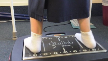
SAN FRANCISCO – Assessing renal health using a modified Cockroft-Gault equation to measure creatinine clearance was more sensitive than using the Modification of Diet in Renal Disease equation to estimate glomerular filtration rate when estimating risk for osteoporosis and fracture, an award-winning study of 400 postmenopausal Puerto Rican women showed.
The study found a high prevalence of mild renal dysfunction (stage 2 chronic kidney disease) in 54%-59% of women, depending on the equation used. With the Cockroft-Gault equation adjusted for body surface area, a determination of mild renal dysfunction was associated with significantly decreased bone mineral density and with a doubling in risk for vertebral or nonvertebral fractures. When the Modification of Diet in Renal Disease (MDRD) equation was used, however, no significant associations were found between mild renal dysfunction and fracture risk, Dr. Loida A. González-Rodriguez reported.
Previous data have shown that severe renal dysfunction is associated with reduced bone mineral density and fractures, and that a creatinine clearance below 65 mL/min per 1.73 m2 is associated with a higher risk for falls and hip fractures in elderly people. Less is known about the effects of mild renal dysfunction on bone mineral density.
"We are postulating that this Cockroft-Gault equation is better to estimate bone," because it includes factors such as weight and age, and is adjusted for body surface area, Dr. González-Rodriguez said in an interview at the annual meeting of the Endocrine Society. She received an award at the meeting for her retrospective secondary analysis of data from the Latin American Vertebral Osteoporosis Study, the first population-based study of vertebral fractures in Latin America.
Many clinicians use the MDRD equation to estimate renal function. Dr. González-Rodriguez of the University of Puerto Rico, San Juan, said she has switched to using the Cockroft-Gault equation, and is trying to get colleagues at her institution to do the same. The MDRD equation will miss some patients who are at risk for osteopenia, osteoporosis, and fracture, she said.
Seventeen percent of patients in the study had normal bone mineral density, 43% had osteopenia, and 41% had osteoporosis. (Percentages were rounded and so exceed 100%.)
When the Cockroft-Gault equation was used to categorize renal function, 9% of patients had stage 1 chronic kidney disease, 54% had stage 2, 35% had stage 3, and 2% had stage 4. When the MDRD equation was used, 2% of patients had stage 1 chronic kidney disease, 59% had stage 2, 38% had stage 3, and 1% had stage 4.
Among patients with stage 2 chronic kidney disease as assessed by the Cockroft-Gault equation, 19% had normal bone mineral density, 49% had osteopenia, and 32% had osteoporosis. Among patients with stage 2 disease assessed using the MDRD equation, 4% had normal bone mineral density, 35% had osteopenia, and 60% had osteoporosis.
Vertebral fractures occurred in 9% and nonvertebral fractures occurred in 18% of patients with stage 2 disease assessed with the Cockroft-Gault equation. When the MDRD equation was used, 9% of patients with stage 2 disease developed vertebral fractures and 24% developed nonvertebral fractures.
Among patients with stage 3 chronic kidney disease assessed using the Cockroft-Gault equation, 18% developed vertebral fractures and 31% developed nonvertebral fractures, compared with vertebral fractures in 16% and nonvertebral fractures in 22% of patients with stage 3 disease assessed using the MDRD equation.
"One of the most important risk factors for vertebral and nonvertebral fractures is osteoporosis," Dr. González-Rodriguez noted. "So, if we can identify earlier the patients that have mild renal dysfunction" using the Cockroft-Gault equation and manage the osteoporosis risk, some fractures may be prevented.
The findings are limited by the retrospective design of the study, a lack of blood pressure measurements to assess arterial hypertension, self-reported nonvertebral fractures, and a lack of measurements of intact parathyroid hormone, 25-hydroxyvitamin D, and microalbuminuria.
Dr. González-Rodriguez reported having no relevant financial disclosures. The National Center for Research Resources and the National Institute on Minority Health and Health Disparities funded the study.
On Twitter @sherryboschert
AT ENDO 2013
Major finding: Fracture risk doubled in women with mild renal dysfunction as assessed by the Cockroft-Gault equation but not when renal function was assessed using the Modification of Diet in Renal Disease equation.
Data source: A retrospective secondary analysis of data on 400 postmenopausal Puerto Rican women.
Disclosures: Dr. Loida A. González-Rodriguez reported having no relevant financial disclosures. The National Center for Research Resources and the National Institute on Minority Health and Health Disparities funded the study.

Discuss quality of life with acromegaly patients
SAN FRANCISCO – Patients with acromegaly struggle with issues that they often don’t share with their physicians, and they have specific suggestions about actions that health care providers can take to help them cope with their disease, a qualitative study of 19 patients found.
Most described a long journey to a correct diagnosis, and other data suggest that an acromegaly diagnosis typically is delayed by 4-10 years. The delay is frustrating for patients but is followed by relief when they finally have a name for their disease, Michelle H. Gurel, R.N., reported at the Endocrine Society’s Annual Meeting.
She and her associates conducted online interviews with 10 patients recruited through AcromegalyCommunity.com and live interviews with 9 patients at the 2012 Acromegaly Community Conference.

The patients said they were shocked at the idea of needing "brain surgery" (pituitary surgery) when discussing treatment with their physicians, and that they felt unsatisfied or left out of the treatment decision process.
Acromegaly is a rare chronic disease characterized by abnormal skeletal growth and soft tissue enlargement caused by excessive secretion of growth hormone by a pituitary adenoma.
Patients also hesitated to raise quality-of-life issues with their physicians and said they were concerned that health care providers did not care about quality of life. "Patients said that they were uncomfortable bringing this up; so you have to open the discussion," said Ms. Gurel of the neuroendocrine unit at Massachusetts General Hospital, Boston.
For a successful physician-patient partnership, patients want to feel that they are being listened to and treated as a person first and as someone with a disease second, they said. Health care providers should take the time to explain in detail the disease and treatment options, covering a slew of questions that patients had in four main categories: What is acromegaly? Will I ever feel like myself again? What is treatment like, and what will I experience? How do I survive financially?
Patients want to know why they are experiencing symptoms, feel bad, and no longer look like themselves, they said. They wonder if their levels of growth hormone and insulin-like growth factor 1 will return to normal, if symptoms will resolve, if the pain will stop, and if the disease can be cured. They question whether they will ever look like themselves again, feel energetic, live a normal life, and be able to forget, even for a moment, that they have acromegaly.
When discussing treatment, patients want to know why "brain surgery" is needed and how it will feel, as well as the pros and cons of radiation. They wonder why they have to take medications, and how to cope with side effects. Most of the medical therapies are expensive, raising questions about whether insurance will cover the costs, how to afford copays, and what happens if patients lose or change insurance.
Patients said they do want to be asked detailed questions about their quality of life during follow-up visits. They want health care providers who are educated about acromegaly and up-to-date in their knowledge of treatments, which they found mainly at Centers of Excellence and at pituitary centers, Ms. Gurel said. Patients wanted physicians to be aggressive in treatment without downplaying the patient’s physical, emotional, and psychological pain and suffering. "They felt stronger support from health care providers if they thought the physician cared," she said.
Nonphysician staff also are key to a successful partnership, patients said. Having a well-run office with courteous, friendly, and competent staff who can help patients with financial questions is important, from a patient’s point of view.
Participants in the study had a mean age of 43 years, and 12 of the 19 were female, although acromegaly generally affects men and women at the same rate. Among 10 patients who described their presenting symptoms, the most common were acral changes or changes in height, in six patients (60%). The disease presented with joint or muscle pain, diabetes, weight gain, headaches, and amenorrhea or an irregular menstrual cycle in 30% of patients each, and hypertension or visual changes affected 20% each at presentation. (Some patients had more than one symptom.)
Twelve of the 19 patients were currently being treated with somatostatin analogues (63%) and 3 with growth hormone receptor antagonists (16%), while 4 patients were not receiving medical therapy (21%) because they had been cured after surgery and/or radiation or because they had not yet chosen a medical therapy. Most medical therapies for acromegaly are expensive and are administered through injection, Ms. Gurel noted.
"(Patients) felt stronger support from health care providers if they thought the physician cared."
Treatment histories from 10 patients showed that 9 had undergone surgery since diagnosis, 2 had radiotherapy, 3 were treated with one medication, 4 had tried two medications, 1 patient had been treated with three drugs, and 2 patients had gone through four medications. (Patients could report more than one treatment category.)
Because patients felt left out of the treatment decision process, they felt motivated to talk to other patients, and many used online resources for support such as AcromegalyCommunity.com or other disease-specific websites. Patients who were not connected to a patient support group reported feeling lonely and helpless. Many of the patients expressed a desire to help improve knowledge about acromegaly in order to shorten the time to diagnosis for future patients.
Acromegaly typically appears in the fourth decade of life, with an annual incidence of 3 or 4 patients per million and a prevalence of 40-90 cases per million people. People with acromegaly are more likely to develop hypertension and heart disease, cardiovascular events and headaches, arthritis and acral changes, sleep apnea, and insulin-resistant diabetes. The disease is associated with premature death, with a doubling or tripling of mortality risk.
Most patients with acromegaly are treated with surgery. Medical therapies include the somatostatin analogues lanreotide and octreotide, the growth hormone receptor antagonist pegvisomant, or the dopamine agonist cabergoline.
The study was funded by Ipsen Biopharmaceuticals, which makes lanreotide (Somatuline). Ms. Gurel reported having no other financial disclosures.
On Twitter @sherryboschert
SAN FRANCISCO – Patients with acromegaly struggle with issues that they often don’t share with their physicians, and they have specific suggestions about actions that health care providers can take to help them cope with their disease, a qualitative study of 19 patients found.
Most described a long journey to a correct diagnosis, and other data suggest that an acromegaly diagnosis typically is delayed by 4-10 years. The delay is frustrating for patients but is followed by relief when they finally have a name for their disease, Michelle H. Gurel, R.N., reported at the Endocrine Society’s Annual Meeting.
She and her associates conducted online interviews with 10 patients recruited through AcromegalyCommunity.com and live interviews with 9 patients at the 2012 Acromegaly Community Conference.
The patients said they were shocked at the idea of needing "brain surgery" (pituitary surgery) when discussing treatment with their physicians, and that they felt unsatisfied or left out of the treatment decision process.
Acromegaly is a rare chronic disease characterized by abnormal skeletal growth and soft tissue enlargement caused by excessive secretion of growth hormone by a pituitary adenoma.
Patients also hesitated to raise quality-of-life issues with their physicians and said they were concerned that health care providers did not care about quality of life. "Patients said that they were uncomfortable bringing this up; so you have to open the discussion," said Ms. Gurel of the neuroendocrine unit at Massachusetts General Hospital, Boston.
For a successful physician-patient partnership, patients want to feel that they are being listened to and treated as a person first and as someone with a disease second, they said. Health care providers should take the time to explain in detail the disease and treatment options, covering a slew of questions that patients had in four main categories: What is acromegaly? Will I ever feel like myself again? What is treatment like, and what will I experience? How do I survive financially?
Patients want to know why they are experiencing symptoms, feel bad, and no longer look like themselves, they said. They wonder if their levels of growth hormone and insulin-like growth factor 1 will return to normal, if symptoms will resolve, if the pain will stop, and if the disease can be cured. They question whether they will ever look like themselves again, feel energetic, live a normal life, and be able to forget, even for a moment, that they have acromegaly.
When discussing treatment, patients want to know why "brain surgery" is needed and how it will feel, as well as the pros and cons of radiation. They wonder why they have to take medications, and how to cope with side effects. Most of the medical therapies are expensive, raising questions about whether insurance will cover the costs, how to afford copays, and what happens if patients lose or change insurance.
Patients said they do want to be asked detailed questions about their quality of life during follow-up visits. They want health care providers who are educated about acromegaly and up-to-date in their knowledge of treatments, which they found mainly at Centers of Excellence and at pituitary centers, Ms. Gurel said. Patients wanted physicians to be aggressive in treatment without downplaying the patient’s physical, emotional, and psychological pain and suffering. "They felt stronger support from health care providers if they thought the physician cared," she said.
Nonphysician staff also are key to a successful partnership, patients said. Having a well-run office with courteous, friendly, and competent staff who can help patients with financial questions is important, from a patient’s point of view.
Participants in the study had a mean age of 43 years, and 12 of the 19 were female, although acromegaly generally affects men and women at the same rate. Among 10 patients who described their presenting symptoms, the most common were acral changes or changes in height, in six patients (60%). The disease presented with joint or muscle pain, diabetes, weight gain, headaches, and amenorrhea or an irregular menstrual cycle in 30% of patients each, and hypertension or visual changes affected 20% each at presentation. (Some patients had more than one symptom.)
Twelve of the 19 patients were currently being treated with somatostatin analogues (63%) and 3 with growth hormone receptor antagonists (16%), while 4 patients were not receiving medical therapy (21%) because they had been cured after surgery and/or radiation or because they had not yet chosen a medical therapy. Most medical therapies for acromegaly are expensive and are administered through injection, Ms. Gurel noted.
"(Patients) felt stronger support from health care providers if they thought the physician cared."
Treatment histories from 10 patients showed that 9 had undergone surgery since diagnosis, 2 had radiotherapy, 3 were treated with one medication, 4 had tried two medications, 1 patient had been treated with three drugs, and 2 patients had gone through four medications. (Patients could report more than one treatment category.)
Because patients felt left out of the treatment decision process, they felt motivated to talk to other patients, and many used online resources for support such as AcromegalyCommunity.com or other disease-specific websites. Patients who were not connected to a patient support group reported feeling lonely and helpless. Many of the patients expressed a desire to help improve knowledge about acromegaly in order to shorten the time to diagnosis for future patients.
Acromegaly typically appears in the fourth decade of life, with an annual incidence of 3 or 4 patients per million and a prevalence of 40-90 cases per million people. People with acromegaly are more likely to develop hypertension and heart disease, cardiovascular events and headaches, arthritis and acral changes, sleep apnea, and insulin-resistant diabetes. The disease is associated with premature death, with a doubling or tripling of mortality risk.
Most patients with acromegaly are treated with surgery. Medical therapies include the somatostatin analogues lanreotide and octreotide, the growth hormone receptor antagonist pegvisomant, or the dopamine agonist cabergoline.
The study was funded by Ipsen Biopharmaceuticals, which makes lanreotide (Somatuline). Ms. Gurel reported having no other financial disclosures.
On Twitter @sherryboschert
AT ENDO 2013

Melatonin receptor agonist resets blind patients’ internal clocks
SAN FRANCISCO – Tasimelteon, an experimental melatonin receptor agonist, reset the internal clock of blind patients to a 24-hour cycle in a randomized trial by Vanda Pharmaceuticals, its maker.
The 84 totally blind subjects had non-24-hour sleep-wake disorder, meaning that their circadian rhythms were out of synch with the external world, causing sleep and daytime performance problems. That’s not uncommon in blindness; without the regulating effects of perceived light, internal clocks revert to their intrinsic rhythm of about 24.5 hours.
Forty-two patients were randomized to 20 mg of tasimelteon 1 hour before bedtime for 6 months, and 42 others to placebo. Entrainment to a 24-hour cycle was gauged by the timing of peak urinary excretions of cortisol and 6-sulfatoxymelatonin, a melatonin metabolite.

Eight patients on tasimelteon synched up to the 24-hour clock, and 10 both entrained and had improvements in various sleep and function measures. One placebo patient entrained during the trial, and none had improvements in sleep or function.
In a follow-up study, 10 entrained tasimelteon patients were randomized to continue the drug, and 10 others to switch to placebo. One tasimelteon patient, but eight placebo patients, reverted to a non-24-hour cycle. "Maintenance is required to maintain entrainment and clinical benefit," said lead investigator Steven Lockley, Ph.D., a neuroscientist in the division of sleep medicine at Brigham and Women’s Hospital in Boston.
"The medication is able to replace the time cue usually provided by light and synchronize the circadian clock in totally blind people. None of the traditional medications used to treat sleep disorders or sleepiness have this ability," he said at the Endocrine Society’s annual meeting.
Vanda filed with the Food and Drug Administration in May for an indication to treat non-24-hour sleep-wake disorder in totally blind patients. The drug might also prove useful for other circadian problems, Dr. Lockley said.
Side effects included nausea, headache, and sleepiness, but were uncommon and not significantly more frequent than with placebo. Subjects were aged 21-84 years, and 34 were women.
Dr. Lockley is an investigator for Vanda Pharmaceuticals, which funded the work. Another investigator is a consultant to the company, and the remaining six are employees.
SAN FRANCISCO – Tasimelteon, an experimental melatonin receptor agonist, reset the internal clock of blind patients to a 24-hour cycle in a randomized trial by Vanda Pharmaceuticals, its maker.
The 84 totally blind subjects had non-24-hour sleep-wake disorder, meaning that their circadian rhythms were out of synch with the external world, causing sleep and daytime performance problems. That’s not uncommon in blindness; without the regulating effects of perceived light, internal clocks revert to their intrinsic rhythm of about 24.5 hours.
Forty-two patients were randomized to 20 mg of tasimelteon 1 hour before bedtime for 6 months, and 42 others to placebo. Entrainment to a 24-hour cycle was gauged by the timing of peak urinary excretions of cortisol and 6-sulfatoxymelatonin, a melatonin metabolite.
Eight patients on tasimelteon synched up to the 24-hour clock, and 10 both entrained and had improvements in various sleep and function measures. One placebo patient entrained during the trial, and none had improvements in sleep or function.
In a follow-up study, 10 entrained tasimelteon patients were randomized to continue the drug, and 10 others to switch to placebo. One tasimelteon patient, but eight placebo patients, reverted to a non-24-hour cycle. "Maintenance is required to maintain entrainment and clinical benefit," said lead investigator Steven Lockley, Ph.D., a neuroscientist in the division of sleep medicine at Brigham and Women’s Hospital in Boston.
"The medication is able to replace the time cue usually provided by light and synchronize the circadian clock in totally blind people. None of the traditional medications used to treat sleep disorders or sleepiness have this ability," he said at the Endocrine Society’s annual meeting.
Vanda filed with the Food and Drug Administration in May for an indication to treat non-24-hour sleep-wake disorder in totally blind patients. The drug might also prove useful for other circadian problems, Dr. Lockley said.
Side effects included nausea, headache, and sleepiness, but were uncommon and not significantly more frequent than with placebo. Subjects were aged 21-84 years, and 34 were women.
Dr. Lockley is an investigator for Vanda Pharmaceuticals, which funded the work. Another investigator is a consultant to the company, and the remaining six are employees.
AT ENDO 2013
Major finding: Tasimelteon, an experimental melatonin receptor agonist, entrains about half of totally blind patients to a 24-hour circadian rhythm.
Data Source: Randomized, blinded, placebo-controlled trial.
Disclosures: The lead researcher is an investigator for Vanda Pharmaceuticals, which makes the drug and funded the study. Another investigator is a consultant to the company, and the remaining six are employees.

No short-term serious side effects from hormones for transsexuals
SAN FRANCISCO – Hormonal therapies for transgender patients that block puberty and/or deliver opposite-sex hormones did not adversely affect bone development in adolescents and caused no serious side effects in teens or adults, two studies found.
The separate European studies, presented at the Endocrine Society Annual Meeting, looked at 127 adolescents aged 12-18 years in one study and 104 adults in the other study who were followed for 1 year after starting hormonal therapy.
The adolescents started gonadotropin-releasing hormone analogues (GnRHa) at ages 12-16 years to block production of sex hormones that initiate puberty, and at age 16 years they began receiving cross-sex hormones (either estrogen or testosterone) to induce sexual characteristics of the opposite sex to which they were transitioning. Adolescence is a key period for developing bone mass, so investigators followed patients’ bone density and other measures to see if there were any long-term adverse effects.

All patients had normal whole-body and hip-bone mineral density before hormonal therapy started. Bone density gradually increased in younger patients during GnRHa treatment, but less than would be expected without GnRHa. In patients who started GnRHa at relatively older ages, bone density decreased slightly, but both groups caught up to normal or near-normal bone density once they got cross-sex hormones and developed physiologic puberty. Patients who started the medical intervention with GnRHa at a young age and, thus, an early pubertal stage, showed the best bone density, reported Dr. Henriette Delemarre-van de Waal and her associates.
"The medical intervention in young transsexuals appears to be safe and effective," said Dr. Delemarre-van de Waal, professor of pediatric endocrinology at Leiden (Netherlands) University.
Patients reported satisfaction and no regrets about GnRHa therapy, but many commented that they would have liked to take cross-sex hormones earlier than age 16, she added at a press briefing on her study. Current Dutch law and Endocrine Society guidelines for treating transgender persons, which Dr. Delemarre-van de Waal helped develop, recommend waiting until age 16 to start cross-sex hormones. Efforts are underway to talk with the Dutch government about lowering the starting age of cross-sex hormonal therapy for selected patients, she said in an interview. Puberty in European girls starts at 10-11 years of age, so starting GnRHa before age 12 and cross-sex hormones before age 16 might optimize bone mass development, "but I think we need a little more data," she said.
Neither lipid levels nor insulin sensitivity per Tanner stage were adversely affected by puberty suppression or cross-sex hormones. GnRHa therapy delayed growth not only in bone age but in height, which was a concern especially for female-to-male transitioning patients who hoped to achieve the average taller height of males. Individualizing the addition of the anabolic steroid oxandrolone to cross-sex therapy helped achieve appropriate height in these patients, and estrogen therapy in male-to-female transgender patients suppressed growth velocity in height as desired by those patients.
A separate ongoing, prospective study analyzed data from four established gender-treatment teams in Belgium, the Netherlands, Norway, and Italy on 104 transgender adults in order to increase the limited knowledge about side effects from hormonal therapy. Females transitioning to males received standardized treatment with intramuscular testosterone undecanoate for 12 weeks. Males transitioning to females who were 45 years of age or younger were treated with standardized regimens of cyproterone acetate and estradiol valerate. Older male-to-female transsexuals received cyproterone acetate and a transdermal estradiol patch.
A year after starting therapy, no patients had developed adverse events serious enough to stop therapy, reported Dr. Katrien Wierckx of Ghent (Belgium) University and her associates. There were no deaths, cardiovascular events, osteoporotic fractures, venous thrombosis, pulmonary embolism, or prolactinomas.
Female-to-male transmen reported a significant increase in sex drive, voice instability, and androgenetic alopecia. Investigators observed significant increases in acne scores and total body lean mass, lower total body fat mass, and development of a less favorable lipid profile with lower high-density lipoprotein (HDL) levels and higher triglyceride levels, Dr. Wierckx said.
Male-to-female transwomen reported a significant decrease in sex drive and significant increases in breast tenderness, emotionality, and hot flashes. The hot flashes surprised investigators because this side effect had not been reported in previous studies. Measurements showed significant gains in fat mass, losses in muscle mass, and decreased insulin sensitivity. They developed a more favorable lipid profile with significantly reduced levels of total and low-density lipoprotein (LDL) cholesterol and triglycerides.
The findings suggest that current treatment regimens for transsexual women and men carry a low risk for short-term adverse events, Dr. Wierckx said. "These results are somewhat in contrast with most previously published studies, which observed a high incidence of venous thrombosis or pulmonary embolism during the first year of treatment," she said. This may suggest that treatment regimens avoiding the use of high-dose estradiol and high-dose cyproterone acetate have less detrimental effects on the coagulation system, she suggested.
Plenty of questions remain about the long-term effects of hormonal therapy for sex transitions. "There are a lot of concerns. It’s still a controversial subject," said Dr. Delemarre-van de Waal. Effects on brain development are a key concern, but preliminary data from serial MRIs and functional MRIs of transgender patients have not produced any alarming findings, she said.
Both speakers reported having no financial disclosures.
On Twitter @sherryboschert
SAN FRANCISCO – Hormonal therapies for transgender patients that block puberty and/or deliver opposite-sex hormones did not adversely affect bone development in adolescents and caused no serious side effects in teens or adults, two studies found.
The separate European studies, presented at the Endocrine Society Annual Meeting, looked at 127 adolescents aged 12-18 years in one study and 104 adults in the other study who were followed for 1 year after starting hormonal therapy.
The adolescents started gonadotropin-releasing hormone analogues (GnRHa) at ages 12-16 years to block production of sex hormones that initiate puberty, and at age 16 years they began receiving cross-sex hormones (either estrogen or testosterone) to induce sexual characteristics of the opposite sex to which they were transitioning. Adolescence is a key period for developing bone mass, so investigators followed patients’ bone density and other measures to see if there were any long-term adverse effects.
All patients had normal whole-body and hip-bone mineral density before hormonal therapy started. Bone density gradually increased in younger patients during GnRHa treatment, but less than would be expected without GnRHa. In patients who started GnRHa at relatively older ages, bone density decreased slightly, but both groups caught up to normal or near-normal bone density once they got cross-sex hormones and developed physiologic puberty. Patients who started the medical intervention with GnRHa at a young age and, thus, an early pubertal stage, showed the best bone density, reported Dr. Henriette Delemarre-van de Waal and her associates.
"The medical intervention in young transsexuals appears to be safe and effective," said Dr. Delemarre-van de Waal, professor of pediatric endocrinology at Leiden (Netherlands) University.
Patients reported satisfaction and no regrets about GnRHa therapy, but many commented that they would have liked to take cross-sex hormones earlier than age 16, she added at a press briefing on her study. Current Dutch law and Endocrine Society guidelines for treating transgender persons, which Dr. Delemarre-van de Waal helped develop, recommend waiting until age 16 to start cross-sex hormones. Efforts are underway to talk with the Dutch government about lowering the starting age of cross-sex hormonal therapy for selected patients, she said in an interview. Puberty in European girls starts at 10-11 years of age, so starting GnRHa before age 12 and cross-sex hormones before age 16 might optimize bone mass development, "but I think we need a little more data," she said.
Neither lipid levels nor insulin sensitivity per Tanner stage were adversely affected by puberty suppression or cross-sex hormones. GnRHa therapy delayed growth not only in bone age but in height, which was a concern especially for female-to-male transitioning patients who hoped to achieve the average taller height of males. Individualizing the addition of the anabolic steroid oxandrolone to cross-sex therapy helped achieve appropriate height in these patients, and estrogen therapy in male-to-female transgender patients suppressed growth velocity in height as desired by those patients.
A separate ongoing, prospective study analyzed data from four established gender-treatment teams in Belgium, the Netherlands, Norway, and Italy on 104 transgender adults in order to increase the limited knowledge about side effects from hormonal therapy. Females transitioning to males received standardized treatment with intramuscular testosterone undecanoate for 12 weeks. Males transitioning to females who were 45 years of age or younger were treated with standardized regimens of cyproterone acetate and estradiol valerate. Older male-to-female transsexuals received cyproterone acetate and a transdermal estradiol patch.
A year after starting therapy, no patients had developed adverse events serious enough to stop therapy, reported Dr. Katrien Wierckx of Ghent (Belgium) University and her associates. There were no deaths, cardiovascular events, osteoporotic fractures, venous thrombosis, pulmonary embolism, or prolactinomas.
Female-to-male transmen reported a significant increase in sex drive, voice instability, and androgenetic alopecia. Investigators observed significant increases in acne scores and total body lean mass, lower total body fat mass, and development of a less favorable lipid profile with lower high-density lipoprotein (HDL) levels and higher triglyceride levels, Dr. Wierckx said.
Male-to-female transwomen reported a significant decrease in sex drive and significant increases in breast tenderness, emotionality, and hot flashes. The hot flashes surprised investigators because this side effect had not been reported in previous studies. Measurements showed significant gains in fat mass, losses in muscle mass, and decreased insulin sensitivity. They developed a more favorable lipid profile with significantly reduced levels of total and low-density lipoprotein (LDL) cholesterol and triglycerides.
The findings suggest that current treatment regimens for transsexual women and men carry a low risk for short-term adverse events, Dr. Wierckx said. "These results are somewhat in contrast with most previously published studies, which observed a high incidence of venous thrombosis or pulmonary embolism during the first year of treatment," she said. This may suggest that treatment regimens avoiding the use of high-dose estradiol and high-dose cyproterone acetate have less detrimental effects on the coagulation system, she suggested.
Plenty of questions remain about the long-term effects of hormonal therapy for sex transitions. "There are a lot of concerns. It’s still a controversial subject," said Dr. Delemarre-van de Waal. Effects on brain development are a key concern, but preliminary data from serial MRIs and functional MRIs of transgender patients have not produced any alarming findings, she said.
Both speakers reported having no financial disclosures.
On Twitter @sherryboschert
AT ENDO 2013
Major finding: Hormonal therapy for transgender patients did not adversely affect bone density in adolescents and caused no serious unwanted side effects in teens or adults in short-term follow-up.
Data source: Two separate studies of 127 transgender adolescents and 104 transgender adults.
Disclosures: Both speakers reported having no financial disclosures.

Vibration platform strengthened bones, improved walking in moderate cerebral palsy
SAN FRANCISCO – Regular sessions on vibration platforms improved bone density, muscle mass, and walking ability in children with cerebral palsy in a small study from New Zealand.
Five boys and eight girls aged 14-19 years with moderate cerebral palsy had four sessions per week in which they stood on the platforms for 3 minutes, took a 3-minute break, and repeated the cycle twice more.
After 5 months, their mean total leg bone mineral density (BMD) increased from 1.06 g/cm2 to 1.08 g/cm2, their total spine BMD increased from 1.01 g/cm2 to 1.02 g/cm2, and their total lower-tibia volumetric BMD increased from 607 mg/cm3 to 690 mm/cm3. These modest changes were similar to those seen with osteoporosis medications.

The subjects’ mean midtibia muscle cross-sectional area increased from 3,163 mm2 to 3,440 mm2, and their mean 6-minute walk distance increased from 286 to 314 m. The results were all significant. One girl was able to walk up stairs for the first time after the trial, and was still able to do so 6 months later, according to lead investigator Silmara Gusso, Ph.D., a research fellow at the University of Auckland, New Zealand.
Parents reported fewer falls at home, better balance, and more regular bowel function. Several said they wanted to buy one of the devices, a Galileo home vibration platform. Physiotherapists said they wanted to try them on children with other musculoskeletal challenges, Dr. Gusso said at the Endocrine Society’s Annual Meeting.
"We were quite surprised to see the changes over such a short period of time ... [and] very impressed with the feedback we got from families, physiotherapists, and the kids themselves," who "opened huge smiles" when they stepped on the platforms. It was all "much more than we expected. We are quite excited about it," she said.
Vibration platforms aren’t new; they’ve been used before to help with osteoporosis, surgery recoveries, and athletic injuries.

The trial may be the first, however, to try them for cerebral palsy. The idea was to see if they’d help with osteopenia and muscle mass, something that’s hard to address with standard physiotherapy, which focuses on strength, stretching, balance, and gentle walking, Dr. Gusso said.
The platforms were set to tip side to side at 1,200 oscillations per minute (20 Hz), "which is pretty hard exercise" even for someone without cerebral palsy. The children had a brace they could hold on to for balance. Their weekly physiotherapy sessions continued throughout the study.
The research will be expanded to include children with other disabilities. "It’s a small and simple device that can bring extra benefits" to standard approaches, Dr. Gusso said.
Dr. Gusso and her team said they had nothing to disclose. The work was funded by the Jubilee Trust, the Australasia Paediatric Endocrine Group, and others.
SAN FRANCISCO – Regular sessions on vibration platforms improved bone density, muscle mass, and walking ability in children with cerebral palsy in a small study from New Zealand.
Five boys and eight girls aged 14-19 years with moderate cerebral palsy had four sessions per week in which they stood on the platforms for 3 minutes, took a 3-minute break, and repeated the cycle twice more.
After 5 months, their mean total leg bone mineral density (BMD) increased from 1.06 g/cm2 to 1.08 g/cm2, their total spine BMD increased from 1.01 g/cm2 to 1.02 g/cm2, and their total lower-tibia volumetric BMD increased from 607 mg/cm3 to 690 mm/cm3. These modest changes were similar to those seen with osteoporosis medications.
The subjects’ mean midtibia muscle cross-sectional area increased from 3,163 mm2 to 3,440 mm2, and their mean 6-minute walk distance increased from 286 to 314 m. The results were all significant. One girl was able to walk up stairs for the first time after the trial, and was still able to do so 6 months later, according to lead investigator Silmara Gusso, Ph.D., a research fellow at the University of Auckland, New Zealand.
Parents reported fewer falls at home, better balance, and more regular bowel function. Several said they wanted to buy one of the devices, a Galileo home vibration platform. Physiotherapists said they wanted to try them on children with other musculoskeletal challenges, Dr. Gusso said at the Endocrine Society’s Annual Meeting.
"We were quite surprised to see the changes over such a short period of time ... [and] very impressed with the feedback we got from families, physiotherapists, and the kids themselves," who "opened huge smiles" when they stepped on the platforms. It was all "much more than we expected. We are quite excited about it," she said.
Vibration platforms aren’t new; they’ve been used before to help with osteoporosis, surgery recoveries, and athletic injuries.
The trial may be the first, however, to try them for cerebral palsy. The idea was to see if they’d help with osteopenia and muscle mass, something that’s hard to address with standard physiotherapy, which focuses on strength, stretching, balance, and gentle walking, Dr. Gusso said.
The platforms were set to tip side to side at 1,200 oscillations per minute (20 Hz), "which is pretty hard exercise" even for someone without cerebral palsy. The children had a brace they could hold on to for balance. Their weekly physiotherapy sessions continued throughout the study.
The research will be expanded to include children with other disabilities. "It’s a small and simple device that can bring extra benefits" to standard approaches, Dr. Gusso said.
Dr. Gusso and her team said they had nothing to disclose. The work was funded by the Jubilee Trust, the Australasia Paediatric Endocrine Group, and others.
AT ENDO 2013
Major finding: After 5 months of vibration platform therapy, children with moderate cerebral palsy could walk about 30 m more in 6 minutes.
Data source: Uncontrolled trial involving 13 subjects.
Disclosures: Dr. Gusso and her team said they had nothing to disclose. The work was funded by the Jubilee Trust, the Australasia Paediatric Endocrine Group, and others.