User login
Idarucizumab may reverse dabigatran anticoagulation in intracranial hemorrhage
LOS ANGELES – It only took a few minutes for idarucizumab to normalize blood-clotting parameters in 18 patients with dabigatran-associated intracranial hemorrhages, according to interim results from an ongoing phase III trial presented at the International Stroke Conference.
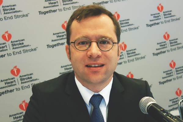
“When I put patients on [dabigatran], they always ask me what happens if they bleed or need surgery. … Until now, I haven’t been able to tell them with any confidence that I have a way of reversing it. Now I think I can. ... It makes a big difference” in their comfort, said lead investigator Dr. Richard Bernstein, a Northwestern University neurology professor and director of the stroke program at Northwestern Memorial Hospital, in Chicago.
“We would love to know if hematoma expansion was limited [and outcomes improved] by giving this reversal agent,” but the study so far is too small. “We hope to have a larger cohort of brain hemorrhage patients to answer these questions,” he said.
Approved in October 2015, idarucizumab (Praxbind) was fast tracked by the Food and Drug Administration to reverse the blockbuster atrial fibrillation anticoagulant dabigatran (Pradaxa); the labeling for idarucizumab doesn’t mention intracranial hemorrhage patients specifically. Boehringer Ingelheim makes both drugs, and funded Dr. Bernstein’s work.
Eleven of the 18 patients were men, and the average age in the study was about 80 years. The patients had either subdural hematomas or bleeding into the brain itself. They were culled from the 90 subjects analyzed so far in the idarucizumab trial, dubbed RE-VERSE AD (Reversal Effects of Idarucizumab in Patients on Active Dabigatran).
The team followed label dosing: 5 g total given as two separate 2.5-g infusions. Blood samples were taken in between to check how well idarucizumab worked. The whole process took no more than 15 minutes.
The first 2.5 g completely reversed dabigatran in all 18 patients, based on their dilute thrombin or ecarin clotting times. Patients “remained reversed out to 12 hours, and all but one out to 24 hours,” Dr. Bernstein said at the conference, sponsored by the American Heart Association.
Idarucizumab is a monoclonal antibody fragment that binds dabigatran more powerfully than dabigatran binds thrombin. In vitro studies have found no prothrombotic effects. “It has no endogenous target, so the drug has no effect on any other clotting factors that we can tell. We did have, I think, five thrombotic events in our cohort, most of them many days after the dabigatran was reversed. It may have just been a reversion to [patient] clotting risks,” he said.
When – or if – to restart dabigatran “is a clinical question.” If bleeding is controlled or patients are stable after surgery, you can go back on the next day,” he said.
Idarucizumab’s labeling notes that 5% or more of patients developed hypokalemia, delirium, constipation, pyrexia, and pneumonia. It wasn’t clear these events were drug related. Patients had dabigatran reversed either for serious bleeding or emergency surgery.
Dr. Bernstein is a speaker and adviser for Boehringer Ingelheim, and reported honoraria from the company.
AT THE INTERNATIONAL STROKE CONFERENCE
Key clinical point: An ongoing investigation suggests that idarucizumab can reliably stop intracranial hemorrhage associated with dabigatran anticoagulation.
Major finding: It took only a few minutes for idarucizumab to normalize blood-clotting parameters in 18 patients with dabigatran intracranial hemorrhage.
Source: Interim results from an ongoing phase III trial.
Disclosures: The work was funded by Boehringer Ingelheim, maker of both dabigatran and idarucizumab. The lead investigator is a speaker and adviser for Boehringer, and reported honoraria from the company.
High false positives found for Medtronic’s implantable AF detectors
LOS ANGELES – Medtronic’s Reveal LINQ implantable loop recorders misidentified 84% of rhythm anomalies as atrial fibrillation in 52 stroke patients at Emory University in Atlanta, according to a presentation at the International Stroke Conference.
Two electrophysiologists reviewed a random sample of 166 rhythm strips from those patients that were identified by Reveal as atrial fibrillation (AF) over a 2-month period; 140 (84%) were false positives. Eighty (57%) of the false positives were premature atrial complexes, 31 (22%) were due to T wave over-sensing, 14 (10%) to noise, 7 (5%) to premature ventricular complexes, 4 (2.9%) to under-sensing, and 4 (2.9%) to sinus arrhythmias.
There wasn’t a mix-and-match of true and false positives in the same patient; false and true positives were consistent in patients over the study period.
The take-home message from the study is that the high sensitivity of Medtronic’s implantable loop recorders means that they are good at detecting possible AF, but their findings must be reviewed and confirmed before being acted upon.
“There are high rates of false positives, but the results can be easily adjudicated by electrophysiologists as evidenced by our observer agreement,” which was 100%. “These strips need to be adjudicated by somebody, and not taken at face value,” said investigator Dr. Spencer Maddox, an Emory resident.
The devices look mainly at RR intervals and the presence or absence of P waves. Runs of 2 minutes are required for AF. One of the issues in the study was that T waves were identified as QRS complexes, which changed the RR interval and trigged the device to report AF, he said.
Medtronic could reduce the false positive rate by, for instance, extending the run required for AF, but it would be a bad idea. “The goal here is to not miss atrial fibrillation,” Dr. Maddox said. “There are false positives, but as long as you go back and reread, I think that’s fine. I wouldn’t mess with the sensitivity of the device.”
The company said the same thing when asked for comment on the study.
“The AF detection algorithm ... is tuned to favor sensitivity because manual review can subsequently rule out any false positives. This is one reason it is important to have trained cardiologists or electrophysiologists involved in reviewing reports of irregular heart rhythms to determine a diagnosis of AF,” said Medtronic spokesman Ryan Mathre, who also noted that company data suggest a lower false positive rate (Cerebrovasc Dis. 2015;40:175-81).
Even so, confirmation doesn’t always happen, “and that’s the scary part. In practice almost all the time,” the report comes in “and it’s acted on,” said Dr. Robert Hart, a neurology professor at McMaster University in Hamilton, Ont., and comoderator of the study presentation.
The Emory patients were all recovering from ischemic strokes or TIAs. They were about 70 years old on average, about 60% were men, and almost all were white. A total of 38 had implantable loop recorders for cryptogenic strokes; the devices detected true AF in 4 (11%) after a mean of 92 days. Early Holter monitoring missed it.
Emory generally prescribes anticoagulants if AF is confirmed, but, as several audience members noted, the burden of AF that requires anticoagulation is unclear. “It’s an area with a lot of thought now, but no good answers,” Dr. Maddox said at the conference, sponsored by the American Heart Association.
The investigators had no disclosures, and there was no outside funding for the work.
AT The INTERNATIONAL STROKE CONFERENCE
Key clinical point: The high sensitivity of Medtronic’s implantable loop recorders means that they’re good at detecting possible AF, but their findings must be reviewed and confirmed before being acted upon.
Major finding: Two electrophysiologists reviewed a random sample of 166 rhythm strips identified by Reveal as atrial fibrillation; 140 (84%) were false positives.
Data source: Fifty-two ischemic stroke and TIA patients.
Disclosures: The investigators had no disclosures. There was no outside funding for the work.
Lower the CT to check the heart for embolic sources in acute stroke
LOS ANGELES – Enlarge the field of CT angiography to include the heart in acute ischemic stroke patients; you’ll quickly identify sources of cardiogenic emboli and other problems that will otherwise be missed, according to investigators from the National University Hospital, Singapore.
It adds only a few seconds to the scan, with no extra contrast or meaningful increase in radiation. There’s no need to gate the heart with beta-blockers.
Among 20 acute ischemic stroke patients presenting within 4.5 hours of symptom onset, Dr. Leonard Yeo and his coinvestigators found one with a localized dissection in the ascending aorta, another with a ventricular thrombus, and a third with an atrial appendage blood clot. Both thrombus cases were confirmed by transesophageal echocardiography and started on anticoagulation the next day. The 2-phase, 64-slice nongated cardiac CT angiographies (CTA) were done in the same sitting as the brain CTA.
“Scans with 1-mm thick slices are best for screening for thrombus and structural abnormalities that cause embolism. Remarkably, [even without gating], the detail is excellent. There’s very little downside [to this, and] it maximizes your return on scans that are already a part of most acute stroke protocols,” said Dr. Yeo, a neurologist at the hospital.
“Since most of our patients get a CTA during acute stroke, it made sense to check the heart for embolic sources.” There isn’t any time to give a beta-blocker, so “these were nongated” scans, Dr. Yeo said during his presentation at the International Stroke Conference, sponsored by the American Heart Association.
If it’s confirmed that nongated heart CTAs provide useful information, “we will probably all be doing this in the future. Everybody does CTs for the head in acute stroke, so all you do is go down a little lower” without any more contrast. “Within an hour of somebody presenting, you know what they have,” said Dr. Robert Hart, a neurology professor at McMaster University in Hamilton, Ont., and co-moderator of Dr. Yeo’s presentation.
In most places, acute ischemic stroke patients only get an ECG. Transesophageal echocardiography (TEE) is also good for checking the heart, but it usually comes later. It “excels at detecting abnormalities with medium embolic risk,” such as patent foramen ovale and septal aneurysm. “However, for these medium-risk cardiac sources of embolism, the optimal choice of therapy is not clear. Unlike high-risk sources which require anticoagulation, TEE does not provide therapeutic gains in terms of clinical decision making,” Dr. Yeo said.
Nongated cardiac CTAs during acute stroke, he added, also check chamber, valve, pericardial, and great vessel morphology, as well as abnormal chambers-vessel communications and “left ventricular aneurysms that can rupture with [tissue plasminogen activator], with catastrophic consequences.”
The mean age in the study was 64 years old, and about 60% of the subjects were men. None of the patients were dead at 3 months, and by then eight (40%) had modified Rankin Scale scores of 0-1. Patients were excluded if they had contraindications to IV contrast, or were unable to provide informed consent. CTA images were read by the treating neurologist and radiologist.
The work was funded by the Singapore Ministry of Health’s National Medical Research Council. The investigators have no relevant disclosures.
AT THE INTERNATIONAL STROKE CONFERENCE
Key clinical point: Nongated heart CTAs may provide useful information in acute ischemic stroke.
Major finding: Among 20 acute ischemic stroke patients presenting within 4.5 hours of symptom onset, one had a localized dissection in the ascending aorta, another had a ventricular thrombus, and a third had an atrial appendage blood clot.
Data source: Prospective investigation of 20 patients.
Disclosures: The work was funded by the Singapore Ministry of Health’s National Medical Research Council. The investigators have no relevant disclosures.
VIDEO: Ischemic-stroke thrombectomy use widens and refines
LOS ANGELES – The use of endovascular thrombectomy in the United States to treat appropriate patients with acute ischemic stroke mushroomed during the past year, following several early-2015 reports that collectively documented the dramatic clinical benefit of the treatment.
As endovascular thrombectomy use grows, stroke centers are also refining and reshaping delivery of the treatment in concert with administration of intravenous tissue plasminogen activator (TPA; alteplase; Activase), which remains a key partner in producing best outcomes for acute ischemic-stroke patients with a proximal occlusion of a large cerebral artery. Collapsing delivery of the two treatments into a more seamless and streamlined process shaves critical minutes to treatment delivery, an approach called parallel processing. Recent findings have also emboldened stroke specialists to seriously consider simplifying the brain imaging that stroke patients receive prior to these treatments, a step that could further cut time to intervention while also making thrombectomy even more widely available.
Use of thrombectomy surges
The biggest endovascular thrombectomy news of the past year is how it has taken off for treating selected patients with acute ischemic stroke. “The rollout over the past year has been explosive. Everything pretty much shut down after the negative trial results in 2013, but now more hospitals are offering thrombectomy,” said Dr. Thomas A. Kent, professor of neurology and director of stroke research and education at Baylor College of Medicine in Houston, in an interview at the International Stroke Conference sponsored by the American Heart Association.
The best documentation of this surge came in a poster presented at the conference by researchers at the University of California, San Francisco. They analyzed data on treatment of 357,973 patients with acute ischemic stroke who were hospitalized at any one of 161 U.S. academic medical centers during October 2009-July 2015 and included in the University Healthsystem Consortium database. They tracked the percentage of patients treated endovascularly during each calendar quarter of the study period.
During 2009-2013, use of endovascular treatment rose steadily but gradually, from 1.5% of stroke patients in 2009 to 3.1% during the fourth quarter of 2012. Then, following three reports of no benefit from endovascular treatment presented at the International Stroke Conference in February 2013 – the IMS III, MR RESCUE, and SYNTHESIS trials – the endovascular rate dropped immediately and quickly bottomed out at a level of 2.6% that remained steady through the third quarter of 2014. But when the positive endovascular results from the MR CLEAN study became public in the final week of 2014, endovascular use began to quickly rise again, and then began to skyrocket during the first quarter of 2015 with three additional positive trial results reported during the Stroke Conference in February 2015. By the end of the second quarter of 2015, usage stood at 4.7%, representing a projected year-over-year increase of about 150% for all of 2015, compared with 2014, reported Dr. Anthony S. Kim, a vascular neurologist and medical director of the Stroke Center at the University of California, San Francisco, and his associates.
To put these percentages in perspective, experts estimate that roughly 10%-15% of all stroke patients qualify for thrombectomy intervention.
Their data also showed that the percentage of hospitals included in the database that performed endovascular therapies for stroke rose steadily from about 40% of centers in 2009 to nearly 60% by mid-2015.
“Endovascular therapy with newer-generation devices is increasingly part of standard treatment for acute ischemic stroke,” they said in their poster. In addition, they cited a “new urgency to evaluate regional access to embolectomy [another name for thrombectomy] nationally and to identify system-based solutions to improve access in underserved areas.”
Several stroke experts interviewed at the conference added their own anecdotal view of thrombectomy’s rapidly expanding use for appropriate acute ischemic stroke patients during 2015, and the need for continued effort to broaden its U.S. availability.
“The number of thrombectomies fell off after the negative 2013 trials and stayed flat until a year ago, but then jumped up. It has been very dramatic,” said Dr. Wade S. Smith, professor of neurology and director of the neurovascular service at the University of California, San Francisco.
“Thrombectomy use tremendously increased since February 2015,” said Dr. Mark J. Alberts, professor of neurology and medical director of the neurology service at the University of Texas Southwestern Medical Center in Dallas, in a video interview during the conference. But despite this growth, “the major challenge [today] is geography;” that is, reaching patients in suburban and rural areas who are not as close to the primarily urban medical centers that currently offer the procedure.
“We now have about 100 certified comprehensive stroke centers in the U.S.,” and by definition comprehensive stroke centers have the capability of treating patients with endovascular thrombectomy, noted Dr. Jeffrey Saver, professor of neurology and director of the stroke unit at the University of California, Los Angeles.
“Certification of these centers did not begin until about 2-3 years ago. But we probably need 300-400 of these centers” to provide thrombectomy to most U.S. stroke patients, he said. “A lot of additional hospitals are close to certification. I anticipate that over the next 1-2 years we will be in the neighborhood of having the number of centers we need,” Dr. Saver said in an interview.
Making thrombectomy better
In addition to expanding availability, the specifics of how endovascular thrombectomy gets delivered is evolving. A major trend is movement toward a “parallel processing” model, in which patients with an acute clinical presentation of a stroke amenable for endovascular treatment simultaneously undergo CT angiography to confirm and localize the large-artery clot causing their stroke, receive intravenous TPA, and undergo preparation for the endovascular access needed to remove the clot.
A pooled analysis of the recent, positive endovascular thrombectomy trials that was presented at the conference showed how quick you need to be to obtain a benefit from the procedures. “This gives us a starting point to further improve the target metrics for imaging and puncture times,” Dr. Saver said. “We want to shorten door-to-needle times for TPA and door-to-puncture times for thrombectomy, and the processes that need to be addressed for rapid delivery of both of these are very similar. We need for patients to only make a pit stop in the ED; we need to have the catheterization team ready to go in the thrombectomy suite within 30 minutes; and we need to emphasize speed in access to the target clot rather than time-consuming diagnostic angiography.”
“We now face the issue of how to best integrate TPA treatment and clot removal.” Dr. Kent said. “People are still trying to work that out. With parallel processing there is some overuse of resources: Some patients recover with TPA alone and don’t need thrombectomy. We are getting closer to the cardiology model of MI treatment. It’s now clear that there needs to be a simple, safe, effective way to do both TPA treatment and thrombectomy. We need to model ourselves on the cardiology experience.”
“If you can deal with the TPA decision in the same room without moving patients from room to room, from a scanner to a catheterization suite, you can really shorten the time to treatment,” Dr. Smith explained. “This is identical to the model that cardiologists have developed. We should now consider taking stroke patients directly to the angiography room in addition to administering TPA. We still need cross-sectional imaging, but the quality of the image from an angiography suite is probably sufficient to make a TPA decision. So you can start TPA while you are getting arterial access. The idea is simultaneous approaches to the patient instead of serial.”
“The whole system moves at the same time to eliminate wasted time,” Dr. Alberts summed up.
One of the big questions that has come up in this effort to speed up treatment and carve the quickest route to endovascular thrombectomy is whether TPA remains necessary. The skeptics’ position is, why waste time administering TPA if you’re also going to take out the offending clot?
The answer, at least for now, is that all signs indicate that giving TPA helps and is worth delivering.
“The 2015 thrombectomy trials had big differences among them in the dosage of TPA administered, and in the percentage of patients who received TPA. When 100% of patients received TPA they had the best outcomes,” Dr. Kent said. “There was a clear synergistic relationship between thrombectomy and TPA. There has been a trend to think about sending patients straight to thrombectomy and skipping TPA, but my colleagues and I think that we need to hold off on doing that. For now, if a patient is eligible to receive TPA they should get it and then quickly move to endovascular therapy. We are not yet ready to know it’s okay to go straight to endovascular treatment. In SWIFT-PRIME, it was pretty clear that the good outcomes were attributable to both [thrombectomy plus TPA]. Treating patients with TPA helps soften the clot to make it easier to remove, and improves flow through collateral arteries.”
“Our data in Memphis show that patients do better with thrombectomy plus intravenous TPA than on TPA alone,” agreed Dr. Lucas Elijovich, a neurologist at the University of Tennessee Health Science Center in Memphis, in an interview.
Simpler imaging also saves time
Although it’s not yet proven, another new wrinkle in working up acute ischemic-stroke patients for TPA and thrombectomy treatment is the idea that simpler and more widely available CT imaging, especially CT angiography of cerebral arteries, may suffice for confirming and localizing the culprit clot.
This concept received a significant boost at the International Stroke Conference in data reported from the Pragmatic Ischaemic Stroke Thrombectomy Evaluation (PISTE) trial, yet another study that compared treatment with TPA alone with TPA plus endovascular thrombectomy, this time in 65 randomized patients treated at any of 11 U.K. centers. PISTE had a low enrollment level because the trial stopped prematurely, in July 2015, following the news that several fully completed trials had collectively established the superiority of endovascular thrombectomy plus TPA, thereby making it unethical to continue yet another randomized study.
This premature stoppage prevented PISTE from itself producing a statistically significant difference for its primary efficacy endpoint in favor of the combined treatment, although the results did show a nominal advantage to using thrombectomy plus TPA over TPA alone that was fully consistent with the other studies, Dr. Keith W. Muir reported at the conference.
But what made the PISTE results especially notable was that the trial achieved this consistent outcome with a “simpler” imaging protocol for patients during their workup that used only CT angiography, avoiding the cerebral CT perfusion imaging or MRI used in several of the other TPA-plus-thrombectomy versus TPA-only trials, noted Dr. Muir, professor of neuroscience and head of the stroke imaging group at the University of Glasgow.
“PISTE raises the question of how much imaging is necessary,” Dr. Kent commented.
“The PISTE results are exciting. A lot of us believe that all we need to know is that there is a blockage in a target vessel,” Dr. Smith said. “If we have that information, then we can identify a population of patients who will benefit from [thrombectomy]. CT angiography is simple and can easily fit into work flows.”
“PISTE used a very simple imaging system that makes thrombectomy even more applicable and generalizable to less resourced health systems,” Dr. Saver said. “Although the results from PISTE were not internally statistically significant because the trial ended early, the results were consistent with the external studies of thrombectomy, so it provides further evidence for benefit from thrombectomy.” And because the consistent results were achieved with simpler imaging it suggests simpler imaging may be all that’s needed.
“That’s a major question to wrestle with,” Dr. Saver suggested. “We need addition trials with a head-to-head comparison of simpler and more sophisticated imaging so we can tailor treatment to patients who would benefit from simpler and faster imaging.”
Dr. Kent had no disclosures. Dr. Kim has received research funding from SanBio and Biogen. Dr. Smith served on the data safety and monitoring board for a trial funded by Stryker. Dr. Alberts has been a consultant to Genentech. Dr. Saver has been a consultant to Stryker, Neuravi, Cognition Medical, Boehringer Ingelheim, and Medtronic. Dr. Elijovich has been a consultant to Stryker and Codman and received research support from Siemens. Dr. Muir has received research support from ReNeuron and unrestricted grants from Codman and Covidien.
The video associated with this article is no longer available on this site. Please view all of our videos on the MDedge YouTube channel
On Twitter @mitchelzoler
EXPERT ANALYSIS FROM THE INTERNATIONAL STROKE CONFERENCE
Seeking an alteplase alternative for stroke
Where is a biosimilar when you need one?
A few weeks ago, I reported on an advisory committee of the Food and Drug Administration overwhelmingly recommending that the agency approve a biosimilar form of infliximab. If the FDA follows this recommendation, and every indication was that it would, it would become the second biosimilar drug approved for U.S. marketing, and a top agency official recently noted during Congressional testimony that the FDA biosimilar program had 59 additional agents working their way through the agency’s development program. A 2014 report cited more than 700 biosimilars under development worldwide.
But when analysts and journalists list the top reference drugs for biosimilars in the pipeline, one reference biologic agent conspicuously absent is alteplase, the thrombolytic also known as tissue plasminogen activator (TPA), which is approved for lysing clots that cause MIs and acute ischemic strokes.
Alteplase, marketed under the brand name Activase, is the only thrombolytic drug with U.S. approval for treating acute ischemic stroke. Although results from several studies published in 2015 rapidly boosted endovascular thrombectomy to physically remove clots causing acute ischemic strokes, this new intervention has by no means relegated intravenous alteplase to the sidelines. Experts I spoke with in February at the International Stroke Conference unanimously endorsed treatment with alteplase as the unshaken keystone of acute ischemic stroke treatment (for appropriate patients), even in the thrombectomy era. Thrombolysis is seen as a complement to thrombectomy rather than a treatment eclipsed by thrombectomy.
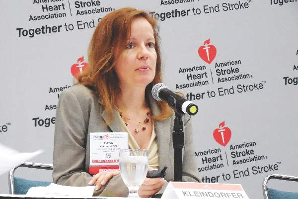
Although alteplase treatment remains central it’s also become increasingly expensive. According to a report at the Stroke Conference by Dr. Dawn Kleindorfer, a neurologist and co–medical director of the comprehensive stroke center at the University of Cincinnati, the price paid by Medicare for alteplase given to acute ischemic stroke patients more than doubled during 2005-2014. Upward pricing became especially dramatic starting in 2010, she noted, with a price tag of about $6,400/100 mg vial (enough for roughly one dose) by mid-2014. The cost for alteplase jumped from 27% of the amount hospitals got reimbursed by Medicare for treating a patient with acute ischemic stroke in 2006 to 53% of the reimbursement in 2013, Dr. Kleindorfer reported.
“I think this might impact the ability of hospitals to provide health care because the [alteplase] cost is so high and now more than half the reimbursement,” she warned. Despite its high price, alteplase “remains cost effective,” she noted, and use of the drug in recent years has increased, not dropped.
Dr. Kleindorfer was at a complete loss to explain why alteplase’s price has risen so much since 2010, and she noted that the drug received FDA approval in 1996 and is now off patent for the acute ischemic stroke indication (as well as the acute MI indication). Despite that, no biosimilar has appeared or seems to be under announced development, nor has any other thrombolytic drug received FDA approval for the stroke indication.
In fact, the history of thrombolytics for acute ischemic stroke is notably checkered. For example, an alternative thrombolytic, desmoteplase, showed early signs of promise for treating acute ischemic stroke, but the Danish drug company Lundbeck halted development of desmoteplase in late 2014 after the drug failed to achieve target primary endpoints in pivotal trials. Another thrombolytic, reteplase (Retavase), has had FDA approval for treating acute MI since 1996 but has never received approval for acute ischemic stroke. A spokesperson for Chiesi, the company that currently markets Retavase, declined to comment on why an indication for stroke was never pursued. A third thrombolytic similar to alteplase, tenecteplase, showed promise for treating acute ischemic stroke in a relatively recent phase II study, where it actually showed superiority to alteplase in a head-to-head comparison, but tenecteplase is owned and marketed by Genentech as TNKase (for acute MI only), the same company that also markets alteplase and so has little incentive to develop tenecteplase as an alternative stroke treatment.
“Even a doubling of the price [for TPA, alteplase] doesn’t change whether the drug is cost effective, but this effectively reduces hospital reimbursements that support acute stroke programs,” commented Dr. S. Claiborne Johnston in an interview. “This will ultimately impact the enthusiasm for and use of TPA,” predicted Dr. Johnston, dean of the Dell Medical School of the University of Texas in Austin and a researcher who has analyzed the economics of thrombolytic drugs for acute ischemic stroke.
“The lack of competition [to alteplase for treating acute ischemic stroke] is an example of how our current biopharma marketplace is dysfunctional,” Dr. Johnston told me. “Trials of new agents are difficult, expensive, and protracted, and there is some question whether any agent that works better [than alteplase] at lysing a clot would also produce a higher hemorrhage risk. We really need a biosimilar, but the path to develop this is not as easy as for [generic] drugs.” Although the higher price alteplase now commands might make development of a biosimilar even more attractive, Dr. Johnston said he was unaware of any company that so far is venturing down that path.
On Twitter @mitchelzoler
ISC: Cryptogenic stroke linked to PSVT in absence of atrial fibrillation
LOS ANGELES – Paroxysmal supraventricular tachycardia is associated with subsequent ischemic stroke in patients without documented atrial fibrillation, according to a claims analysis of 42,152 Medicare enrollees at least 66 years old.
Atrial fibrillation accounts for perhaps 30% of cryptogenic strokes, “so clearly there’s something more to the story than just atrial fibrillation in” the other 70%, said investigator Dr. Hooman Kamel, a neurologist at Weill Cornell Medical College, New York. “Most cryptogenic strokes seem like they are embolic. The question is what are the undiscovered sources of embolism?”
Dr. Kamel and his colleagues focused on paroxysmal supraventricular tachycardia (PSVT) even though it’s generally considered benign. But “PSVT is increasingly recognized as a marker for underlying atrial dysfunction, especially in older patients. In some cases, the abnormal atrial substrate could cause thromboembolism even before atrial fibrillation [AF] appears,” he said at the International Stroke Conference.
To ensure regular heart rhythm monitoring, the study was limited to patients with implanted pacemakers or defibrillators. Patients with AF or stroke before or at the time of device implantation were excluded.
After a median of 1.8 years of follow-up, 2,245 patients (5.3%) were diagnosed with PSVT, and 1,007 (2.4%) had an ischemic stroke. The incidence of stroke without PSVT diagnosis was 0.95% per year, but 2.17% per year with a preceding PSVT diagnosis (P less than .001). Adjusting for age, gender, income, hypertension, diabetes, heart failure, and other potential confounders, the team found that a diagnosis of PSVT was associated with a doubling of ischemic stroke risk (HR, 2.0; 95% CI, 1.3-3.0), and an almost quadrupling of the risk for embolic stroke (HR, 3.6; 95% CI, 1.1-11.8).
“A lot more work needs to be done to nail this down, but potentially we are broadening the pool of atrial markers for stroke risk. These results build on recent findings that disturbances of atrial rhythm and function other than AF may” lead to stroke, Dr. Kamel said.
It’s way too soon to consider atrial ablation for PSVT to reduce stroke risk, he said, but his team is interrogating its administrative data for clues of its utility. “The idea of ablation for stroke is really interesting. I think ablation should help reduce the risk of stroke. It’s a really important question, and we don’t know the answer yet. There’s a lot more to be learned, [but] there does seem to be a definite progression from PSVT to AF,” Dr. Kamel said.
The National Institutes of Health funded the work. Dr. Kamel is a speaker for Genentech.
AT THE INTERNATIONAL STROKE CONFERENCE
Key clinical point: Paroxysmal supraventricular tachycardia could be an atrial marker for increased stroke risk when atrial fibrillation is not present, but additional research needs to confirm the finding.
Major finding: The incidence of stroke without PSVT diagnosis was 0.95% per year, but 2.17% per year with a preceding PSVT diagnosis (P less than .001).
Data source: Retrospective cohort of 42,152 Medicare enrollees.
Disclosures: The National Institutes of Health funded the work. The presenter is a speaker for Genentech.
ISC: Pick up Extra AF With Extended Holter Monitoring
LOS ANGELES – Atrial fibrillation is three times more likely to be detected within 6 months of an ischemic stroke if, instead of the usual 24 or so hours of Holter ECG monitoring, patients are monitored for 10 days poststroke, and then again for 10 days at 3 and 6 months, according to German investigators.
“Enhanced and prolonged monitoring should be considered for all stroke patients to improve the detection of atrial fibrillation,” said investigator Dr. Rolf Wachter, a cardiologist at the University of Göttingen (Germany).
Patients in the trial had no history of atrial fibrillation (AF) and were at least 60 years old; 200 were randomized to the extended-monitoring protocol, and 198 to standard of care, which included a median of 24 hours of Holter monitoring. The median time from symptom onset to randomization was 3 days. All patients were enrolled by day 5.
The study wasn’t limited to cryptogenic strokes; although the first stroke may not have been related to atrial fibrillation, a recurrent stroke could be, so “our approach was to look for AF in all stroke patients irrespective of etiology,” Dr. Wachter said at the International Stroke Conference.
By 6 months, AF was detected in 27 patients (13.5 %) in the extended-monitoring group versus 9 (4.5 %) in the control group (absolute difference, 9%; 95% confidence interval, 3.5%-14.6%; P = .002). The findings were largely the same when results were broken down by age, sex, National Institutes of Health Stroke Scale scores, and other metrics. AF was detected in the majority of both groups within a month.
Dr. Wachter did not elaborate on the frequency and duration of AF, but inclusion criteria required at least one episode lasting 30 seconds or longer.
The work touches on key issues facing neurologists and cardiologists today: How aggressive should post-stroke AF monitoring be, and when should treatment start?
Every AF patient in the study was orally anticoagulated, and almost all remained on their anticoagulant at 1 year. Their oral anticoagulant wasn’t reported, and the study wasn’t powered to detect differences in clinical outcomes. Even so, “we saw very positive trends in the right direction” for prolonged monitoring, he said.
Five patients in the intervention arm (2.5%) had a recurrent stroke and three (1.5%) had transient ischemic events at 1 year, versus nine (4.5%) recurrent strokes and five (2.5%) TIAs in the control arm. Six intervention patients (3%) and nine (4.5%) in the control group, had died.
A total of 60% of the subjects were men, and the mean age in the study was 73 years. The mean NIH Stroke Scale score was 3 in the intervention group and 2 in the control group. The mean CHADS2 (heart failure, hypertension, age, diabetes, stroke) score – a marker of thromboembolic risk – was 3.5 in both. Patients in the extended-monitoring group wore their Holter monitors for a median of 9.5 days at all three time points, but the initial compliance of 100% dropped to about 75% by the third session.
The work was funded by Boehringer Ingelheim, maker of the oral anticoagulant dabigatran (Pradaxa). Dr. Wachter is a speaker and consultant for the company.
There is widespread belief among most stroke neurologists that when you have a stroke that looks embolic and you don’t have a source, you need to monitor for longer than we have done traditionally. The nuances are really how to do that. This is a great approach; it’s a lot easier than wearing a 30-day Holter monitor or having an implanted device, but we have to compare it to other approaches and see what the heart endpoints really are to know if it’s the best approach.
I think everybody in the stroke community appreciates that the more you monitor, the more you are likely to find AF. How long you should monitor, by what technique, and whether monitoring should be continuous or intermittent are unanswered questions. The other question is how much AF is significant. Is a 30-second run significant, or does it need to be minutes at a time? These questions need to be answered before there’s a wholesale buy-in for this kind of monitoring for every patient.
Dr. Kyra Becker is a professor in the departments of neurology and neurological surgery, and director of Vascular Neurology Services at the University of Washington’s Comprehensive Stroke Center in Seattle. She was not involved in the study.
LOS ANGELES – Atrial fibrillation is three times more likely to be detected within 6 months of an ischemic stroke if, instead of the usual 24 or so hours of Holter ECG monitoring, patients are monitored for 10 days poststroke, and then again for 10 days at 3 and 6 months, according to German investigators.
“Enhanced and prolonged monitoring should be considered for all stroke patients to improve the detection of atrial fibrillation,” said investigator Dr. Rolf Wachter, a cardiologist at the University of Göttingen (Germany).
Patients in the trial had no history of atrial fibrillation (AF) and were at least 60 years old; 200 were randomized to the extended-monitoring protocol, and 198 to standard of care, which included a median of 24 hours of Holter monitoring. The median time from symptom onset to randomization was 3 days. All patients were enrolled by day 5.
The study wasn’t limited to cryptogenic strokes; although the first stroke may not have been related to atrial fibrillation, a recurrent stroke could be, so “our approach was to look for AF in all stroke patients irrespective of etiology,” Dr. Wachter said at the International Stroke Conference.
By 6 months, AF was detected in 27 patients (13.5 %) in the extended-monitoring group versus 9 (4.5 %) in the control group (absolute difference, 9%; 95% confidence interval, 3.5%-14.6%; P = .002). The findings were largely the same when results were broken down by age, sex, National Institutes of Health Stroke Scale scores, and other metrics. AF was detected in the majority of both groups within a month.
Dr. Wachter did not elaborate on the frequency and duration of AF, but inclusion criteria required at least one episode lasting 30 seconds or longer.
The work touches on key issues facing neurologists and cardiologists today: How aggressive should post-stroke AF monitoring be, and when should treatment start?
Every AF patient in the study was orally anticoagulated, and almost all remained on their anticoagulant at 1 year. Their oral anticoagulant wasn’t reported, and the study wasn’t powered to detect differences in clinical outcomes. Even so, “we saw very positive trends in the right direction” for prolonged monitoring, he said.
Five patients in the intervention arm (2.5%) had a recurrent stroke and three (1.5%) had transient ischemic events at 1 year, versus nine (4.5%) recurrent strokes and five (2.5%) TIAs in the control arm. Six intervention patients (3%) and nine (4.5%) in the control group, had died.
A total of 60% of the subjects were men, and the mean age in the study was 73 years. The mean NIH Stroke Scale score was 3 in the intervention group and 2 in the control group. The mean CHADS2 (heart failure, hypertension, age, diabetes, stroke) score – a marker of thromboembolic risk – was 3.5 in both. Patients in the extended-monitoring group wore their Holter monitors for a median of 9.5 days at all three time points, but the initial compliance of 100% dropped to about 75% by the third session.
The work was funded by Boehringer Ingelheim, maker of the oral anticoagulant dabigatran (Pradaxa). Dr. Wachter is a speaker and consultant for the company.
AT THE INTERNATIONAL STROKE CONFERENCE
ISC: Pick up extra AF with extended Holter monitoring
LOS ANGELES – Atrial fibrillation is three times more likely to be detected within 6 months of an ischemic stroke if, instead of the usual 24 or so hours of Holter ECG monitoring, patients are monitored for 10 days poststroke, and then again for 10 days at 3 and 6 months, according to German investigators.
“Enhanced and prolonged monitoring should be considered for all stroke patients to improve the detection of atrial fibrillation,” said investigator Dr. Rolf Wachter, a cardiologist at the University of Göttingen (Germany).
Patients in the trial had no history of atrial fibrillation (AF) and were at least 60 years old; 200 were randomized to the extended-monitoring protocol, and 198 to standard of care, which included a median of 24 hours of Holter monitoring. The median time from symptom onset to randomization was 3 days. All patients were enrolled by day 5.
The study wasn’t limited to cryptogenic strokes; although the first stroke may not have been related to atrial fibrillation, a recurrent stroke could be, so “our approach was to look for AF in all stroke patients irrespective of etiology,” Dr. Wachter said at the International Stroke Conference.
By 6 months, AF was detected in 27 patients (13.5 %) in the extended-monitoring group versus 9 (4.5 %) in the control group (absolute difference, 9%; 95% confidence interval, 3.5%-14.6%; P = .002). The findings were largely the same when results were broken down by age, sex, National Institutes of Health Stroke Scale scores, and other metrics. AF was detected in the majority of both groups within a month.
Dr. Wachter did not elaborate on the frequency and duration of AF, but inclusion criteria required at least one episode lasting 30 seconds or longer.
The work touches on key issues facing neurologists and cardiologists today: How aggressive should post-stroke AF monitoring be, and when should treatment start?
Every AF patient in the study was orally anticoagulated, and almost all remained on their anticoagulant at 1 year. Their oral anticoagulant wasn’t reported, and the study wasn’t powered to detect differences in clinical outcomes. Even so, “we saw very positive trends in the right direction” for prolonged monitoring, he said.
Five patients in the intervention arm (2.5%) had a recurrent stroke and three (1.5%) had transient ischemic events at 1 year, versus nine (4.5%) recurrent strokes and five (2.5%) TIAs in the control arm. Six intervention patients (3%) and nine (4.5%) in the control group, had died.
A total of 60% of the subjects were men, and the mean age in the study was 73 years. The mean NIH Stroke Scale score was 3 in the intervention group and 2 in the control group. The mean CHADS2 (heart failure, hypertension, age, diabetes, stroke) score – a marker of thromboembolic risk – was 3.5 in both. Patients in the extended-monitoring group wore their Holter monitors for a median of 9.5 days at all three time points, but the initial compliance of 100% dropped to about 75% by the third session.
The work was funded by Boehringer Ingelheim, maker of the oral anticoagulant dabigatran (Pradaxa). Dr. Wachter is a speaker and consultant for the company.
There is widespread belief among most stroke neurologists that when you have a stroke that looks embolic and you don’t have a source, you need to monitor for longer than we have done traditionally. The nuances are really how to do that. This is a great approach; it’s a lot easier than wearing a 30-day Holter monitor or having an implanted device, but we have to compare it to other approaches and see what the heart endpoints really are to know if it’s the best approach.
I think everybody in the stroke community appreciates that the more you monitor, the more you are likely to find AF. How long you should monitor, by what technique, and whether monitoring should be continuous or intermittent are unanswered questions. The other question is how much AF is significant. Is a 30-second run significant, or does it need to be minutes at a time? These questions need to be answered before there’s a wholesale buy-in for this kind of monitoring for every patient.
Dr. Kyra Becker is a professor in the departments of neurology and neurological surgery, and director of Vascular Neurology Services at the University of Washington’s Comprehensive Stroke Center in Seattle. She was not involved in the study.
LOS ANGELES – Atrial fibrillation is three times more likely to be detected within 6 months of an ischemic stroke if, instead of the usual 24 or so hours of Holter ECG monitoring, patients are monitored for 10 days poststroke, and then again for 10 days at 3 and 6 months, according to German investigators.
“Enhanced and prolonged monitoring should be considered for all stroke patients to improve the detection of atrial fibrillation,” said investigator Dr. Rolf Wachter, a cardiologist at the University of Göttingen (Germany).
Patients in the trial had no history of atrial fibrillation (AF) and were at least 60 years old; 200 were randomized to the extended-monitoring protocol, and 198 to standard of care, which included a median of 24 hours of Holter monitoring. The median time from symptom onset to randomization was 3 days. All patients were enrolled by day 5.
The study wasn’t limited to cryptogenic strokes; although the first stroke may not have been related to atrial fibrillation, a recurrent stroke could be, so “our approach was to look for AF in all stroke patients irrespective of etiology,” Dr. Wachter said at the International Stroke Conference.
By 6 months, AF was detected in 27 patients (13.5 %) in the extended-monitoring group versus 9 (4.5 %) in the control group (absolute difference, 9%; 95% confidence interval, 3.5%-14.6%; P = .002). The findings were largely the same when results were broken down by age, sex, National Institutes of Health Stroke Scale scores, and other metrics. AF was detected in the majority of both groups within a month.
Dr. Wachter did not elaborate on the frequency and duration of AF, but inclusion criteria required at least one episode lasting 30 seconds or longer.
The work touches on key issues facing neurologists and cardiologists today: How aggressive should post-stroke AF monitoring be, and when should treatment start?
Every AF patient in the study was orally anticoagulated, and almost all remained on their anticoagulant at 1 year. Their oral anticoagulant wasn’t reported, and the study wasn’t powered to detect differences in clinical outcomes. Even so, “we saw very positive trends in the right direction” for prolonged monitoring, he said.
Five patients in the intervention arm (2.5%) had a recurrent stroke and three (1.5%) had transient ischemic events at 1 year, versus nine (4.5%) recurrent strokes and five (2.5%) TIAs in the control arm. Six intervention patients (3%) and nine (4.5%) in the control group, had died.
A total of 60% of the subjects were men, and the mean age in the study was 73 years. The mean NIH Stroke Scale score was 3 in the intervention group and 2 in the control group. The mean CHADS2 (heart failure, hypertension, age, diabetes, stroke) score – a marker of thromboembolic risk – was 3.5 in both. Patients in the extended-monitoring group wore their Holter monitors for a median of 9.5 days at all three time points, but the initial compliance of 100% dropped to about 75% by the third session.
The work was funded by Boehringer Ingelheim, maker of the oral anticoagulant dabigatran (Pradaxa). Dr. Wachter is a speaker and consultant for the company.
AT THE INTERNATIONAL STROKE CONFERENCE
Key clinical point: Repeated Holter monitoring over 6 months catches more atrial fibrillation than one-time, poststroke monitoring.
Major finding: By 6 months, AF was detected in 27 patients (13.5 %) in the extended-monitoring group, versus 9 (4.5 %) in the control group (absolute difference, 9%; 95% confidence interval, 3.5%-14.6%; P = .002).
Data source: Randomized trial with 398 ischemic stroke patients.
Disclosures: The work was funded by Boehringer Ingelheim, maker of the oral anticoagulant dabigatran (Pradaxa). The presenter is a speaker and consultant for the company.
VIDEO: Hands-off yields best brain arteriovenous malformation outcomes
LOS ANGELES – Hang a do-not-disturb sign on brain arteriovenous malformations.
Patients who underwent invasive interventions to repair an unruptured arteriovenous malformation (AVM) in their brain faced a greater than two-fold increased rate of death or stroke during an average 4 years of follow-up, compared with patients who received medical treatment only with no active intervention, Dr. Christian Stapf reported at the International Stroke Conference.
When analyzed on an intention-to-treat basis, for every five AVM patients treated by endovascular surgery, conventional surgery, or radiotherapy, one additional patient died or had a stroke, compared with the death or stroke rate among control patients who received only medical management. When analyzed based on the treatments that patients actually received, the number-needed-to-harm fell to one excess death or stroke for every three AVM patients who underwent an invasive procedure, compared with control patients, reported Dr. Stapf, a professor in the department of neurosciences at the University of Montreal.
The results from A Randomized Trial of Unruptured Brain AVMs (ARUBA) “show us that we clearly have not been as good as we thought in helping patients against their stroke risk,” said Dr. Stapf in a video interview during the meeting. “Given that the risk of death or stroke was reduced three- to fivefold with no [invasive] treatment and leaving the AVM alone makes us think that we can’t recommend preventive intervention with currently-used techniques. Living with the AVM seems like the far better option.”
The ARUBA study, run at 39 centers in nine countries including 13 U.S. centers, randomized 226 patients with unruptured AVMs before the study’s data safety and monitoring board stopped study enrollment prematurely in April 2013. The study group included 110 patients randomized to receive medical interventions only and 116 randomized to medical intervention plus “best possible” AVM eradication. The exact type of eradication for each patient was left up to local clinicians, who tailored the intervention to address the size, location, and anatomy of each AVM. Medical management included steps such as treatment with antiepileptic drugs to treat seizures, various treatments for headaches, and physiotherapy for patients with neurologic deficits.
The study’s primary endpoint was the combined rate of death or stroke, which occurred in 41 of the 116 patients (35%) randomized to receive an invasive intervention and in 15 of the 110 (14%) randomized to medical treatment only during an average follow-up of 50 months, with many patients followed for 5 years.
When analyzed by the treatment patients actually received, 106 underwent an invasive intervention and 43 of these patients (41%) died or had a stroke, and 120 patients received medical treatments only, of whom 13 (11%) died or had a stroke.
A secondary endpoint was the rate of death or disability after 5-year follow-up, with disability defined as a modified Rankin Scale score of 2 or more. This occurred in 38% of the 45 patients who underwent AVM eradication and had this follow-up available, and in 18% of 51 patients who had medial treatment only and received this follow-up.
Interim results from the study came out 2 years ago, with an average follow-up of 33 months (Lancet. 2014 Feb 15;383[9917]:614-21), but the trial was designed to have 5-year follow-up, largely accomplished in the new data reported by Dr. Stapf.
Many clinicians had already abandoned invasive interventions to treat brain AVMs following release of the interim results, and Dr. Stapf predicted that this trend will further strengthen now that the final results are in and confirm the earlier indication of hazard. Until the ARUBA results became available, clinicians had presumed invasive interventions to resolve or minimize malformations were beneficial based on intuition. ARUBA is the first systematic comparison of procedures versus a hands-off approach for brain AVMs, he said.
“Neurologists will now be less likely to refer patients for intervention, and interventionalists will be less enthusiastic to perform procedures,” Dr. Stapf said during the meeting, sponsored by the American Heart Association. In addition, anyone now performing an intervention in routine practice would need to consider the possible legal implications if the patient were to have a bad outcome. Dr. Stapf also noted that some professional societies are now considering recommendations against routine interventions. He conceded that some invasive interventions might still occur for selected cases on an investigational basis, but the ARUBA results “set the bar very high against performing any new interventions,” he concluded.
Approximately 3,000 patients annually are newly diagnosed with an unruptured brain AVM in the United States and Canada, he estimated.
ARUBA received no commercial support. Dr. Stapf had no disclosures.
The video associated with this article is no longer available on this site. Please view all of our videos on the MDedge YouTube channel
On Twitter @mitchelzoler
AT THE INTERNATIONAL STROKE CONFERENCE
Key clinical point: The first direct comparison of invasive treatment and medical management only for intracerebral arteriovenous malformations showed substantial hazard from active intervention.
Major finding: The incidence of death or stroke was 35% with active intervention and 14% with medical management only.
Data source: ARUBA, a multicenter, prospective, international, randomized study with 226 patients.
Disclosures: ARUBA received no commercial support. Dr. Stapf had no disclosures.
ISC: Thrombectomy shown highly cost-effective for stroke
LOS ANGELES – Endovascular thrombectomy is not only clinically the best option for many patients with acute, ischemic strokes involving a proximal occlusion in a large cerebral artery; it’s also highly cost effective, based on follow-up analyses of two of the five randomized trials published in 2015 that collectively established thrombectomy as standard of care for these patients.
Thrombectomy plus administration of intravenous tissue plasminogen activator (TPA), compared with TPA only, “is highly cost effective and economically dominant with lower long-term cost and better outcomes,” Theresa I. Shireman, Ph.D., said at the International Stroke Conference.
And in an independent analysis of data from a totally different trial, endovascular thrombectomy on average reduced patients’ acute length of hospitalization, improved their survival and quality of life, and was cost saving when compared with treatment with intravenous TPA only, which had previously been the standard of care, Dr. Bruce C.V. Campbell reported at the meeting.
The analysis presented by Dr. Shireman used data collected in the SWIFT-PRIME trial, which randomized 196 patients at centers in the United States and Europe to treatment with either intravenous TPA plus endovascular thrombectomy or TPA alone. Average total costs during the index hospitalization ran to roughly $46,000 in the combined-treatment arm and about $29,000 in the TPA-only arm, a difference largely driven by a roughly $15,000 average incremental cost for the thrombectomy procedure, said Dr. Shireman, professor of health services research at Brown University in Providence, R.I.
However, the cost-effectiveness of thrombectomy began to kick in soon after. During the 90 days following index hospitalization, patients who underwent thrombectomy had substantial average reductions in their need for inpatient rehabilitation, time spent in skilled nursing facilities, and in outpatient rehabilitation. Overall, total medical costs during the first 90 days post discharge ran on average close to $5,000 less per patient following thrombectomy. In addition, based on their health status after 90 days, patients treated with thrombectomy were projected to have a greater than 1.7-year average life expectancy than those randomized to TPA only, with a projected net gain of 1.74 quality-adjusted life years (QALY) per patient and with a projected average decrease of roughly $23,000 in total lifetime medical costs.
Based on this average increase in QALYs and decreased long-term cost, adding thrombectomy to TPA for routine treatment of the types of patients enrolled in SWIFT-PRIME was economically dominant, Dr. Shireman said at the meeting sponsored by the American Heart Association. She also projected that despite the higher upfront cost for adding thrombectomy to treatment, the eventual savings in long-term care meant that thrombectomy began producing a net saving once patients survived for more than 22 months following their index hospitalization.
Dr. Campbell reported very similar findings in his analysis of data collected from the EXTEND-IA trial, which randomized 70 patients at 10 centers in Australia and New Zealand. During the first 90 days of treatment, including the index hospitalization, treatment with thrombectomy plus TPA saved an average of roughly $4,000 U.S.per patient, compared with TPA only, even though the average incremental cost for adding thrombectomy was nearly $11,000 U.S. The overall increased total 90-day costs with TPA only was largely driven by a substantially longer time spent hospitalized among the TPA-only patients, compared with those treated with thrombectomy plus TPA, said Dr. Campbell, a neurologist and head of hyperacute stroke at Royal Melbourne Hospital.
In addition, adding thrombectomy resulted in a projected average 4-year increase in life expectancy, and an average gain of about 3 QALYs per patient. Thrombectomy “is an incredibly powerful procedure, not just in terms of clinical response but also in terms of economics,” he concluded. Even when judged by the worst-case scenario of the analysis, “there is a 100% probability that the cost-effectiveness per QALY is less than $10,000 U.S., which is incredible value,” Dr. Campbell said.
SWIFT-PRIME was sponsored by Covidien/Medtronic. EXTEND-IA received partial funding through an unrestricted grant from Covidien/Medtronic. Dr. Shireman and Dr. Campbell had no personal disclosures.
On Twitter @mitchelzoler
AT THE INTERNATIONAL STROKE CONFERENCE
Key clinical point: Adding endovascular thrombectomy to TPA treatment for selected patients with acute, ischemic stroke proved highly cost effective on the basis of data collected in two independent randomized trials.
Major finding: In SWIFT-PRIME, thrombectomy saved a projected average of $23,000 in lifetime health care costs and added 1.74 QALYs.
Data source: SWIFT-PRIME, an international, multicenter, randomized trial that enrolled 196 patients.
Disclosures: SWIFT-PRIME was sponsored by Covidien/Medtronic. EXTEND-IA received partial funding through an unrestricted grant from Covidien/Medtronic. Dr. Shireman and Dr. Campbell had no personal disclosures.

