User login
VIDEO: A better way to treat large intraventricular hemorrhages
LOS ANGELES – For intraventricular hemorrhages of at least 20 mL, alteplase (Activase – Genentech) delivered directly into the clot by external ventricular drain almost doubles the odds of achieving a modified Rankin Score of 0-3 by 6 months.
More clot is removed – and patients with large intraventricular hemorrhages (IVHs) do better – with more vigorous alteplase dosing and when more than one drain is used.
The findings come from the Clot Lysis Evaluation of Accelerated Resolution (CLEAR III) trial, which randomized 249 IVH patients to 1 mg alteplase every 8 hours for up to 12 doses, and 251 to saline on the same schedule, delivered by external ventricular drain. The intervention didn’t make much difference with small hemorrhages.
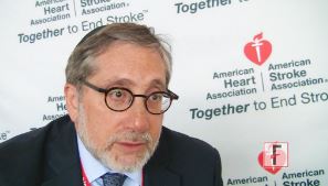
In a video interview at the International Stroke Conference, investigator Dr. Issam Awad, a professor of surgery and neurology and director of neurovascular surgery at the University of Chicago, explained how to do the technique correctly for larger clots, and the expected benefits.
The video associated with this article is no longer available on this site. Please view all of our videos on the MDedge YouTube channel
LOS ANGELES – For intraventricular hemorrhages of at least 20 mL, alteplase (Activase – Genentech) delivered directly into the clot by external ventricular drain almost doubles the odds of achieving a modified Rankin Score of 0-3 by 6 months.
More clot is removed – and patients with large intraventricular hemorrhages (IVHs) do better – with more vigorous alteplase dosing and when more than one drain is used.
The findings come from the Clot Lysis Evaluation of Accelerated Resolution (CLEAR III) trial, which randomized 249 IVH patients to 1 mg alteplase every 8 hours for up to 12 doses, and 251 to saline on the same schedule, delivered by external ventricular drain. The intervention didn’t make much difference with small hemorrhages.
In a video interview at the International Stroke Conference, investigator Dr. Issam Awad, a professor of surgery and neurology and director of neurovascular surgery at the University of Chicago, explained how to do the technique correctly for larger clots, and the expected benefits.
The video associated with this article is no longer available on this site. Please view all of our videos on the MDedge YouTube channel
AT THE INTERNATIONAL STROKE CONFERENCE
VIDEO: Intracranial warfarin bleeds smaller with prothrombin complex instead of FFP
LOS ANGELES – The international normalized ratio fell to 1.2 or less within 3 hours among 18 of 27 (67%) patients who received four-factor prothrombin complex concentrate (octaplex [Octapharma]) for warfarin-related intracranial hemorrhages, but only 2 of 23 (9%) who received fresh frozen plasma, according to a randomized trial from Germany.
Hematoma expansion was reduced by 16.9 mL (P = .026) at 3 hours and 16.4 mL (P = .018) at 24 hours in the prothrombin complex concentrate (PCC) group.
All the patients presented within 12 hours of symptom onset with an INR of at least 2; they received fresh frozen plasma (FFP) or four-factor PCC within an hour of their cerebral CT. There were eight deaths in the FFP group, including five due to hematoma expansion. The five deaths in the PCC group occurred after day 5, and one was thought to be because of hematoma expansion. Patients were 76 years old, on average, and the majority were men; both groups received vitamin K.
The findings make a case for PCC at a time when it’s unclear how best to handle warfarin-related intracranial bleeds, and whether the extra cost of PCC is worth it. Investigator Dr. Thorsten Steiner, a professor of neurology at the University of Heidelberg (Germany), addressed the relevant issues, including PCC safety, in a video interview at the International Stroke Conference sponsored by the American Heart Association. The investigator-initiated trial was funded by Octapharma, and Dr. Steiner reported receiving a research grant from the company.
The video associated with this article is no longer available on this site. Please view all of our videos on the MDedge YouTube channel
AT THE INTERNATIONAL STROKE CONFERENCE
ISC: Imaging supplants clocks for targeting stroke reperfusion
LOS ANGELES – Can brain imaging surpass the clock for identifying acute ischemic stroke patients who will benefit from thrombolytic or thrombectomy treatment?
That’s what experts now envision, based on early findings from several studies. Although the evidence is not yet definitive enough to justify using imaging as a replacement for time-from-stroke-onset in routine practice, the results so far are encouraging and have led to the start or planning of several phase III trials that will try to nail down a role for imaging, either CT or MR, to identify acute ischemic stroke patients who qualify for reperfusion therapy.

“What we’re trying to do is move away from using the clock as a surrogate marker and instead use imaging as the surrogate,” Dr. Jenny P. Tsai said in an interview at the International Stroke Conference.
The linchpin of this new approach is that clocking time elapsed from the onset of stroke symptoms to initiation of thrombolytic or endovascular treatment makes no allowance for patient-to-patient variations in collateral cerebral circulation, a factor that appears to make a big difference in whether patients can be many more hours removed from the start of their stroke and still have salvageable brain tissue. And relying on time since stroke symptom onset gives a seriously flawed estimate of a stroke’s duration when patients have an unwitnessed stroke.
The alternative is to use either CT perfusion imaging or a combination of MR perfusion and diffusion-weighted imaging “to get effective reperfusion treatments to patients who present at later time windows,” said Dr. Gregory W. Albers, professor of neurology at Stanford (Calif.) University and director of the Stanford Stroke Center. He called these two new approaches to gauging a patient’s suitability for reperfusion therapy “the next big thing in imaging” for stroke.
Using CT perfusion to assess target mismatch
Dr. Tsai presented an analysis of thrombectomy reperfusion done in 181 patients enrolled in the CT Perfusion to Predict Response to Recanalization in Ischemic Stroke Project (CRISP), which included a total of 201 acute ischemic stroke patients with large cerebral-artery occlusions treated at six U.S. centers. Her analysis excluded nine patients who presented more than 18 hours after their stroke onset, six patients who did not have successful CT perfusion assessment, and five additional patients excluded for other reasons. The 181 patients analyzed included 125 with a target mismatch in CT perfusion, indicating that salvageable tissue remained in the area of their stroke. Among these 125 patients, 111 underwent successful reperfusion by thrombectomy.
The researchers identified good treatment outcomes as patients with a modified Rankin Scale score of 2 or less 90 days after treatment. A multivariate analysis showed that among these 111 patients, achievement of a good 90-day outcome had no statistically significant relationship with time-to-treatment out to at least the first 8 hours following stroke onset, reported Dr. Tsai, a neurologist at the Stanford Stroke Center. The data also showed a nonsignificant relationship between good outcomes and time from stroke onset to treatment beyond 8 hours, but the confidence interval for this nonsignificant relationship became very wide at later times as the analysis focused on fewer and fewer patients.
“In patients with large-artery occlusions who have a target mismatch profile [on perfusion CT] and achieve successful reperfusion there is no significant association between onset-to-reperfusion-time and the probability of a good functional outcome,” suggesting that “CT perfusion is a biomarker of good outcomes beyond 6 hours” after stroke onset, Dr. Tsai concluded. CT perfusion has the advantage of being more widely available than MRI is, she added. Last year, her associates at Stanford reported similar findings using perfusion-diffusion mismatch in the cerebral area around a stroke visualized with MRI (Neurology. 2015 Aug 25;85[8]:708-15).
“MRI is not readily available” at all U.S. centers that treat stroke patients, while CT perfusion imaging is much more ubiquitous, but the findings reported by Dr. Tsai require confirmation in the several prospective, randomized trials now underway and testing this approach, Dr. Albers said.
Using MRI in unwitnessed strokes
A second challenge for using reperfusion therapy in stroke patients are patients who have unwitnessed strokes, with a completely unknown elapsed time from onset to presentation. The ability of MRI to help identify patients with unwitnessed stroke who can safely receive intravenous thrombolytic therapy with alteplase (tissue plasminogen activator, Activase) underwent testing in the MR WITNESS (Study of Intravenous Thrombolysis With Alteplase in MRI-Selected Patients) trial.
The study enrolled 80 patients with an unwitnessed acute, ischemic stroke at 10 U.S. centers. Patients had to be in a position to receive alteplase within 4.5 hours of when their stroke was first identified; 57 (71%) of the participants had wake-up strokes. All patients underwent two types of MRI to identify them as potential candidates for safe administration of alteplase: diffusion weighted imaging, to identify that a stroke had occurred, and fluid-attenuated inversion recovery (FLAIR) assessment, to identify strokes that had occurred during the prior 4 hours.
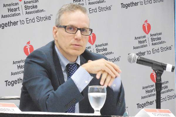
This phase II study’s primary safety endpoint was the incidence of symptomatic intracerebral hemorrhage following alteplase treatment, which occurred in 1 of the 80 patients (1.25%), not a statistically significant difference when compared with the historical standard, the 2.4% rate seen in stroke patients treated with intravenous alteplase 3 to 4.5 hours after their known stroke onset in the ECASS III (European Cooperative Acute Stroke Study) (N Engl J Med. 2008 Sept 25;359[13]:1317-29). The rate of asymptomatic intracerebral hemorrhage was not significantly different between the new study and ECASS III, reported Dr. Lee H. Schwamm, director of TeleStroke & Acute Stroke Services at the Massachusetts General Hospital in Boston.
Based on this result, “we know this approach is safe, and we saw a signal of efficacy, but we really don’t know how effective it will be” until this approach to assessing unwitnessed-stroke patients by imaging undergoes testing in a phase III trial, cautioned Dr. Schwamm at the meeting, sponsored by the American Heart Association.
Future work will also examine whether similar results can be obtained by CT imaging, which would “open this approach to every U.S. emergency department,” Dr. Schwamm said. Although the MR diffusion-weighted imaging and FLAIR analyses used in the current study do not require anything more than standard MRI equipment and software, it remains less available than CT imaging at most U.S. hospitals, he said.
The video associated with this article is no longer available on this site. Please view all of our videos on the MDedge YouTube channel
On Twitter @mitchelzoler
Traditionally, we have used a clock to identify patients who are eligible to receive reperfusion therapy, but now researchers are trying to extend patient eligibility with imaging. The Stanford (Calif.) team is trying to identify good candidates for reperfusion treatment who present with acute ischemic strokes beyond the traditional time window of 6 hours. The researchers at Massachusetts General Hospital, Boston, and their collaborators are trying to apply a similar approach to patients who have unwitnessed strokes and so have an unknown elapsed time from the start of their stroke.
The rationale behind the Stanford work is that some patients will have salvageable neurons beyond the traditional treatment time window and that this tissue can be identified by MR and CT perfusion imaging.
 |
| Mitchel L. Zoler/Frontline Medical News Dr. Bruce I. Ovbiagele |
The workers in Boston and their collaborators used MR diffusion weighted imaging to confirm that a patient has had a stroke, and then use MR fluid attenuated inversion recovery (FLAIR) to determine if the stroke had occurred within the previous 4 hours. If a stroke has not been going on long enough to produce a positive FLAIR image, it means that the patient is still eligible for thrombolytic therapy. This could be huge for U.S. clinical practice because so many patients have unwitnessed strokes. We definitely need a larger efficacy study, but the results Dr. Schwamm reported are highly encouraging.
CT is more widely available right now in U.S. practice than is MRI, so ideally we would like to be able to use CT imaging.
Dr. Bruce I. Ovbiagele is professor and chairman of neurology at the Medical University of South Carolina in Charleston. He had no disclosures. He made these comments in an interview.
Traditionally, we have used a clock to identify patients who are eligible to receive reperfusion therapy, but now researchers are trying to extend patient eligibility with imaging. The Stanford (Calif.) team is trying to identify good candidates for reperfusion treatment who present with acute ischemic strokes beyond the traditional time window of 6 hours. The researchers at Massachusetts General Hospital, Boston, and their collaborators are trying to apply a similar approach to patients who have unwitnessed strokes and so have an unknown elapsed time from the start of their stroke.
The rationale behind the Stanford work is that some patients will have salvageable neurons beyond the traditional treatment time window and that this tissue can be identified by MR and CT perfusion imaging.
|
| Mitchel L. Zoler/Frontline Medical News Dr. Bruce I. Ovbiagele |
The workers in Boston and their collaborators used MR diffusion weighted imaging to confirm that a patient has had a stroke, and then use MR fluid attenuated inversion recovery (FLAIR) to determine if the stroke had occurred within the previous 4 hours. If a stroke has not been going on long enough to produce a positive FLAIR image, it means that the patient is still eligible for thrombolytic therapy. This could be huge for U.S. clinical practice because so many patients have unwitnessed strokes. We definitely need a larger efficacy study, but the results Dr. Schwamm reported are highly encouraging.
CT is more widely available right now in U.S. practice than is MRI, so ideally we would like to be able to use CT imaging.
Dr. Bruce I. Ovbiagele is professor and chairman of neurology at the Medical University of South Carolina in Charleston. He had no disclosures. He made these comments in an interview.
LOS ANGELES – Can brain imaging surpass the clock for identifying acute ischemic stroke patients who will benefit from thrombolytic or thrombectomy treatment?
That’s what experts now envision, based on early findings from several studies. Although the evidence is not yet definitive enough to justify using imaging as a replacement for time-from-stroke-onset in routine practice, the results so far are encouraging and have led to the start or planning of several phase III trials that will try to nail down a role for imaging, either CT or MR, to identify acute ischemic stroke patients who qualify for reperfusion therapy.
“What we’re trying to do is move away from using the clock as a surrogate marker and instead use imaging as the surrogate,” Dr. Jenny P. Tsai said in an interview at the International Stroke Conference.
The linchpin of this new approach is that clocking time elapsed from the onset of stroke symptoms to initiation of thrombolytic or endovascular treatment makes no allowance for patient-to-patient variations in collateral cerebral circulation, a factor that appears to make a big difference in whether patients can be many more hours removed from the start of their stroke and still have salvageable brain tissue. And relying on time since stroke symptom onset gives a seriously flawed estimate of a stroke’s duration when patients have an unwitnessed stroke.
The alternative is to use either CT perfusion imaging or a combination of MR perfusion and diffusion-weighted imaging “to get effective reperfusion treatments to patients who present at later time windows,” said Dr. Gregory W. Albers, professor of neurology at Stanford (Calif.) University and director of the Stanford Stroke Center. He called these two new approaches to gauging a patient’s suitability for reperfusion therapy “the next big thing in imaging” for stroke.
Using CT perfusion to assess target mismatch
Dr. Tsai presented an analysis of thrombectomy reperfusion done in 181 patients enrolled in the CT Perfusion to Predict Response to Recanalization in Ischemic Stroke Project (CRISP), which included a total of 201 acute ischemic stroke patients with large cerebral-artery occlusions treated at six U.S. centers. Her analysis excluded nine patients who presented more than 18 hours after their stroke onset, six patients who did not have successful CT perfusion assessment, and five additional patients excluded for other reasons. The 181 patients analyzed included 125 with a target mismatch in CT perfusion, indicating that salvageable tissue remained in the area of their stroke. Among these 125 patients, 111 underwent successful reperfusion by thrombectomy.
The researchers identified good treatment outcomes as patients with a modified Rankin Scale score of 2 or less 90 days after treatment. A multivariate analysis showed that among these 111 patients, achievement of a good 90-day outcome had no statistically significant relationship with time-to-treatment out to at least the first 8 hours following stroke onset, reported Dr. Tsai, a neurologist at the Stanford Stroke Center. The data also showed a nonsignificant relationship between good outcomes and time from stroke onset to treatment beyond 8 hours, but the confidence interval for this nonsignificant relationship became very wide at later times as the analysis focused on fewer and fewer patients.
“In patients with large-artery occlusions who have a target mismatch profile [on perfusion CT] and achieve successful reperfusion there is no significant association between onset-to-reperfusion-time and the probability of a good functional outcome,” suggesting that “CT perfusion is a biomarker of good outcomes beyond 6 hours” after stroke onset, Dr. Tsai concluded. CT perfusion has the advantage of being more widely available than MRI is, she added. Last year, her associates at Stanford reported similar findings using perfusion-diffusion mismatch in the cerebral area around a stroke visualized with MRI (Neurology. 2015 Aug 25;85[8]:708-15).
“MRI is not readily available” at all U.S. centers that treat stroke patients, while CT perfusion imaging is much more ubiquitous, but the findings reported by Dr. Tsai require confirmation in the several prospective, randomized trials now underway and testing this approach, Dr. Albers said.
Using MRI in unwitnessed strokes
A second challenge for using reperfusion therapy in stroke patients are patients who have unwitnessed strokes, with a completely unknown elapsed time from onset to presentation. The ability of MRI to help identify patients with unwitnessed stroke who can safely receive intravenous thrombolytic therapy with alteplase (tissue plasminogen activator, Activase) underwent testing in the MR WITNESS (Study of Intravenous Thrombolysis With Alteplase in MRI-Selected Patients) trial.
The study enrolled 80 patients with an unwitnessed acute, ischemic stroke at 10 U.S. centers. Patients had to be in a position to receive alteplase within 4.5 hours of when their stroke was first identified; 57 (71%) of the participants had wake-up strokes. All patients underwent two types of MRI to identify them as potential candidates for safe administration of alteplase: diffusion weighted imaging, to identify that a stroke had occurred, and fluid-attenuated inversion recovery (FLAIR) assessment, to identify strokes that had occurred during the prior 4 hours.
This phase II study’s primary safety endpoint was the incidence of symptomatic intracerebral hemorrhage following alteplase treatment, which occurred in 1 of the 80 patients (1.25%), not a statistically significant difference when compared with the historical standard, the 2.4% rate seen in stroke patients treated with intravenous alteplase 3 to 4.5 hours after their known stroke onset in the ECASS III (European Cooperative Acute Stroke Study) (N Engl J Med. 2008 Sept 25;359[13]:1317-29). The rate of asymptomatic intracerebral hemorrhage was not significantly different between the new study and ECASS III, reported Dr. Lee H. Schwamm, director of TeleStroke & Acute Stroke Services at the Massachusetts General Hospital in Boston.
Based on this result, “we know this approach is safe, and we saw a signal of efficacy, but we really don’t know how effective it will be” until this approach to assessing unwitnessed-stroke patients by imaging undergoes testing in a phase III trial, cautioned Dr. Schwamm at the meeting, sponsored by the American Heart Association.
Future work will also examine whether similar results can be obtained by CT imaging, which would “open this approach to every U.S. emergency department,” Dr. Schwamm said. Although the MR diffusion-weighted imaging and FLAIR analyses used in the current study do not require anything more than standard MRI equipment and software, it remains less available than CT imaging at most U.S. hospitals, he said.
The video associated with this article is no longer available on this site. Please view all of our videos on the MDedge YouTube channel
On Twitter @mitchelzoler
AT THE INTERNATIONAL STROKE CONFERENCE
Key clinical point: New CT and MR techniques provide alternatives to time-from-stroke-onset to select acute ischemic stroke patients for reperfusion therapies.
Major finding: CT perfusion identified ischemic stroke patients who could undergo effective thrombectomy more than 6 hours after their stroke onset.
Data source: The CRISP study, which enrolled 201 patients at six U.S. centers.
Disclosures: Dr. Tsai and Dr. Schwamm had no disclosures. Dr. Albers has been a consultant to iSchemaView and Covidien/Medtronic and has an ownership interest in iSchemaView.

VIDEO: Rapidly improving stroke patients still at risk for bad outcomes
LOS ANGELES – Stroke patients might seem to improve on their way to the hospital, but it’s hard to know if they really are.
Investigators at State University of New York, Brooklyn, have taken a step toward identifying risk factors for poor outcomes when patients seem to be getting better. In a post hoc analysis of the Field Administration of Stroke Therapy-Magnesium (FAST-MAG) trial, they found that in general, clinical outcomes were better with rapid neurologic improvement on the Los Angeles Motor Scale, but about half who had improved 2 points by the time they reached the hospital – and more than a third who improved 4 points – were not discharged home.
Investigator Dr. Steven Levine, a professor of neurology and emergency medicine at SUNY Brooklyn, is working on the risk factors, and he shared what’s known so far in an interview at the International Stroke Conference, sponsored by the American Heart Association. The work is part of Genentech’s efforts to identify patients who benefit from the company’s tissue plasminogen activator, alteplase, despite arriving with low stroke scale scores. Dr. Levine is an adviser to Genentech.
The video associated with this article is no longer available on this site. Please view all of our videos on the MDedge YouTube channel
AT THE INTERNATIONAL STROKE CONFERENCE
ISC: Carotid Surgery, Stenting Offer Patients Balanced Alternatives
LOS ANGELES – The equipoise between carotid stenting and endarterectomy received a further boost in 10-year results from the landmark Carotid Revascularization Endarterectomy versus Stenting Trial (CREST) that compared the two options head-to-head.
Reported the day after results from another big trial that pitted carotid stenting against surgery, the Asymptomatic Carotid Trial (ACT I), the new long-term results from the CREST study mean that deciding among the options relies largely on patient preference although individual clinical characteristics might favor one approach or the other, experts said.

The big remaining unknown and wild card is whether doing no procedural intervention at all and relying entirely on optimal, contemporary medical treatment works just as well as endarterectomy or carotid stenting. The role for stand-alone medical therapy against carotid surgery or stenting (on top of medical therapy) is currently undergoing a formal, direct comparison in the randomized Carotid Revascularization and Medical Management for Asymptomatic Carotid Stenosis Trial (CREST-2).
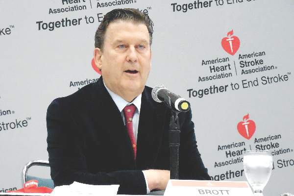
Taking the 5-year outcome results from ACT I and the 10-year outcome results from CREST both into account, “we now have a lot of evidence that both carotid stenting and surgery are safe and durable. The results support both options” for either patients with symptomatic carotid artery stenosis or asymptomatic patients with carotid stenosis as extensive as in the patients enrolled in these trials, said Dr. Thomas G. Brott at the International Stroke Conference.
“In routine practice, we lay out the options of endarterectomy, carotid stenting, or no intervention with just medical treatment to patients and let them decide,” noted Dr. Brott, professor of neurology and director of research at the Mayo Clinic in Jacksonville, Fla.
CREST randomized 2,502 symptomatic or asymptomatic patients with significant carotid stenosis during 2000-2008 at 117 U.S. and Canadian centers. From this group, 1,607 consented and were available for long-term follow-up, done at a median of 7.4 years and as long as 10 years after follow-up.
The study’s primary, long-term endpoint was stroke, MI, or death during the periprocedural period (30 days after treatment or 36 days after enrollment depending on when the procedural intervention occurred) plus the rate of ipsilateral stroke during up to 10 years of follow-up. This combined endpoint occurred in 10% of the patients who underwent endarterectomy and in 12% of those who had stenting, a difference that was not statistically significant, Dr. Brott reported. Concurrent with his presentation at the meeting, sponsored by the American Heart Association, the results also were published online (N Engl J Med. 2016 Feb 18. doi: 10.1056/NEJMoa1505215).
The results included a secondary endpoint that showed a significant benefit for endarterectomy. The tally of periprocedural strokes or deaths plus ipsilateral strokes during 10-year follow-up was 8% for the surgical group and 11% for those who received a stent, a 37% excess hazard with stenting.
Dr. Brott attributed this secondary difference between the two arms of the study to a statistically significant excess of stroke or death during the periprocedural period in the patients treated by stenting, and more specifically an excess of strokes. The rate of total periprocedural strokes was 4% with stenting and 2% with endarterectomy, a statistically significant difference. Although an embolic protection device was used “when feasible” during stenting, this protection can be fallible, Dr. Brott noted. In contrast, the results from the ACT I trial showed no statistically significant difference in the rate of periprocedural total strokes between the stented and endarterectomy patients.
Dr. Brott had no relevant disclosures. The CREST trial received partial funding from Abbott Vascular.
The 10-year CREST results are good news for patients with carotid disease because they show the durability of both interventions we can offer patients. Having these data and the results from ACT I allows physicians to have an informed discussion with patients about their treatment options. I also hope that with these results from both trials, reimbursement will cease to be a deciding factor and that both surgery and stenting will be on a level playing field for insurance coverage.
Although on a population level stenting and surgery appear to produce comparable results, individual patient characteristics can make one option more appropriate. These include the morphology of a patient’s carotid arteries and stenotic lesions that can make stenting a technical challenge, and a patient’s medical condition and comorbidities which could put them at higher risk for general anesthesia and surgery. Also, a big concern for many patients is how long they will require hospitalization.
 |
Dr. Mark J. Alberts |
A major unresolved question now about treating carotid disease is whether medical treatment alone is an equally good third alternative for asymptomatic patients. We are in a relatively new era of medical therapy, with more options for smoking cessation, better and more diverse drugs for blood pressure and hyperglycemia control, and wider use of high-dose statins. Some patients are eager to avoid any intervention and already opt for medical management only, but only after CREST-2 is completed will we know whether they will truly fare as well as patients who have a procedure performed.
Another issue that needs to be considered when extrapolating the results from CREST and ACT I to routine practice is that the surgeons and interventionalists who performed the procedures in these trials were highly selected and experienced. One cannot assume that the results in these trials will be replicated by any surgeon or interventionalist in the community. I suggest that patients investigate the track record of their community hospitals and operators by consulting the performance information that is increasingly posted on the Internet.
Dr. Mark J. Alberts is professor of neurology and medical director of the neurology service at the University of Texas Southwestern Medical Center in Dallas. He had no disclosures. He made these comments in an interview.
The 10-year CREST results are good news for patients with carotid disease because they show the durability of both interventions we can offer patients. Having these data and the results from ACT I allows physicians to have an informed discussion with patients about their treatment options. I also hope that with these results from both trials, reimbursement will cease to be a deciding factor and that both surgery and stenting will be on a level playing field for insurance coverage.
Although on a population level stenting and surgery appear to produce comparable results, individual patient characteristics can make one option more appropriate. These include the morphology of a patient’s carotid arteries and stenotic lesions that can make stenting a technical challenge, and a patient’s medical condition and comorbidities which could put them at higher risk for general anesthesia and surgery. Also, a big concern for many patients is how long they will require hospitalization.
|
Dr. Mark J. Alberts |
A major unresolved question now about treating carotid disease is whether medical treatment alone is an equally good third alternative for asymptomatic patients. We are in a relatively new era of medical therapy, with more options for smoking cessation, better and more diverse drugs for blood pressure and hyperglycemia control, and wider use of high-dose statins. Some patients are eager to avoid any intervention and already opt for medical management only, but only after CREST-2 is completed will we know whether they will truly fare as well as patients who have a procedure performed.
Another issue that needs to be considered when extrapolating the results from CREST and ACT I to routine practice is that the surgeons and interventionalists who performed the procedures in these trials were highly selected and experienced. One cannot assume that the results in these trials will be replicated by any surgeon or interventionalist in the community. I suggest that patients investigate the track record of their community hospitals and operators by consulting the performance information that is increasingly posted on the Internet.
Dr. Mark J. Alberts is professor of neurology and medical director of the neurology service at the University of Texas Southwestern Medical Center in Dallas. He had no disclosures. He made these comments in an interview.
LOS ANGELES – The equipoise between carotid stenting and endarterectomy received a further boost in 10-year results from the landmark Carotid Revascularization Endarterectomy versus Stenting Trial (CREST) that compared the two options head-to-head.
Reported the day after results from another big trial that pitted carotid stenting against surgery, the Asymptomatic Carotid Trial (ACT I), the new long-term results from the CREST study mean that deciding among the options relies largely on patient preference although individual clinical characteristics might favor one approach or the other, experts said.
The big remaining unknown and wild card is whether doing no procedural intervention at all and relying entirely on optimal, contemporary medical treatment works just as well as endarterectomy or carotid stenting. The role for stand-alone medical therapy against carotid surgery or stenting (on top of medical therapy) is currently undergoing a formal, direct comparison in the randomized Carotid Revascularization and Medical Management for Asymptomatic Carotid Stenosis Trial (CREST-2).
Taking the 5-year outcome results from ACT I and the 10-year outcome results from CREST both into account, “we now have a lot of evidence that both carotid stenting and surgery are safe and durable. The results support both options” for either patients with symptomatic carotid artery stenosis or asymptomatic patients with carotid stenosis as extensive as in the patients enrolled in these trials, said Dr. Thomas G. Brott at the International Stroke Conference.
“In routine practice, we lay out the options of endarterectomy, carotid stenting, or no intervention with just medical treatment to patients and let them decide,” noted Dr. Brott, professor of neurology and director of research at the Mayo Clinic in Jacksonville, Fla.
CREST randomized 2,502 symptomatic or asymptomatic patients with significant carotid stenosis during 2000-2008 at 117 U.S. and Canadian centers. From this group, 1,607 consented and were available for long-term follow-up, done at a median of 7.4 years and as long as 10 years after follow-up.
The study’s primary, long-term endpoint was stroke, MI, or death during the periprocedural period (30 days after treatment or 36 days after enrollment depending on when the procedural intervention occurred) plus the rate of ipsilateral stroke during up to 10 years of follow-up. This combined endpoint occurred in 10% of the patients who underwent endarterectomy and in 12% of those who had stenting, a difference that was not statistically significant, Dr. Brott reported. Concurrent with his presentation at the meeting, sponsored by the American Heart Association, the results also were published online (N Engl J Med. 2016 Feb 18. doi: 10.1056/NEJMoa1505215).
The results included a secondary endpoint that showed a significant benefit for endarterectomy. The tally of periprocedural strokes or deaths plus ipsilateral strokes during 10-year follow-up was 8% for the surgical group and 11% for those who received a stent, a 37% excess hazard with stenting.
Dr. Brott attributed this secondary difference between the two arms of the study to a statistically significant excess of stroke or death during the periprocedural period in the patients treated by stenting, and more specifically an excess of strokes. The rate of total periprocedural strokes was 4% with stenting and 2% with endarterectomy, a statistically significant difference. Although an embolic protection device was used “when feasible” during stenting, this protection can be fallible, Dr. Brott noted. In contrast, the results from the ACT I trial showed no statistically significant difference in the rate of periprocedural total strokes between the stented and endarterectomy patients.
Dr. Brott had no relevant disclosures. The CREST trial received partial funding from Abbott Vascular.
AT THE INTERNATIONAL STROKE CONFERENCE

ISC: Carotid surgery, stenting offer patients balanced alternatives
LOS ANGELES – The equipoise between carotid stenting and endarterectomy received a further boost in 10-year results from the landmark Carotid Revascularization Endarterectomy versus Stenting Trial (CREST) that compared the two options head-to-head.
Reported the day after results from another big trial that pitted carotid stenting against surgery, the Asymptomatic Carotid Trial (ACT I), the new long-term results from the CREST study mean that deciding among the options relies largely on patient preference although individual clinical characteristics might favor one approach or the other, experts said.

The big remaining unknown and wild card is whether doing no procedural intervention at all and relying entirely on optimal, contemporary medical treatment works just as well as endarterectomy or carotid stenting. The role for stand-alone medical therapy against carotid surgery or stenting (on top of medical therapy) is currently undergoing a formal, direct comparison in the randomized Carotid Revascularization and Medical Management for Asymptomatic Carotid Stenosis Trial (CREST-2).
Taking the 5-year outcome results from ACT I and the 10-year outcome results from CREST both into account, “we now have a lot of evidence that both carotid stenting and surgery are safe and durable. The results support both options” for either patients with symptomatic carotid artery stenosis or asymptomatic patients with carotid stenosis as extensive as in the patients enrolled in these trials, said Dr. Thomas G. Brott at the International Stroke Conference.
“In routine practice, we lay out the options of endarterectomy, carotid stenting, or no intervention with just medical treatment to patients and let them decide,” noted Dr. Brott, professor of neurology and director of research at the Mayo Clinic in Jacksonville, Fla.
CREST randomized 2,502 symptomatic or asymptomatic patients with significant carotid stenosis during 2000-2008 at 117 U.S. and Canadian centers. From this group, 1,607 consented and were available for long-term follow-up, done at a median of 7.4 years and as long as 10 years after follow-up.
The study’s primary, long-term endpoint was stroke, MI, or death during the periprocedural period (30 days after treatment or 36 days after enrollment depending on when the procedural intervention occurred) plus the rate of ipsilateral stroke during up to 10 years of follow-up. This combined endpoint occurred in 10% of the patients who underwent endarterectomy and in 12% of those who had stenting, a difference that was not statistically significant, Dr. Brott reported. Concurrent with his presentation at the meeting, sponsored by the American Heart Association, the results also were published online (N Engl J Med. 2016 Feb 18. doi: 10.1056/NEJMoa1505215).
The results included a secondary endpoint that showed a significant benefit for endarterectomy. The tally of periprocedural strokes or deaths plus ipsilateral strokes during 10-year follow-up was 8% for the surgical group and 11% for those who received a stent, a 37% excess hazard with stenting.
Dr. Brott attributed this secondary difference between the two arms of the study to a statistically significant excess of stroke or death during the periprocedural period in the patients treated by stenting, and more specifically an excess of strokes. The rate of total periprocedural strokes was 4% with stenting and 2% with endarterectomy, a statistically significant difference. Although an embolic protection device was used “when feasible” during stenting, this protection can be fallible, Dr. Brott noted. In contrast, the results from the ACT I trial showed no statistically significant difference in the rate of periprocedural total strokes between the stented and endarterectomy patients.
Dr. Brott had no relevant disclosures. The CREST trial received partial funding from Abbott Vascular.
On Twitter @mitchelzoler
The 10-year CREST results are good news for patients with carotid disease because they show the durability of both interventions we can offer patients. Having these data and the results from ACT I allows physicians to have an informed discussion with patients about their treatment options. I also hope that with these results from both trials, reimbursement will cease to be a deciding factor and that both surgery and stenting will be on a level playing field for insurance coverage.
Although on a population level stenting and surgery appear to produce comparable results, individual patient characteristics can make one option more appropriate. These include the morphology of a patient’s carotid arteries and stenotic lesions that can make stenting a technical challenge, and a patient’s medical condition and comorbidities which could put them at higher risk for general anesthesia and surgery. Also, a big concern for many patients is how long they will require hospitalization.
|
Dr. Mark J. Alberts |
A major unresolved question now about treating carotid disease is whether medical treatment alone is an equally good third alternative for asymptomatic patients. We are in a relatively new era of medical therapy, with more options for smoking cessation, better and more diverse drugs for blood pressure and hyperglycemia control, and wider use of high-dose statins. Some patients are eager to avoid any intervention and already opt for medical management only, but only after CREST-2 is completed will we know whether they will truly fare as well as patients who have a procedure performed.
Another issue that needs to be considered when extrapolating the results from CREST and ACT I to routine practice is that the surgeons and interventionalists who performed the procedures in these trials were highly selected and experienced. One cannot assume that the results in these trials will be replicated by any surgeon or interventionalist in the community. I suggest that patients investigate the track record of their community hospitals and operators by consulting the performance information that is increasingly posted on the Internet.
Dr. Mark J. Alberts is professor of neurology and medical director of the neurology service at the University of Texas Southwestern Medical Center in Dallas. He had no disclosures. He made these comments in an interview.
LOS ANGELES – The equipoise between carotid stenting and endarterectomy received a further boost in 10-year results from the landmark Carotid Revascularization Endarterectomy versus Stenting Trial (CREST) that compared the two options head-to-head.
Reported the day after results from another big trial that pitted carotid stenting against surgery, the Asymptomatic Carotid Trial (ACT I), the new long-term results from the CREST study mean that deciding among the options relies largely on patient preference although individual clinical characteristics might favor one approach or the other, experts said.
The big remaining unknown and wild card is whether doing no procedural intervention at all and relying entirely on optimal, contemporary medical treatment works just as well as endarterectomy or carotid stenting. The role for stand-alone medical therapy against carotid surgery or stenting (on top of medical therapy) is currently undergoing a formal, direct comparison in the randomized Carotid Revascularization and Medical Management for Asymptomatic Carotid Stenosis Trial (CREST-2).
Taking the 5-year outcome results from ACT I and the 10-year outcome results from CREST both into account, “we now have a lot of evidence that both carotid stenting and surgery are safe and durable. The results support both options” for either patients with symptomatic carotid artery stenosis or asymptomatic patients with carotid stenosis as extensive as in the patients enrolled in these trials, said Dr. Thomas G. Brott at the International Stroke Conference.
“In routine practice, we lay out the options of endarterectomy, carotid stenting, or no intervention with just medical treatment to patients and let them decide,” noted Dr. Brott, professor of neurology and director of research at the Mayo Clinic in Jacksonville, Fla.
CREST randomized 2,502 symptomatic or asymptomatic patients with significant carotid stenosis during 2000-2008 at 117 U.S. and Canadian centers. From this group, 1,607 consented and were available for long-term follow-up, done at a median of 7.4 years and as long as 10 years after follow-up.
The study’s primary, long-term endpoint was stroke, MI, or death during the periprocedural period (30 days after treatment or 36 days after enrollment depending on when the procedural intervention occurred) plus the rate of ipsilateral stroke during up to 10 years of follow-up. This combined endpoint occurred in 10% of the patients who underwent endarterectomy and in 12% of those who had stenting, a difference that was not statistically significant, Dr. Brott reported. Concurrent with his presentation at the meeting, sponsored by the American Heart Association, the results also were published online (N Engl J Med. 2016 Feb 18. doi: 10.1056/NEJMoa1505215).
The results included a secondary endpoint that showed a significant benefit for endarterectomy. The tally of periprocedural strokes or deaths plus ipsilateral strokes during 10-year follow-up was 8% for the surgical group and 11% for those who received a stent, a 37% excess hazard with stenting.
Dr. Brott attributed this secondary difference between the two arms of the study to a statistically significant excess of stroke or death during the periprocedural period in the patients treated by stenting, and more specifically an excess of strokes. The rate of total periprocedural strokes was 4% with stenting and 2% with endarterectomy, a statistically significant difference. Although an embolic protection device was used “when feasible” during stenting, this protection can be fallible, Dr. Brott noted. In contrast, the results from the ACT I trial showed no statistically significant difference in the rate of periprocedural total strokes between the stented and endarterectomy patients.
Dr. Brott had no relevant disclosures. The CREST trial received partial funding from Abbott Vascular.
On Twitter @mitchelzoler
AT THE INTERNATIONAL STROKE CONFERENCE
Key clinical point: Long-term follow-up of the CREST trial out to 10 years showed no statistically significant difference between endarterectomy or carotid stenting for patients with carotid artery stenosis.
Major finding: The primary, long-term endpoint occurred in 10% of endarterectomy patients and 12% of stented patients, a nonsignificant difference.
Data source: The CREST trial, which followed 1,607 patients for up to 10 years after their randomized intervention.
Disclosures: Dr. Brott had no relevant disclosures. The CREST trial received partial funding from Abbott Vascular.
VIDEO: Stenting in asymptomatic patients noninferior to endarterectomy at 5 years
LOS ANGELES – In asymptomatic patients under 80 years old, carotid stenting and endarterectomy perform equally as well for severe carotid stenosis out to 5 years, according to a randomized trial published online in the New England Journal of Medicine.
Overall, 1,032 patients were stented, and 343 had endarterectomies in the trial, called Asymptomatic Carotid Trial I (ACT I). If stenting didn’t look safe on postrandomization angiography, patients were given the option of medical management or crossover into the surgical group. The subjects all had bifurcation carotid stenosis blocking at least 70% of the lumen. None were at high risk for surgical complications. “Asymptomatic” meant they hadn’t had a stroke, transient ischemic attack, or amaurosis fugax in the 6 months before enrollment. Stenting and endarterectomy were done by physicians and centers well experienced in the techniques (N Engl J Med. 2016 Feb 17. doi: 10.1056/NEJMoa1515706).
At 1 year, stenting was noninferior to endarterectomy for the primary composite endpoint of death, stroke, or myocardial infarction within 30 days after the procedure or ipsilateral stroke within 1 year; the event rate was 3.8% among stent patients and 3.4% among endarterectomy patients (P = .01 for noninferiority, with a noninferiority margin of 3 percentage points).
The cumulative 5-year stroke-free survival rate was 93.1% in the stenting group and 94.7% in the endarterectomy group (P = .44).
For now, the results mean that sometimes choosing between carotid endarterectomy or stenting (or medical management) has as much to do with patient and physician preference as medical science, raising the difficult question of how to choose. In a video interview at the International Stroke Conference, investigator Dr. Lawrence Wechsler, professor of neurology/neurosurgery and chair of the department of neurology at the University of Pittsburgh, shared his thoughts on that and the other implications of the study. The work was funded by Abbott Vascular.
The video associated with this article is no longer available on this site. Please view all of our videos on the MDedge YouTube channel
AT The INTERNATIONAL STROKE CONFERENCE

VIDEO: Octogenarians benefit from ischemic stroke thrombectomy
LOS ANGELES – Clot removal to recanalize the occluded intracerebral arteries of acute ischemic stroke patients was as effective for producing good outcomes in patients aged 80 years or older as it was in younger patients, according to results from a pooled analysis of 1,287 patients in five separate but similar randomized trials.
This unprecedented evidence for the safety and efficacy of thrombectomy (also known as embolectomy) in octogenarians experiencing an acute occlusive, large-vessel, proximal anterior-circulation stroke was one of several new findings from the pooled analysis that should help further push thrombectomy to the forefront of acute care for patients undergoing this type of ischemic stroke, predicted Dr. Wade S. Smith in a video interview at the International Stroke Conference.
“By looking at all the data, we have much more refined information on the robustness of the treatment across age groups, which is quite important, especially patients in the 80-plus age group,” commented Dr. Smith, professor of neurology and chief of the neurovascular division at the University of California, San Francisco.
Until now, during the year following the reports in early 2015 from all five studies, “age had been a limiting factor” in applying the practicing-changing intervention of thrombectomy to patients, he noted.
“This [the new pooled analysis] will change that,” Dr. Smith predicted. “It does not apply to patients who were infirm prior to their stroke – but for patients who were otherwise healthy, with a modified Rankin scale level of 0 or 1 at initial presentation, it appears that they benefit [from thrombectomy] regardless of their age.” In the pooled analysis, 198 of the 1,287 total patients (15%) were at least 80 years old.
“It removes age discrimination. A healthy 80-year-old may do extremely well with this treatment,” Dr. Smith said.
The consistency of benefit across a wide range of stroke severity that showed up in the trials as four distinct strata of NIH Stroke Scale scores prior to treatment was another important finding that could not previously be definitively made by analyzing each of the five trials individually.
In patients with stroke-severity scores that ranged from 10 or less (the least severely affected) to patients with scores of 21 or greater, all had post-thrombectomy improvements that clustered around the overall average number-needed-to-treat of 2.6 patients to reduce the disability of one patient at follow-up by at least one level on the modified Rankin scale.
Other notable findings from the pooled analysis were that thrombectomy also produced a consistent benefit to patients across every other subgroup examined, including sex, specific occlusion site, whether or not patients also received thrombolytic treatment with tissue plasminogen activator, and time to thrombectomy treatment (5 or fewer hours from stroke onset or more than 5 hours), reported Dr. Michael D. Hill and Dr. Tudor G. Jovin in a joint presentation at the meeting, sponsored by the American Heart Association.
Their pooled analysis, known as HERMES, pooled data from the MR CLEAN, ESCAPE, REVASCAT, SWIFT PRIME, and EXTEND IA trials, all run during 2010-2014.
“Endovascular treatment is a highly effective treatment across all subgroups,” concluded Dr. Hill and Dr. Jovin as they completed their talk. “These data may provide additional support for endovascular treatment in subgroups not addressed in the individual trials.”
Concurrent with their report at the meeting, the results appeared in a paper published online (Lancet. 2016 Feb 18;doi: 10.1016/S0140-6736(16)00163-X).
Both Dr. Jovin and Dr. Hill shared the enthusiasm of Dr. Smith and others in the packed meeting room about the age finding.
“Older patients seemed to benefit even more” from thrombectomy, compared with younger patients, noted Dr. Jovin, chief of the stroke division at the University of Pittsburgh and a coinvestigator on SWIFT PRIME. “There is no reason to deny this treatment to appropriately selected patients based on age,” he said.
“There is no upper age limit,” agreed Dr. Hill, professor of neurology and director of the stroke unit at the University of Calgary (Alta.) and a coinvestigator on the ESCAPE trial. “If it’s an otherwise healthy 90-year-old who is living independently, you can surely consider them for this treatment.”
HERMES received fundings through an unrestricted grant from Medtronic. Dr. Hill and Dr. Jovin had no personal disclosures. Dr. Smith served on the data safety and monitoring board for a trial funded by Stryker.
The video associated with this article is no longer available on this site. Please view all of our videos on the MDedge YouTube channel
On Twitter @mitchelzoler
AT THE INTERNATIONAL STROKE CONFERENCE
Key clinical point: A pooled analysis of five recent trials of thrombectomy for acute ischemic stroke should help further propel its widespread U.S. adoption.
Major finding: Intracerebral, transcatheter clot removal in acute ischemic stroke was equally effective in octogenarian and younger patients.
Data source: HERMES, a pooled analysis of data from 1,287 acute ischemic stroke patients randomized in five separate but similar trials.
Disclosures: HERMES received fundings through an unrestricted grant from Medtronic. Dr. Hill and Dr. Jovin had no personal disclosures. Dr. Smith served on the data safety and monitoring board for a trial funded by Stryker.

VIDEO: Post-stroke pioglitazone modestly protective against secondary vascular events
LOS ANGELES – Nondiabetic, insulin-resistant patients who started pioglitazone within 6 months of an ischemic stroke or transient ischemic attack had almost a 3% absolute risk reduction in secondary strokes and myocardial infarctions after 5 years, in a randomized, clinical trial published online Feb. 17 in the New England Journal of Medicine.
Stroke or MI – the study’s primary combined outcome – occurred in 175 of 1,939 (9.0%) pioglitazone (Actos) patients, but 228 of 1,937 (11.8%) placebo patients (hazard ratio, 0.76; P = 0.007). There was no significant difference in all-cause mortality (HR, 0.93; P = 0.52).
Seventy-three pioglitazone patients (3.8%) developed diabetes, compared with 149 (7.7%) in the placebo group (HR, 0.48; P less than 0.001). That wasn’t a surprise; pioglitazone has been shown to protect insulin resistant patients against diabetes, the study investigators noted (N Engl J Med. 2016 Feb 17; doi: 10.1056/NEJMoa1506930).
Known side effects showed up as well. Pioglitazone was associated with a greater frequency of weight gain exceeding 4.5 kg (52.2% versus 33.7%, P less than 0.001), edema (35.6% versus 24.9%, P less than 0.001), and bone fracture requiring surgery or hospitalization (5.1% versus 3.2%, P = 0.003).
Heart failure – another known risk with the drug – did not show up in the trial; people with heart failure histories or other risk factors were excluded.
The median baseline modified Rankin Scale in both groups was 1, and the median NIH Stroke Scale score was 0. The pioglitazone target dose was 45 mg daily.
Insulin resistance is a risk factor for heart attack and stroke, which may help explain the thiazolidinedione’s apparent protective effects. It was defined in the trial as a score greater than 3.0 on the homeostasis model assessment of insulin resistance (HOMA-IR) index.
So, should pioglitazone be a part of routine post-stroke care?
In a video interview at the International Stroke Conference, lead investigator Dr. Walter N. Kernan, a professor of general medicine at Yale University, New Haven, Conn., shared his thoughts.
The video associated with this article is no longer available on this site. Please view all of our videos on the MDedge YouTube channel
AT THE INTERNATIONAL STROKE CONFERENCE
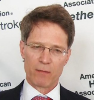
VIDEO: Migraine plus aura doubles ischemic stroke rate
LOS ANGELES – Adults who have migraine with aura had twice the rate of ischemic strokes, compared with people with migraine without aura in a review of 11,663 older Americans followed for an average of 18 years.
Migraine with aura appeared to increase the rate of cardioembolic strokes particularly, and the findings highlighted that middle-aged and older adults diagnosed with migraine with aura require special attention to interventions that could reduce their cardiovascular disease risks, Dr. Souvik Sen said in a video interview during the International Stroke Conference.
While results from prior studies have implicated migraine with aura as a risk factor for ischemic stroke, the new analysis was notable for involving an especially large number of community-dwelling older Americans, suggesting that the finding is broadly applicable to the general population of people with an average age of 58-60 years, said Dr. Sen, professor of clinical neurology and chairman of the department of neurology at the University of South Carolina in Columbia.
“It’s been known that migraine can increase the risk of stroke and that certain subgroups of migraine patients are especially important, such as patients with aura,” commented Dr. Ralph Sacco, professor and chairman of neurology at the University of Miami. “When I see patients with migraine I think about treating their migraine effectively and getting it under control, and putting even greater focus on controlling stroke risk factors. I also warn women with migraine about their increased stroke risk from using estrogen-containing oral contraceptives or hormone replacement,” Dr. Sacco noted.
Dr. Sen noted that the new analysis included people enrolled in the Atherosclerosis Risk in Communities (ARIC) study, which enrolled a random sample of more than 15,000 participants from four U.S. communities in 1987. The analysis focused on 11,663 people with migraine information collected during the third ARIC examination during 1993-1995, which identified 458 people (4%) who had migraine with aura, 1,158 (10%) who had migraine without aura, and 10,047 (86%) who were migraine free. During 18 years of follow-up the incidence of ischemic stroke among those with migraine with aura was twofold higher than among those with migraine without aura after adjustment for baseline rate of stroke risk factors, a statistically significant difference, he reported at the meeting sponsored by the American Heart Association. This difference seemed largely driven by a significant increase in the rate of cardioembolic strokes among the people with migraine with aura.
One hypothesis explaining the increased stroke rate in patients with aura is that, in patients with this presentation of migraine, the neurologic activity of migraine appears to spread to cerebral blood vessels and may affect blood flow within those vessels, Dr. Sen explained. It is also possible that migraine with aura may be a risk marker for or may predispose people to more extensive cardiovascular disease including atrial fibrillation.
The primary clinical implication of the finding is that people with migraine with aura should especially undergo a thorough assessment for cardiovascular disease risk factors, and should receive especially aggressive risk factor management, including controlling hypertension and smoking cessation, assessing ECG and atrial fibrillation, and avoiding estrogen treatment, Dr. Sen said.
The video associated with this article is no longer available on this site. Please view all of our videos on the MDedge YouTube channel
On Twitter @mitchelzoler
AT THE INTERNATIONAL STROKE CONFERENCE
Key clinical point: Older adults with migraine and aura had twice the ischemic strokes, compared with patients with aura-free migraines.
Major finding: Strokes occurred twofold more often in patients with migraine with aura, compared with patients with migraine alone after adjustments.
Data source: Review of 11,663 older adult Americans enrolled in the Atherosclerosis Risk in Communities study.
Disclosures: Dr. Sen had no disclosures.