User login
Ankylosing spondylitis patients develop multiple comorbidities after diagnosis
DENVER – Evidence continues to mount that ankylosing spondylitis patients are at increased risk for developing various comorbidities, compared with the general adult population.
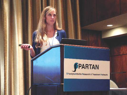
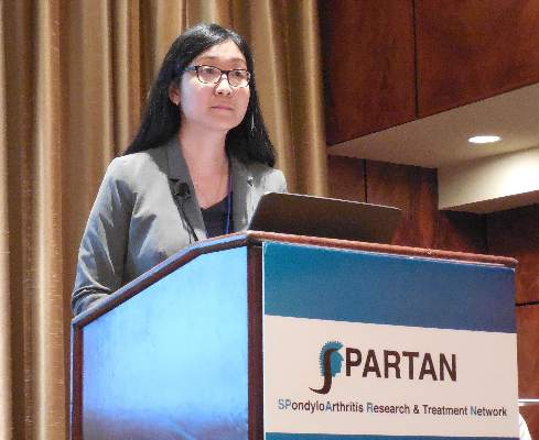
Patients newly diagnosed with ankylosing spondylitis (AS) had twice the rate of new-onset depression during the first 3 years following diagnosis, compared with matched people from the general population in a study of more than 21,000 American adults. Patients with newly diagnosed AS also had a 60% higher rate of developing a new cardiovascular disease, compared with the matched general population, Jessica A. Walsh, MD, said at the annual meeting of the Spondyloarthritis Research and Treatment Network.
“We need to figure out what to do about all the comorbidities. Rheumatologists need to either screen their AS patients for comorbidities, or they need to be sure their patients are plugged in with another physician who will screen them,” said Dr. Walsh, director of the spondyloarthritis clinic at the University of Utah in Salt Lake City.
Her analysis used data from the Truven Health MarketScan databases for U.S. patients covered by Medicare or commercial health insurance, and included 6,370 patients with newly diagnosed AS and 14,988 adults matched by age and sex. The analysis only included AS patients who were free from comorbidities during the 2 years prior to their AS diagnosis. The average age of the people in the study was 52 years, and 54% were men.
During an average follow-up of 2.9 years following initial AS diagnosis, the most common comorbidity among the AS patients was uveitis, which occurred nearly 15-fold more frequently among the AS patients than in the controls. Other common incident comorbidities included inflammatory bowel disease, nearly sixfold more common among the AS patients, and osteoporosis, which was nearly threefold more common during follow-up after AS diagnosis, compared with the controls.
Other comorbidities with increased incidence in the AS patients included sleep apnea (80% more common among the AS patients during follow-up), asthma (50% more often), hypertension (44% more common), malignancy (23% more common), diabetes (20% more common), and dyslipidemia (11% more common). All these incidence rates represented statistically significant increases in the AS patients, compared with the controls.
A related analysis reported by Dr. Walsh also used data from the Truven Health databases for a somewhat larger group of AS patients, 6,679 followed for 1 year after their AS diagnosis, and 19,951 matched controls. The AS patients had a significantly higher rate of hospital admissions – 12%, compared with 6% among the controls – and a significantly higher rate of emergency department visits, at 23%, compared with 15% among the controls. The AS patients also had double the rate of physician office visits and prescribed medications.
“Obviously, the AS patients are not as healthy,” Dr. Walsh said in an interview. “We adjusted for their comorbidities, but that did not affect the hospitalization rates. We need to look into this more; I don’t know why the AS patients are being hospitalized. Typically AS itself does not lead to hospitalization, so I suspect it’s because of comorbidities, or perhaps because of adverse events from treatment.”
Dr. Walsh is a consultant to AbbVie and Novartis.
On Twitter @mitchelzoler
AT THE 2016 SPARTAN ANNUAL MEETING
Key clinical point: In the 3 years after initial ankylosing spondylitis diagnosis, the incidence of several comorbidities rises significantly above those in the general population.
Major finding: The incidence of new-onset depression among newly diagnosed ankylosing spondylitis patients was twice as high as it was among matched controls.
Data source: Observational data collected by Truven Health MarketScan, with a total of 21,358 patients and controls.
Disclosures: Dr. Walsh is a consultant to AbbVie and Novartis.
MRI now anchors spondyloarthritis diagnosis
DENVER – MRI of the sacroiliac joint now serves as the primary imaging driver for a diagnosis of spondyloarthritis, especially early spondyloarthritis, and has largely supplanted radiographic assessment, Walter P. Maksymowych, MD, said at an educational symposium organized by the Spondyloarthritis Research and Treatment Network.
“MRI is reliable for early diagnosis; radiographic assessment of early spondyloarthritis is problematic,” said Dr. Maksymowych, a rheumatologist and professor of medicine at the University of Alberta in Edmonton. “The EULAR recommendations now say that early diagnosis of spondyloarthritis needs to focus on MRI.”
While the recommendations from the European League Against Rheumatism (EULAR) that came out last year on using imaging for diagnosing and managing spondyloarthritis (SpA) cite radiography of the patient’s sacroiliac joint as the recommended first imaging method to use to diagnose sacroiliitis as part of a diagnosis of axial SpA, the EULAR recommendations place MRI very close behind.
The recommendations list MRI as an “alternative” first-line approach for diagnostic imaging. For patients who appear negative for axial SpA on radiographic imaging but who remain suspected of having SpA the EULAR panel “recommended” MRI of the sacroiliac joints as a backup method for imaging assessment and to definitively rule out SpA. No other imaging method received EULAR’s endorsement for SpA diagnosis aside from these two approaches.
The recommendations direct the MRI examination to include assessment of inflammation based on bone marrow edema, and also structural abnormalities including bone erosion, new bone formation, sclerosis and fat infiltration. The 2015 EULAR recommendations also cite roles for MRI in diagnosing peripheral SpA, monitoring disease activity in axial and peripheral SpA, monioring structural changes in axial and peripheral SpA, predicting outcome and treatment effect in axial SpA, assessing spinal fracture in patients with axial SpA and to assess osteoporosis in selected axial SpA patients.
The MRI imaging sequences particularly useful for SpA diagnosis are “short tau inversion recovery” (STIR), a “water sensitive” sequence that involves suppression of the fat MRI signal, and a T1 weighted sequence that is a “fat sensitive” scan, explained Dr. Maksymowych at the symposium, also organized by the Group for the Research and Assessment of Psoriasis and Psoriatic Arthritis (GRAPPA). He strongly encouraged clinicians to apply a standardized approach to imaging in every patient suspected of having axial SpA.
The definition of an MRI imaging result that is positive for SpA remains a consensus of expert opinion without a firm evidence base. The currently accepted MRI marker of SpA involves identifying several areas of bone marrow edema in a single T1 and STIR MRI image-slice through the sacroiliac joint, or focal bone marrow edema visible in at least two consecutive sacroiliac joint T1 and STIR image slices.
The confluence of a structural lesion at the same site as bone marrow edema provides further confirmatory information, he said. “A structural lesions at the site of bone marrow edema enhances your confidence in the diagnosis,” he said.
“The specificity of MRI for SpA is quite high,” Dr. Maksymowych noted, with specificity rates often in the range of 85%-95%. Sensitivity rates are lower, often 50%-70%. Sensitivity further improves by factoring in the presence of bone erosions. Diagnostic confidence also increases as the number of lesions visualized increases.
MRI has become so integral to the assessment of SpA that “to understand SpA you need to understand the language of MRI,” Dr. Maksymowych concluded.
Dr. Maksymowych has received honoraria from eight drug companies and research grant support from Abbvie and Pfizer.
On Twitter @mitchelzoler
EXPERT ANALYSIS FROM A SPARTAN-GRAPPA SYMPOSIUM
RISE registry stockpiles U.S. rheumatology data
DENVER – During early 2015, exactly a quarter of all patients seen in U.S. rheumatology practices were diagnosed with rheumatoid arthritis, and among them two-thirds received treatment with a disease modifying nonbiologic drug, based on data from more than 200,000 patients who enrolled in an American College of Rheumatology registry during that time.
That’s an example of some of the data now available in an EHR-based registry run by the ACR and open to participation by any U.S.-based rheumatologist, Kaleb Michaud, PhD, said at the annual meeting of the Spondyloarthritis Research and Treatment Network.
The ACR’s Rheumatology Informatics System for Effectiveness (RISE) is an EHR-based registry of passive reporting that as of July 2016 had enrolled 614,738 U.S. patients treated by 1,284 providers in 213 practices, said Dr. Michaud, codirector of the National Data Bank for Rheumatic Diseases in Wichita, Kan., who works with the ACR on RISE as a volunteer.
Patient participation in RISE more than doubled during the past year, rising from 302,000 participating patients in September 2015. In addition to enrolling even more providers with more patients to participate, RISE’s organizers is also now seeking investigators who can use the data collected in RISE to address useful research questions, he said.
RISE has been designated a qualified clinical data registry (QCDR) by the Centers for Medicare & Medicaid Services. If providers using RISE sign an agreement stating that they want to participate for quality reporting purposes, it then happens automatically. Participating practices also meet Medicare’s meaningful use measure of reporting to a specialty registry. Although most rheumatologists with academic positions already have these opportunities, for community rheumatologists this access is “a perq,” Dr. Michaud said.
As just an example of the data trove accumulating in RISE, Dr, Michaud rattled off some quick figures culled from analysis of the 239,302 RISE patients in 55 U.S. practices who entered the registry during October 2014-September 2015. The patients averaged 59 years of age, three quarters were women, 61% were white, 48% had commercial insurance and 30% were covered through Medicare.
Their most recent diagnosis was rheumatoid arthritis in 25%, unspecified myalgia or myositis in 21%, Sjögren’s syndrome in 7%, systemic lupus erythematosus in 6%, psoriatic arthritis in 6%, spondyloarthritides in 4%, gout in 4%, and several other disorders at rates of 1% or less.
Among the rheumatoid arthritis patients, two-thirds were on a nonbiologic disease modifying drug, led by methotrexate in 65% of this subgroup and hydroxychloroquine in a third of the subgroup. A third of the entire study group was on treatment with a biologic or small-molecule drug. Etanercept (Enbrel) and adalimumab (Humira) led in this category with 8% of patients on each of these two drugs, followed by infliximab (Remicade) in 6%. Other usage rates in this category included abatacept (Orencia) in 4%, and tocilizumab (Actemra) and tofacitinib (Xeljanz) each used in about 2% of rheumatoid arthritis patients.
On Twitter @mitchelzoler
EXPERT ANALYSIS FROM THE 2016 SPARTAN ANNUAL MEETING
Revised axial spondyloarthritis classification criteria remain elusive
DENVER – The revised axial spondyloarthritis classification criteria that U.S. rheumatologists say they desperately need remain elusive, with no firm path to creation.
The 2009 classification criteria for axial spondyloarthritis (SpA) developed by the Assessment of Spondyloarthritis international Society (ASAS) was a landmark in creating a definition for axial SpA to use when enrolling patients into clinical trials and to also potentially use for diagnosis.
But the 2009 classification criteria also have several shortcomings, especially from a U.S. perspective, that have limited the utility of the criteria in U.S. practice and studies.
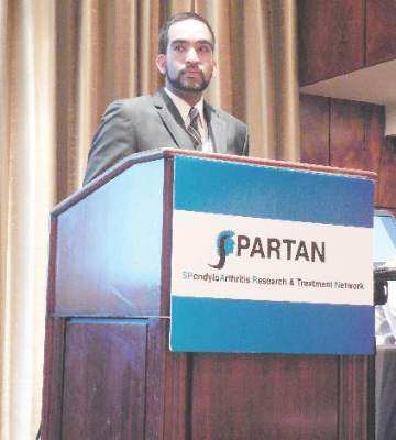
Several years of discussions among North American rheumatologists about the classification criteria have produced consensus about what is wrong with them: The clinical elements of the criteria are not specific enough with sensitivity and specificity rates that are each about 80%-85%; the imaging component of the criteria is not totally up to date and as one example only considers osteitis and not other MRI abnormalities such as fatty metaplasia and T1 structural lesions; the criteria were entirely based on results from studies with Europeans, making their applicability to other patient types uncertain; and some terminology in the criteria are confusing, Liron Caplan, MD, said during a discussion of the criteria at the annual meeting of the of the Spondyloarthritis Research and Treatment Network.
But while the SpA experts who gathered at the meeting and who have also been discussing this issue since 2013 are of one mind on the problems, agreement on exactly how to solve them remains unresolved.
“Everything is still on the table,” said Dr. Caplan, a rheumatologist and director of the SpA program at the University of Colorado in Aurora.
A year ago, at SPARTAN’s prior annual meeting, circumstances looked on track for a quicker resolution. In a vote of the membership at the 2015 gathering, 64% backed creation of new classification criteria, and Dr. Caplan proposed quickly preparing a research proposal to present in early 2016 to the American College of Rheumatology and the European League Against Rheumatism for funding.
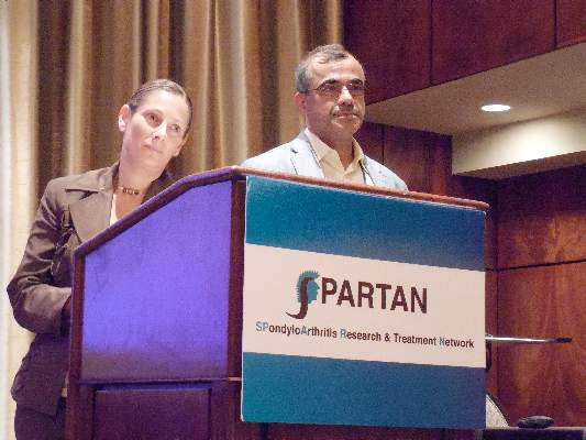
But in the ensuing year, the effort got mired in discussions on how to perform the study needed to gather the evidence base for criteria revisions. Some progress occurred, most notably an agreement between SPARTAN officials and the ASAS leadership to work together on the revision, said Atul A. Deodhar, MD, professor of medicine and medical director of the rheumatology clinics at the Oregon Health & Science University in Portland.
The SPARTAN and ASAS representatives also agreed on several specifics of what’s needed, including a weighting formula for the 10 different clinical criteria of the SpA classification. Currently, in individuals with low back pain lasting 3 months or longer at an age of onset younger than 45 years, any one of these 10 clinical criteria can create a case of SpA either if combined with MRI or radiographic evidence of sacroiliitis, or without any imaging if the individual is HLA-B27 positive and fulfills two items from the 10-item list of clinical criteria.
It’s the lack of radiographic imaging confirmation, so-called “nonradiographic SpA,” that generates the greatest concern about below-par specificity and that may be helped by a weighting adjustment that varies the classification power of each of the 10 clinical items. A more ideal balance might be criteria with sensitivity reduced from the current level to about 70% and with specificity rising to about 90%, Dr. Caplan suggested.
The keystone of any revision will be a large, new study with SpA patients from various locations. Dr. Caplan emphasized that “no firm decisions have yet been made” regarding the study's name or research focus.
On Twitter @mitchelzoler
This article was updated August 11, 2016.
EXPERT ANALYSIS FROM THE 2016 SPARTAN ANNUAL MEETING
Concurrent fibromyalgia intensifies ankylosing spondylitis symptoms
DENVER – Fibromyalgia syndrome commonly occurred in patients with ankylosing spondylitis who were reviewed at one U.S. center, and when the two coexisted ankylosing spondylitis disease severity significantly increased.
In the study’s 62 patients with confirmed ankylosing spondylitis (AS), 27 (44%) also met the 2010 American College of Rheumatology diagnostic criteria for fibromyalgia syndrome, Sherilyn Diomampo, MD, said at the annual meeting of the Spondyloarthritis Research and Treatment Network. The fibromyalgia rate in these AS patients was substantially above prior reports of fibromyalgia prevalence rates in the range of 10%-15%, said Dr. Diomampo, a rheumatologist at MetroHealth Medical Center in Cleveland.
Patients with both disorders also had substantially higher scores across the board for all the measures of AS severity that Dr. Diomampo and her associates evaluated. For example, scores on the Bath AS Disease Activity Index averaged 6.8 in the patients with fibromyalgia and 3.8 in those without fibromyalgia. (A BASDAI score of 4.0 or higher generally suggests suboptimal disease control.) The average score of the AS Disease Activity Score using C-reactive protein (CRP) as the serum marker of inflammation was 4.2 in the subgroup with fibromyalgia and 2.8 in those without. (An ASDAS score of 3.5 or higher denotes high or very high disease activity. A score reduced by at least 1.1 indicates a clinically important improvement.) All these between-group differences were statistically significant.
Other measures of AS severity that were significantly higher with fibromyalgia included patient global self assessment, physician global assessment, average serum levels of CRP, and the average erythrocyte sedimentation rate.
The 2010 ACR diagnostic criteria for fibromyalgia syndrome used by Dr. Diomampo and her associates in their analysis require a patient to have a widespread pain index of at least 7 and symptom severity of at least 5, or alternatively, a widespread pain index of 3-6 and symptom severity of at least 9 (Arthritis Care Res. 2010 May;62[5]:600-10). In addition, for this study, the researchers stipulated that diagnosis of concomitant fibromyalgia meant patients had their fibromyalgia symptoms in place for at least 3 months, and the examining clinician could not attribute the patient’s pain to AS.
Using linear regression models with the widespread pain index and the symptom severity as the dependent variables, the researchers failed to see any statistically significant relationship between either of these two determinants of fibromyalgia severity and five different measures of AS severity, including the BASDAI and the ASDAS. In short, the assessment tools used to measure AS severity showed no ability to also measure the core characteristics of fibromyalgia, Dr. Diomampo said.
Overall, patients in the study averaged about 49 years old. The analysis showed a significantly higher rate of African-American patients in the fibromyalgia group, 63%, compared with a 37% rate of African Americans in those with AS and no fibromyalgia. The presence of acute, anterior uveitis was 27% among those with fibromyalgia and 73% of those with AS and no fibromyalgia. One notable similarity between the two subgroups was the percentage on treatment with a tumor necrosis factor inhibitor: 52% among those with concurrent fibromyalgia and 49% among those with AS alone.
Dr. Diomampo had no disclosures.
On Twitter @mitchelzoler
AT THE 2016 SPARTAN ANNUAL MEETING
Key clinical point: Nearly half of patients with ankylosing spondylitis had concurrent fibromyalgia at one U.S. center, and patients with both had much greater AS severity.
Major finding: The average BASDAI was 6.8 in patients with ankylosing spondylitis and fibromyalgia and 3.8 in patients with AS only.
Data source: Observational data collected on 62 adults with ankylosing spondylitis at one U.S. center.
Disclosures: Dr. Diomampo had no disclosures.
VIDEO: Minimal disease activity criteria sought for axial spondyloarthritis
DENVER – U.S. rheumatologists plan to develop new criteria to define minimal disease activity in patients with axial spondyloarthritis to better gauge in routine clinical practice how well these patients respond to treatment.
A score on the Ankylosing Spondylitis Disease Activity Score (ASDAS) of less than 1.3 is the most commonly used measure today of minimal disease activity, but the ASDAS isn’t suitable for point-of-care assessment in routine practice because of the need for a C-reactive protein level or erythrocyte sedimentation rate.
“In the clinic, ASDAS is very difficult or next to impossible to do” because it needs CRP or ESR, which are “never available at the point of care,” Atul A. Deodhar, MD, said in an interview at the annual meeting of the Spondyloarthritis Research and Treatment Network (SPARTAN). Another limitation of ASDAS is that it focuses exclusively on musculoskeletal measures and does not take into account extra-articular manifestations of axial spondyloarthritis such as those in the eye, gastrointestinal tract, or skin.
“We would like a clinical measurement that might tell us that a patient is appropriately treated and at minimal disease activity,” said Dr. Deodhar, professor of medicine and medical director of the rheumatology clinics at Oregon Health & Science University in Portland.
As outgoing chair of SPARTAN, Dr. Deodhar introduced a proposal that SPARTAN develop new minimal disease activity criteria for patients with axial spondyloarthritis, presenting the rationale for this project at the meeting along with the incoming chair Lianne S. Gensler, MD.
“We want a way to measure disease activity at a stage that is not full remission but with enough of a reduction in disease activity to make a difference,” said Dr. Gensler, director of the Ankylosing Spondylitis Clinic at the University of California, San Francisco. Another frequently used gauge of minimal disease activity in axial spondyloarthritis, the Bath Ankylosing Spondylitis Disease Activity Index (BASDAI), is flawed by being totally based on subjective measurements without input from the attending rheumatologist, Dr. Gensler said.
The SPARTAN leadership will try to partner in this effort with the OMERACT (Outcome Measures in Rheumatology) program, the Assessment of Spondyloarthritis International Society (ASAS), or both, but if necessary SPARTAN will develop new minimal disease activity criteria on its own, Dr. Deodhar said.
Dr. Deodhar has received research support from 10 drug companies. Dr. Gensler has been a consultant to or has received research support from AbbVie, Amgen, Janssen, Novartis, and UCB.
The video associated with this article is no longer available on this site. Please view all of our videos on the MDedge YouTube channel
On Twitter @mitchelzoler
EXPERT ANALYSIS FROM THE 2016 SPARTAN ANNUAL MEETING
TNF inhibitors slow hip deterioration in ankylosing spondylitis
DENVER – Treatment with a tumor necrosis factor inhibitor strongly linked with protection against progressive hip deterioration in observational data from 576 patients with ankylosing spondylitis enrolled in a prospective U.S. cohort.
But tumor necrosis factor (TNF) inhibitor treatment had less than full success for treating spondyloarthritis (SpA) disease activity in results from a separate registry study of 596 U.S. veterans diagnosed with SpA. About two-thirds were on a TNF inhibitor, and among these recipients, about 40% had suboptimal disease control, Delamo Bekele, MBBS, said at the annual meeting of the Spondyloarthritis (SpA) Research and Treatment Network.
This substantial rate of SpA patients with high disease activity despite TNF inhibitor treatment “suggests a need for more aggressive treatment,” with either a higher TNF inhibitor dosage or by treatment with a different biological drug, said Dr. Bekele, a rheumatologist at Howard University Hospital in Washington.
The analysis that assessed the impact of TNF inhibitor treatment on hip status used data from 576 AS patients enrolled in the Prospective Study of Outcomes in AS (PSOAS) at any of five U.S. centers. PSOAS included patients diagnosed with AS for at least 20 years, and the new study focused on patients who had undergone hip imaging at least twice while in the study and who entered with a Bath AS Radiologic Index (BASRI) hip score of less than 4. The analysis considered patients to have experienced progressive hip deterioration if they had a follow-up BASRI score that was at least two points higher than their baseline score. During a median 3 years of follow-up, 25 patients had this level of hip-disease progression.
A multivariate analysis showed that the rate of hip disease progression was cut by 98% among patients on TNF inhibitor treatment, compared with those not on a TNF inhibitor, a highly significant difference between the two subgroups, reported Daphne Scaramangas-Plumley, MD, a rheumatologist at Cedars-Sinai Medical Center in Los Angeles. The only other variable identified that also correlated with the risk of hip progression was a patient’s hip score at baseline: For every additional point of the BASRI hip score the rate of later worsening rose by 60%.
The second study involved 596 U.S. veterans diagnosed with a type of SpA and enrolled in the Program to Understand the Longterm Outcomes in Spondyloarthritis (PULSAR) registry study at any of seven U.S. Department of Veterans Affairs centers. The analysis showed that about 64% of these SpA patients were receiving a TNF inhibitor. Among these patients on a TNF inhibitor, about 40% continued to have a Bath AS Disease Activity Index (BASDAI) score of 4 or higher, a level high enough to flag an inadequate response to current treatment.
Dr. Scaramangas-Plumley and Dr. Bekele had no disclosures.
On Twitter @mitchelzoler
AT THE 2016 SPARTAN ANNUAL MEETING
Key clinical point: Ankylosing spondylitis patients on tumor necrosis factor inhibitor treatment had substantially better maintenance of hip function. In a second study, 40% of spondyloarthritis patients on a TNF inhibitor failed to receive adequate treatment efficacy.
Major finding: Tumor necrosis factor inhibitor treatment cut the rate of hip deterioration by 98%, compared with treatment without a biological drug.
Data source: PSOAS, a prospective observational study of 576 U.S. spondyloarthritis patients, and 596 U.S. veterans with SpA enrolled in the PULSAR registry study.
Disclosures: Dr. Scaramangas-Plumley and Dr. Bekele had no disclosures.
Routine cardiac screening in spondyloarthritis shown unneeded
DENVER – The largest controlled assessment of structural cardiac disease in patients with axial spondyloarthritis (SpA) failed to show any excess above a matched healthy sample, a finding that boosts recent guidelines that recommended against routine cardiac assessments in these patients.
“These findings provide the first evidence to support current recommendations against routine echocardiographic screening in asymptomatic patients with axial spondyloarthritis,” Risheen Reejhsinghani, MD, said at the annual meeting of the Spondyloarthritis Research and Treatment Network.
“If a patient has symptoms of cardiac disease – uncontrolled hypertension, dyspnea, or chest pain – then you would do the appropriate testing as you would for anyone,” said Dr. Reejhsinghani, a cardiologist at the University of California, San Francisco.
Past reports from small and often uncontrolled groups of patients with ankylosing spondylitis (AS) had suggested a possible link between the disease and structural heart disease. The 2015 treatment recommendations for AS and axial SpA from the American College of Rheumatology and other organizations included a “strong” recommendation against screening for cardiac conduction defects or for valvular heart disease (Arthritis Rheum. 2016 Feb;68[2]:282-98).
The prospective study by Dr. Reejhsinghani and her associates enrolled 154 patients diagnosed with axial SpA and 51 age-matched controls recruited from Health eHeart participants, a community-based heart disease study run from San Francisco. Additional matching by hypertension status refined the population to 133 patients with axial SpA matched with 51 healthy controls. The researchers also did another prespecified analysis that compared the 51 controls with 94 age- and hypertension-matched patients who fulfilled the 1984 modified New York criteria for a specific diagnosis of AS. Overall, nearly two-thirds of the people in the study were men, they averaged about 43 years old, and the average duration from AS diagnosis was 18 years.
The researchers performed systematic cardiac examination by transthoracic echo in all 205 participants that examined them for aortic root size, aortic regurgitation, diastolic dysfunction, and size of the aortic annulus, sinotubular junction, and ascending aorta. These examinations identified an unusually large aortic root diameter in about 5% of the cases and 2% of the controls. Aortic insufficiency (regurgitation) occurred in about 40% of the cases and 50% of the controls. An analysis of the cases and controls that was matched for age and hypertension showed a diastolic dysfunction prevalence of 17% in the cases and 27% in the controls. None of the between-group differences were statistically significant. The results were similar for the entire age-matched group studied, the age- and hypertension-matched subgroup, and the AS subgroup.
Future studies need to examine the possible impact that various treatments, including biologics, have on the prevalence of cardiac disorders, Dr. Reejhsinghani said.
On Twitter @mitchelzoler
Some patients with ankylosing spondylitis develop aortic insufficiency and need aortic valve replacements. Historically, clinicians worried about this risk and this led to a debate when an American College of Rheumatology panel recently developed ankylosing spondylitis treatment recommendations. This committee, on which I participated, decided to strongly recommend against routine screening with ECG or echocardiography. When we made that decision, we did not have these new data; we based our recommendation largely on our concerns about the cost efficacy of routine screening of asymptomatic patients.
 |
Dr. David T.Y. Yu |
The new findings reported by Dr. Reejhsinghani show for certain that we do not need routine structural screening on these patients. It’s certainly appropriate to assess a patient with ECG and echo if they have persistent hypertension, dyspnea, or other cardiac symptoms. I would also do further testing in an ankylosing spondylitis patient with a heart murmur, and it is also appropriate to keep in mind a possible increased risk in spondyloarthritis patients for ischemic cardiovascular diseases.
The data reported by Dr. Reejhsinghani are convincing, especially because of the matching she did to control for age and presence of hypertension.
David T.Y. Yu, MD, is a rheumatologist at the University of California, Los Angeles. He made these comments in an interview. He had no disclosures.
Some patients with ankylosing spondylitis develop aortic insufficiency and need aortic valve replacements. Historically, clinicians worried about this risk and this led to a debate when an American College of Rheumatology panel recently developed ankylosing spondylitis treatment recommendations. This committee, on which I participated, decided to strongly recommend against routine screening with ECG or echocardiography. When we made that decision, we did not have these new data; we based our recommendation largely on our concerns about the cost efficacy of routine screening of asymptomatic patients.
 |
Dr. David T.Y. Yu |
The new findings reported by Dr. Reejhsinghani show for certain that we do not need routine structural screening on these patients. It’s certainly appropriate to assess a patient with ECG and echo if they have persistent hypertension, dyspnea, or other cardiac symptoms. I would also do further testing in an ankylosing spondylitis patient with a heart murmur, and it is also appropriate to keep in mind a possible increased risk in spondyloarthritis patients for ischemic cardiovascular diseases.
The data reported by Dr. Reejhsinghani are convincing, especially because of the matching she did to control for age and presence of hypertension.
David T.Y. Yu, MD, is a rheumatologist at the University of California, Los Angeles. He made these comments in an interview. He had no disclosures.
Some patients with ankylosing spondylitis develop aortic insufficiency and need aortic valve replacements. Historically, clinicians worried about this risk and this led to a debate when an American College of Rheumatology panel recently developed ankylosing spondylitis treatment recommendations. This committee, on which I participated, decided to strongly recommend against routine screening with ECG or echocardiography. When we made that decision, we did not have these new data; we based our recommendation largely on our concerns about the cost efficacy of routine screening of asymptomatic patients.
 |
Dr. David T.Y. Yu |
The new findings reported by Dr. Reejhsinghani show for certain that we do not need routine structural screening on these patients. It’s certainly appropriate to assess a patient with ECG and echo if they have persistent hypertension, dyspnea, or other cardiac symptoms. I would also do further testing in an ankylosing spondylitis patient with a heart murmur, and it is also appropriate to keep in mind a possible increased risk in spondyloarthritis patients for ischemic cardiovascular diseases.
The data reported by Dr. Reejhsinghani are convincing, especially because of the matching she did to control for age and presence of hypertension.
David T.Y. Yu, MD, is a rheumatologist at the University of California, Los Angeles. He made these comments in an interview. He had no disclosures.
DENVER – The largest controlled assessment of structural cardiac disease in patients with axial spondyloarthritis (SpA) failed to show any excess above a matched healthy sample, a finding that boosts recent guidelines that recommended against routine cardiac assessments in these patients.
“These findings provide the first evidence to support current recommendations against routine echocardiographic screening in asymptomatic patients with axial spondyloarthritis,” Risheen Reejhsinghani, MD, said at the annual meeting of the Spondyloarthritis Research and Treatment Network.
“If a patient has symptoms of cardiac disease – uncontrolled hypertension, dyspnea, or chest pain – then you would do the appropriate testing as you would for anyone,” said Dr. Reejhsinghani, a cardiologist at the University of California, San Francisco.
Past reports from small and often uncontrolled groups of patients with ankylosing spondylitis (AS) had suggested a possible link between the disease and structural heart disease. The 2015 treatment recommendations for AS and axial SpA from the American College of Rheumatology and other organizations included a “strong” recommendation against screening for cardiac conduction defects or for valvular heart disease (Arthritis Rheum. 2016 Feb;68[2]:282-98).
The prospective study by Dr. Reejhsinghani and her associates enrolled 154 patients diagnosed with axial SpA and 51 age-matched controls recruited from Health eHeart participants, a community-based heart disease study run from San Francisco. Additional matching by hypertension status refined the population to 133 patients with axial SpA matched with 51 healthy controls. The researchers also did another prespecified analysis that compared the 51 controls with 94 age- and hypertension-matched patients who fulfilled the 1984 modified New York criteria for a specific diagnosis of AS. Overall, nearly two-thirds of the people in the study were men, they averaged about 43 years old, and the average duration from AS diagnosis was 18 years.
The researchers performed systematic cardiac examination by transthoracic echo in all 205 participants that examined them for aortic root size, aortic regurgitation, diastolic dysfunction, and size of the aortic annulus, sinotubular junction, and ascending aorta. These examinations identified an unusually large aortic root diameter in about 5% of the cases and 2% of the controls. Aortic insufficiency (regurgitation) occurred in about 40% of the cases and 50% of the controls. An analysis of the cases and controls that was matched for age and hypertension showed a diastolic dysfunction prevalence of 17% in the cases and 27% in the controls. None of the between-group differences were statistically significant. The results were similar for the entire age-matched group studied, the age- and hypertension-matched subgroup, and the AS subgroup.
Future studies need to examine the possible impact that various treatments, including biologics, have on the prevalence of cardiac disorders, Dr. Reejhsinghani said.
On Twitter @mitchelzoler
AT THE 2016 SPARTAN ANNUAL MEETING
Key clinical point: Patients with axial spondyloarthritis or ankylosing spondylitis had no excess prevalence of structural cardiac abnormalities in the largest controlled study yet reported.
Major finding: Aortic insufficiency occurred in about 40% of the axial spondyloarthritis cases and 50% of the matched controls.
Data source: A prospective, matched case and control study with 205 total subjects from a single U.S. center.
Disclosures: Dr. Reejhsinghani had no disclosures.
Weight loss boosts TNFi’s psoriatic arthritis efficacy
DENVER – Weight loss enhances responsiveness of patients with psoriatic arthritis to tumor necrosis factor inhibitors and should be part of routine care when using these drugs in this setting, Lianne Gensler, MD, said at an educational symposium organized by the Spondyloarthritis Research and Treatment Network.
She cited results from a randomized study conducted in Naples, Italy, with 138 patients and published in 2014, which showed the greater the weight loss of patients with psoriatic arthritis (PsA) during their first 6 months on treatment with a tumor necrosis factor (TNF) inhibitor, the greater their rate of achieving minimal disease activity by the end of the first 6 months.
Patients achieving a 5%-10% weight loss in the first 6 months on TNF-inhibitor treatment had a nearly fourfold increased rate of minimally active disease, compared with patients who had anything less than a 5% weight loss (including patients who may have had no weight change or gained weight). Those who lost more than 10% of their starting weight had a nearly sevenfold higher rate of achieving minimal disease activity, compared with those who had anything less than a 5% weight loss (Ann Rheum Dis. 2014 June;73[6]:1157-62).
“I use this result in my routine practice when starting patients on a TNF inhibitor or when patients are not responding to TNF-inhibitor treatment,” said Dr. Gensler, director of the ankylosing spondylitis clinic at the University of California, San Francisco.
“It’s a patient-centered approach to improving outcomes,” she said at the symposium, also organized by the Group for the Research and Assessment of Psoriasis and Psoriatic Arthritis (GRAPPA).
The 2015 guidelines (Arthritis Rheum. 2016 May;68[5]:1060-71) for managing psoriasis and PsA from GRAPPA cite the Naples data to recommend that all PsA patients be encouraged to achieve and maintain a healthy body weight, she noted.
The evidence that weight can affect TNF-inhibitor response in PsA patients first dates to a prior report from the same Naples group, which prospectively followed 270 PsA patients starting a TNF-inhibitor regimen, including 135 obese patients and 135 at normal weight. After 12 months, 36% of patients had minimal disease activity. The obese patients were nearly fivefold more likely to not achieve minimal disease activity during the first 12 months on treatment, compared with the normal-weight patients (Arthritis Care Res. 2013 Jan;65[1]:141-7). Obesity also was linked with a significantly increased risk that patients who achieved minimal disease activity after 1 year would relapse by 2-year follow-up.
“These studies have provided a new reason for [PsA] patients to lose weight,” agreed Atul A. Deodhar, MD, professor of medicine and medical director of the rheumatology clinics at the Oregon Health and Science University in Portland. “Before we counseled patients to lose weight for other reasons. Now there is a rheumatologic reason.”
Smoking cessation is another lifestyle step recently shown to improve TNF-inhibitor response in PsA patients, Dr. Deodhar added. For example, results from a recent Danish study of 1,388 PsA patients enrolled in the Danish national registry showed that smokers had significantly worse responses, compared with nonsmokers, during their first 6 months on a TNF-inhibitor regimen (Ann Rheum Dis. 2015 Dec;74[12]:2130-6).
Both weight loss and smoking cessation “have a powerful effect. I use these results in my practice to counsel patients to stop smoking and lose weight [for] those going on a TNF inhibitor, or when a TNF inhibitor is not working,” Dr. Deodhar said in an interview.
On Twitter @mitchelzoler
EXPERT ANALYSIS FROM A SPARTAN-GRAPPA SYMPOSIUM

