User login
Combo may improve PFS, OS for certain ovarian cancer patients
HONOLULU – Combining avelumab with pegylated liposomal doxorubicin (PLD) may provide a survival benefit in certain patients with platinum-resistant or refractory epithelial ovarian cancer, a phase 3 trial suggests.

In the overall study population, progression-free survival (PFS) and overall survival (OS) rates were not significantly different for patients who received avelumab plus PLD and those who received avelumab or PLD alone.
However, some subgroups did experience survival benefits with the combination, including patients who were positive for programmed death–ligand 1 (PD-L1) and those who had received two or three prior lines of therapy.
Eric Pujade-Lauraine, MD, PhD, of ARCAGY-GINECO in Paris, presented these results from the JAVELIN Ovarian 200 trial at the Society of Gynecologic Oncology’s Annual Meeting on Women’s Cancer.
The trial enrolled 566 patients with platinum-resistant or refractory epithelial ovarian cancer. They were not preselected for PD-L1 expression.
Patients were randomized 1:1:1 to receive avelumab at 10 mg/kg every 2 weeks (n = 188), avelumab plus PLD at 40 mg/m2 every 4 weeks (n = 188), or PLD (n = 190).
Baseline characteristics were similar across the treatment arms. The median age was 61 in the avelumab arm and 60 in the other two arms (range, 26-86 years). In each arm, about 37% of patients had bulky disease, and all but two patients (both in the avelumab arm) had an Eastern Cooperative Oncology Group performance status of 0 or 1.
Nearly half of patients had received one line of prior therapy, and the rest had received two or three prior lines of therapy. About 75% of patients had platinum-resistant disease, and 25% were platinum refractory.
The median duration of study treatment was 10.1 weeks in the avelumab arm and 16.0 weeks in the pegylated liposomal doxorubicin (PLD) arm. In the combination arm, the median treatment duration was 16.9 weeks for avelumab and 16.3 weeks for PLD. In each arm, the most common reason for treatment discontinuation was disease progression.
Safety
Dr. Pujade-Lauraine said no new safety signals were observed with avelumab alone or in combination.
Serious treatment-related adverse events (AEs) occurred in 7.5% of patients in the avelumab arm, 17.6% of patients in the combination arm, and 10.7% of patients in the PLD arm. Discontinuation because of a treatment-related AE occurred in 6.4%, 4.4%, and 7.3% of patients, respectively.
There was one treatment-related AE leading to death in the avelumab arm and one in the PLD arm.
AEs that were more common in the combination arm than in the avelumab and PLD arms (respectively) were fatigue/asthenia (42.3%, 26.7%, and 28.8%), rash (34.1%, 8.0%, and 16.9%), stomatitis (28.0%, 2.1%, and 20.3%), and palmar-plantar erythrodysesthesia syndrome (33.0%, 0.5%, and 22.6%).
Response
The objective response rate was 3.7% in the avelumab arm, 13.3% in the combination arm, and 4.2% in the PLD arm. There were two complete responses; both occurred in the combination arm.
The response rate was significantly higher in the combination arm (odds ratio, 3.458, P = .0018) than in the PLD arm, but there was no significant difference in response rate between the avelumab arm and the PLD arm (OR, 0.890, P = .8280).
The median duration of response was 9.2 months in the avelumab arm, 8.5 months in the combination arm, and 13.1 months in the PLD arm.
Survival
There was a trend toward improved PFS with avelumab plus PLD, but the significance threshold was not met.
The median PFS was 1.9 months in the avelumab arm, 3.7 months in the combination arm, and 3.5 months in the PLD arm. With the PLD arm as a reference, the stratified hazard ratio was 1.68 for the avelumab arm (P greater than .999) and 0.78 for the combination arm (P = .0301).
The median OS was 11.8 months in the avelumab arm, 15.7 months in the combination arm, and 13.1 months in the PLD arm. The HR was 1.14 for the avelumab arm (P = .8253) and 0.89 for the combination arm (P = .2082).
“[A]velumab plus PLD showed clinical activity, but, in this unselected population, the trial did not meet the primary objective [of improving PFS or OS compared to PLD alone],” Dr. Pujade-Lauraine noted. “However, prespecified analyses indicate a potential role of PD-L1 expression as a predictor of clinical benefit.”
When Dr. Pujade-Lauraine and his colleagues looked at patients who were positive for PD-L1 (57%), the researchers found a significant improvement in PFS, but not OS, with avelumab plus PLD.
The median PFS was 1.9 months in the avelumab arm (HR,1.45, P = .0303), 3.7 months in the combination arm (HR, 0.65, P = .0143), and 3.0 months in the PLD arm.
The median OS was 13.7 months in the avelumab arm (HR, 0.83, P = .3580), 17.7 months in the combination arm (HR, 0.72, P = .0842), and 13.1 months in the PLD arm.
The researchers also found that PFS and OS were better with avelumab plus PLD versus PLD alone among patients who had received two or three prior treatment regimens at baseline. The HR was 0.62 for PFS and 0.64 for OS.
Dr. Pujade-Lauraine said further subgroup analyses are ongoing.
This trial was sponsored by Pfizer. Dr. Pujade-Lauraine reported relationships with AstraZeneca, Clovis Oncology, Incyte, Pfizer, Roche, and Tesaro.
SOURCE: Pujade-Lauraine E et al. SGO 2019, Abstract LBA1.
HONOLULU – Combining avelumab with pegylated liposomal doxorubicin (PLD) may provide a survival benefit in certain patients with platinum-resistant or refractory epithelial ovarian cancer, a phase 3 trial suggests.
In the overall study population, progression-free survival (PFS) and overall survival (OS) rates were not significantly different for patients who received avelumab plus PLD and those who received avelumab or PLD alone.
However, some subgroups did experience survival benefits with the combination, including patients who were positive for programmed death–ligand 1 (PD-L1) and those who had received two or three prior lines of therapy.
Eric Pujade-Lauraine, MD, PhD, of ARCAGY-GINECO in Paris, presented these results from the JAVELIN Ovarian 200 trial at the Society of Gynecologic Oncology’s Annual Meeting on Women’s Cancer.
The trial enrolled 566 patients with platinum-resistant or refractory epithelial ovarian cancer. They were not preselected for PD-L1 expression.
Patients were randomized 1:1:1 to receive avelumab at 10 mg/kg every 2 weeks (n = 188), avelumab plus PLD at 40 mg/m2 every 4 weeks (n = 188), or PLD (n = 190).
Baseline characteristics were similar across the treatment arms. The median age was 61 in the avelumab arm and 60 in the other two arms (range, 26-86 years). In each arm, about 37% of patients had bulky disease, and all but two patients (both in the avelumab arm) had an Eastern Cooperative Oncology Group performance status of 0 or 1.
Nearly half of patients had received one line of prior therapy, and the rest had received two or three prior lines of therapy. About 75% of patients had platinum-resistant disease, and 25% were platinum refractory.
The median duration of study treatment was 10.1 weeks in the avelumab arm and 16.0 weeks in the pegylated liposomal doxorubicin (PLD) arm. In the combination arm, the median treatment duration was 16.9 weeks for avelumab and 16.3 weeks for PLD. In each arm, the most common reason for treatment discontinuation was disease progression.
Safety
Dr. Pujade-Lauraine said no new safety signals were observed with avelumab alone or in combination.
Serious treatment-related adverse events (AEs) occurred in 7.5% of patients in the avelumab arm, 17.6% of patients in the combination arm, and 10.7% of patients in the PLD arm. Discontinuation because of a treatment-related AE occurred in 6.4%, 4.4%, and 7.3% of patients, respectively.
There was one treatment-related AE leading to death in the avelumab arm and one in the PLD arm.
AEs that were more common in the combination arm than in the avelumab and PLD arms (respectively) were fatigue/asthenia (42.3%, 26.7%, and 28.8%), rash (34.1%, 8.0%, and 16.9%), stomatitis (28.0%, 2.1%, and 20.3%), and palmar-plantar erythrodysesthesia syndrome (33.0%, 0.5%, and 22.6%).
Response
The objective response rate was 3.7% in the avelumab arm, 13.3% in the combination arm, and 4.2% in the PLD arm. There were two complete responses; both occurred in the combination arm.
The response rate was significantly higher in the combination arm (odds ratio, 3.458, P = .0018) than in the PLD arm, but there was no significant difference in response rate between the avelumab arm and the PLD arm (OR, 0.890, P = .8280).
The median duration of response was 9.2 months in the avelumab arm, 8.5 months in the combination arm, and 13.1 months in the PLD arm.
Survival
There was a trend toward improved PFS with avelumab plus PLD, but the significance threshold was not met.
The median PFS was 1.9 months in the avelumab arm, 3.7 months in the combination arm, and 3.5 months in the PLD arm. With the PLD arm as a reference, the stratified hazard ratio was 1.68 for the avelumab arm (P greater than .999) and 0.78 for the combination arm (P = .0301).
The median OS was 11.8 months in the avelumab arm, 15.7 months in the combination arm, and 13.1 months in the PLD arm. The HR was 1.14 for the avelumab arm (P = .8253) and 0.89 for the combination arm (P = .2082).
“[A]velumab plus PLD showed clinical activity, but, in this unselected population, the trial did not meet the primary objective [of improving PFS or OS compared to PLD alone],” Dr. Pujade-Lauraine noted. “However, prespecified analyses indicate a potential role of PD-L1 expression as a predictor of clinical benefit.”
When Dr. Pujade-Lauraine and his colleagues looked at patients who were positive for PD-L1 (57%), the researchers found a significant improvement in PFS, but not OS, with avelumab plus PLD.
The median PFS was 1.9 months in the avelumab arm (HR,1.45, P = .0303), 3.7 months in the combination arm (HR, 0.65, P = .0143), and 3.0 months in the PLD arm.
The median OS was 13.7 months in the avelumab arm (HR, 0.83, P = .3580), 17.7 months in the combination arm (HR, 0.72, P = .0842), and 13.1 months in the PLD arm.
The researchers also found that PFS and OS were better with avelumab plus PLD versus PLD alone among patients who had received two or three prior treatment regimens at baseline. The HR was 0.62 for PFS and 0.64 for OS.
Dr. Pujade-Lauraine said further subgroup analyses are ongoing.
This trial was sponsored by Pfizer. Dr. Pujade-Lauraine reported relationships with AstraZeneca, Clovis Oncology, Incyte, Pfizer, Roche, and Tesaro.
SOURCE: Pujade-Lauraine E et al. SGO 2019, Abstract LBA1.
REPORTING FROM SGO 2019
Preliminary data show similar declines in worry, low regret with RRSO and RRS/RRO
HONOLULU – Women with BRCA1/2 mutations who undergo risk-reducing salpingo-oophorectomy (RRSO) or risk-reducing salpingectomy followed by delayed risk-reducing oophorectomy (RRS/RRO) experience reduced cancer-related worry after surgery and low levels of decision regret, according to preliminary findings from the Dutch multicenter TUBA trial.
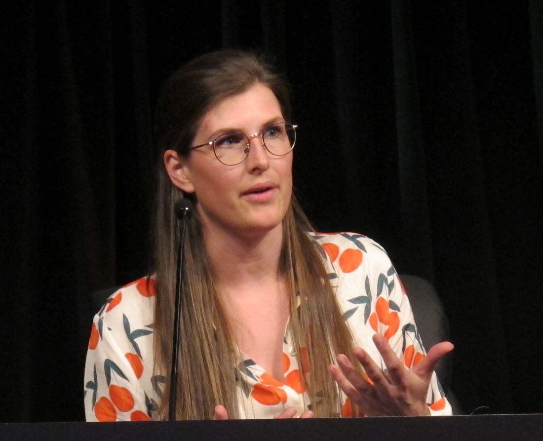
Of 384 women included to date in the ongoing trial, 51% carried a BRCA1 mutation and 49% carried a BRCA2 mutation; 72% chose RRS/RRO. A total of 289 completed the 3-month follow-up and 197 completed 1-year follow-up. At 3 and 12 months the decline on the Cancer Worry Scale was similar in the groups after adjusting for age and other baseline characteristics, with 1.9- and 1.4-point declines from baseline scores of 14.4 and 14.0 at 3 months, and 2.2- and 1.0-point declines at 12 months in the RRSO and RRS/RRO groups, respectively, Miranda P. Steenbeek, MD, reported at the Society of Gynecologic Oncology’s Annual Meeting on Women’s Cancer.
The mean scores on the 100-point Decision Regret Scale at 1-year follow-up were low at 13.4 and 13.0 after RRSO and RRS, respectively.
“But it is interesting that there is one group of women who underwent standard salpingo-oophorectomy, but weren’t using hormone replacement therapy [HRT], as these women had a higher level of decision regret [mean score, 18.8],” said Dr. Steenbeek of Radboud University Nijmegen, the Netherlands. “It is interesting to see how these results will develop over time.”
Current recommendations for preventive surgery in woman at increased risk because of BRCA1/2 mutations call for RRSO around age 40 years. However, an innovative approach using RRS followed by RRO has emerged as recent data indicate the fallopian tube, rather than the ovary, as the origin of high-grade serous ovarian cancer, she explained.
The TUBA trial was launched at 13 Dutch oncology centers in 2015, and plans to enroll 510 BRCA1/2 mutation carriers. Participants choose either standard RRSO within current guidelines (at age 35-40 years for BRCA1 carriers, age 40-45 years for BRCA2 carriers) or the innovative RRS approach followed by RRO at up to 5 years after the current guideline age (at age 40-45 years for BRCA1 carriers, age 45-50 years for BRCA2 carriers), she said.
These early findings show that baseline levels of cancer worry are higher among women choosing RRSO, which might explain the slightly larger postsurgery decline in worry in the RRSO group, Dr. Steenbeek said, noting that the higher level of regret with RRSO without HRT might be related to more severe menopausal complaints in that subset of patients.
“Based on these results we cannot conclude whether or not salpingectomy is a safe alternative to for salpingo-oophorectomy, and therefore it is very important to not perform salpingectomy with delayed oophorectomy outside the safety of a clinical and control of a clinical trial,” she said, adding that a report on the quality of life data from the study is expected in 2020.
Dr. Steenbeek reported having no disclosures.
SOURCE: Steenbeek MP et al. SGO 2019, Abstract LB2.
HONOLULU – Women with BRCA1/2 mutations who undergo risk-reducing salpingo-oophorectomy (RRSO) or risk-reducing salpingectomy followed by delayed risk-reducing oophorectomy (RRS/RRO) experience reduced cancer-related worry after surgery and low levels of decision regret, according to preliminary findings from the Dutch multicenter TUBA trial.
Of 384 women included to date in the ongoing trial, 51% carried a BRCA1 mutation and 49% carried a BRCA2 mutation; 72% chose RRS/RRO. A total of 289 completed the 3-month follow-up and 197 completed 1-year follow-up. At 3 and 12 months the decline on the Cancer Worry Scale was similar in the groups after adjusting for age and other baseline characteristics, with 1.9- and 1.4-point declines from baseline scores of 14.4 and 14.0 at 3 months, and 2.2- and 1.0-point declines at 12 months in the RRSO and RRS/RRO groups, respectively, Miranda P. Steenbeek, MD, reported at the Society of Gynecologic Oncology’s Annual Meeting on Women’s Cancer.
The mean scores on the 100-point Decision Regret Scale at 1-year follow-up were low at 13.4 and 13.0 after RRSO and RRS, respectively.
“But it is interesting that there is one group of women who underwent standard salpingo-oophorectomy, but weren’t using hormone replacement therapy [HRT], as these women had a higher level of decision regret [mean score, 18.8],” said Dr. Steenbeek of Radboud University Nijmegen, the Netherlands. “It is interesting to see how these results will develop over time.”
Current recommendations for preventive surgery in woman at increased risk because of BRCA1/2 mutations call for RRSO around age 40 years. However, an innovative approach using RRS followed by RRO has emerged as recent data indicate the fallopian tube, rather than the ovary, as the origin of high-grade serous ovarian cancer, she explained.
The TUBA trial was launched at 13 Dutch oncology centers in 2015, and plans to enroll 510 BRCA1/2 mutation carriers. Participants choose either standard RRSO within current guidelines (at age 35-40 years for BRCA1 carriers, age 40-45 years for BRCA2 carriers) or the innovative RRS approach followed by RRO at up to 5 years after the current guideline age (at age 40-45 years for BRCA1 carriers, age 45-50 years for BRCA2 carriers), she said.
These early findings show that baseline levels of cancer worry are higher among women choosing RRSO, which might explain the slightly larger postsurgery decline in worry in the RRSO group, Dr. Steenbeek said, noting that the higher level of regret with RRSO without HRT might be related to more severe menopausal complaints in that subset of patients.
“Based on these results we cannot conclude whether or not salpingectomy is a safe alternative to for salpingo-oophorectomy, and therefore it is very important to not perform salpingectomy with delayed oophorectomy outside the safety of a clinical and control of a clinical trial,” she said, adding that a report on the quality of life data from the study is expected in 2020.
Dr. Steenbeek reported having no disclosures.
SOURCE: Steenbeek MP et al. SGO 2019, Abstract LB2.
REPORTING FROM SGO 2019
Financial toxicity may be common in gynecologic cancer patients
HONOLULU – Financial toxicity may be common among gynecologic cancer patients starting a new line of treatment, based on the results of a survey presented at the Society of Gynecologic Oncology’s Annual Meeting on Women’s Cancer.
More than half of the 121 patients surveyed reported mild, moderate, or severe financial toxicity, Margaret Liang, MD, of University of Alabama at Birmingham, said in presenting the findings.
Younger age and lower income were both risk factors for financial toxicity, and having insurance did not protect patients from financial toxicity. Dr. Liang noted that insurance coverage “was not protective for financial toxicity, as the majority of those who screened positive for financial toxicity were insured.” Specifically, 89% of patients with financial toxicity and 98% of patients without it were insured (P = .07).
“This finding supports financial toxicity screening regardless of insurance status and may favor universal screening,” Dr. Liang said.
She and her colleagues conducted the survey of 121 gynecologic cancer patients who had started a new line of systemic therapy in the previous 8 weeks.
The patients’ mean age was 59 years (range, 33-80), and 58% were starting their first line of systemic therapy. Half of the patients had a household income below $40,000, and about one-third of the patients were employed. Most had private (74%) or public (20%) insurance, and 7% of patients were uninsured.
To assess financial toxicity, the researchers used the Comprehensive Score for Financial Toxicity (COST). A score of less than 26 was used as a threshold of financial toxicity. The severity of financial toxicity was graded on a scale of 1 to 4, with a lower score indicating worse toxicity.
Patients were most concerned about having enough money to cover the cost of treatment. Patients reported the lowest mean score—0.97—in response to the statement, “I know that I have enough money in savings, retirement, or assets to cover the costs of my treatment.”
Patients were least concerned about losing their job or income. They reported the highest mean score—3.29—in response to the statement, “I am concerned about keeping my job and income, including working at home.” However, as Dr. Liang pointed out, only one-third of patients were employed at baseline.
In all, 54% of patients reported financial toxicity—37% mild, 16% moderate, and 1% severe.
Dr. Liang and her colleagues found that younger age and lower income were associated with an increased risk for financial toxicity. The mean age was 57 years in patients with financial toxicity and 62 years in patients without it (P = .02). Household incomes were below $40,000 in 63% of patients with financial toxicity and 34% of those without it (P less than .01).
The researchers also found that patients with financial toxicity were significantly more likely to say their cancer diagnosis resulted in lost wages, borrowed money, altered spending habits, the need to sacrifice other things, and not paying bills on time (P less than .01 for all).
On the other hand, patients with financial toxicity were not significantly more likely to become unemployed, file for bankruptcy, sell their house, or get a second job due to their cancer diagnosis.
However, it’s important to note that these data were collected within 8 weeks of patients starting their new line of therapy. The final survey results will include follow-up at 3 months and 6 months.
Dr. Liang said the fact that more than half of patients reported financial toxicity within 8 weeks of starting a new line of therapy suggests early interventions are needed to prevent or reduce financial toxicity. This may include counseling patients on anticipated costs of care, screening for financial toxicity, and linking patients to available financial resources.
Dr. Liang had no financial disclosures.
jensmith@mdedge.com
SOURCE: Liang M et al. SGO 2019. Abstract 8.
REPORTING FROM SGO 2019

Tumor testing cost-effective for triage to germline testing in HGSOC patients
HONOLULU – Tumor testing for the triage of women with high-grade serous ovarian cancer to confirmatory genetic testing for BRCA mutations appears feasible, according to a cost-effectiveness analysis.
In fact, based on a Markov Monte Carlo simulation model developed to compare a tumor-testing approach with a universal germline testing approach, tumor testing yields an incremental cost-effectiveness ratio (ICER) of $127,000 per year of life gained, Janice S. Kwon, MD, reported at the Society of Gynecologic Oncology’s Annual Meeting on Women’s Cancer.

“This is well in excess of [the $50,000 to $100,000 that] would be considered an acceptable threshold in the United States,” said Dr. Kwon of the University of British Columbia, Vancouver, Canada.
“We predict that tumor testing will be a cost-effective method of triage in women with high-grade serous ovarian cancer for confirmatory genetic testing to identify BRCA mutation carriers, assuming high sensitivity and acceptable cost of tumor testing,” she said.
In many areas around the world, germline testing is recommended for all women with high-grade serous ovarian cancer (HGSOC) because they have a 20% chance of carrying a BRCA 1 or 2 mutation.
“However, we all know that the referral rate for genetic testing is far from optimal, and furthermore, there are costs incurred to the healthcare system for resources utilized for genetic counseling and testing,” she said.
Tumor testing for triage is an alternative approach.
“If you consider 100 women with high-grade serous ovarian cancer and follow them through the conventional pathway in which all of them are referred for germline testing, you would expect to find 20 mutation carriers. If you take those same 100 women and apply tumor testing first, then 25 are expected to have a mutation in the tumor, Dr. Kwon said.
“If [all 25] are referred for germline testing, you would expect to find the same number of BRCA mutation carriers but with far less resource utilization.”
The remaining 75 are not expected to have a mutation in the tumor, and they may not need to be referred for confirmatory genetic testing unless there is a compelling family history or panel testing reveals a concerning mutation, she explained.
Since a randomized trial to compare these two strategies is not feasible, Dr. Kwon and her colleagues performed the current cost-effectiveness analysis.
The Markov simulation model was used to estimate the number of BRCA mutation carriers from index cases and their first degree relatives, and the number of cancer cases averted among first degree relatives, assuming they would undergo risk-reducing surgery.
“We conducted extensive sensitivity analyses to account for uncertainly around various parameters and we modeled a time horizon of 50 years,” Dr. Kwon noted. “We know that there are approximately 10,000 new [HGSOC] cases diagnosed in the United States every year, and we assumed that for every woman with [HGSOC], there was at least 1 female first-degree relative who would benefit from genetic testing.”
The model showed that applying tumor testing first would lead to a substantial reduction in the number of women undergoing germline mutation testing, but the number of BRCA mutation carriers identified would be comparable with the two strategies – assuming that the sensitivity of tumor testing is less than 100%, she said.
“As expected, the average lifetime costs associated with germline testing would be less than that for tumor testing, and even though you would expect that more first-degree relatives would be identified as BRCA mutation carriers after universal germline testing for index cases, the life expectancy gain for those first-degree relatives is averaged over the entire cohort at risk, and therefore the average incremental gain or benefit was actually quite small,” she said, noting that this yielded the ICER of $127,000 per year of life gained.
Based on this finding, tumor testing would be the preferred strategy, she added.
Sensitivity analysis around the sensitivity and specificity of tumor testing showed that tumor testing would be cost effective if its sensitivity is above 97%, and that tumor testing is cost-effective as long as it costs less than a third of the cost of germline testing – including genetic counseling.
Dr. Kwon has received research funding from AstraZeneca.
sworcester@mdedge.com
SOURCE: Kwon J et al., SGO 2019: Abstract 5.
HONOLULU – Tumor testing for the triage of women with high-grade serous ovarian cancer to confirmatory genetic testing for BRCA mutations appears feasible, according to a cost-effectiveness analysis.
In fact, based on a Markov Monte Carlo simulation model developed to compare a tumor-testing approach with a universal germline testing approach, tumor testing yields an incremental cost-effectiveness ratio (ICER) of $127,000 per year of life gained, Janice S. Kwon, MD, reported at the Society of Gynecologic Oncology’s Annual Meeting on Women’s Cancer.
“This is well in excess of [the $50,000 to $100,000 that] would be considered an acceptable threshold in the United States,” said Dr. Kwon of the University of British Columbia, Vancouver, Canada.
“We predict that tumor testing will be a cost-effective method of triage in women with high-grade serous ovarian cancer for confirmatory genetic testing to identify BRCA mutation carriers, assuming high sensitivity and acceptable cost of tumor testing,” she said.
In many areas around the world, germline testing is recommended for all women with high-grade serous ovarian cancer (HGSOC) because they have a 20% chance of carrying a BRCA 1 or 2 mutation.
“However, we all know that the referral rate for genetic testing is far from optimal, and furthermore, there are costs incurred to the healthcare system for resources utilized for genetic counseling and testing,” she said.
Tumor testing for triage is an alternative approach.
“If you consider 100 women with high-grade serous ovarian cancer and follow them through the conventional pathway in which all of them are referred for germline testing, you would expect to find 20 mutation carriers. If you take those same 100 women and apply tumor testing first, then 25 are expected to have a mutation in the tumor, Dr. Kwon said.
“If [all 25] are referred for germline testing, you would expect to find the same number of BRCA mutation carriers but with far less resource utilization.”
The remaining 75 are not expected to have a mutation in the tumor, and they may not need to be referred for confirmatory genetic testing unless there is a compelling family history or panel testing reveals a concerning mutation, she explained.
Since a randomized trial to compare these two strategies is not feasible, Dr. Kwon and her colleagues performed the current cost-effectiveness analysis.
The Markov simulation model was used to estimate the number of BRCA mutation carriers from index cases and their first degree relatives, and the number of cancer cases averted among first degree relatives, assuming they would undergo risk-reducing surgery.
“We conducted extensive sensitivity analyses to account for uncertainly around various parameters and we modeled a time horizon of 50 years,” Dr. Kwon noted. “We know that there are approximately 10,000 new [HGSOC] cases diagnosed in the United States every year, and we assumed that for every woman with [HGSOC], there was at least 1 female first-degree relative who would benefit from genetic testing.”
The model showed that applying tumor testing first would lead to a substantial reduction in the number of women undergoing germline mutation testing, but the number of BRCA mutation carriers identified would be comparable with the two strategies – assuming that the sensitivity of tumor testing is less than 100%, she said.
“As expected, the average lifetime costs associated with germline testing would be less than that for tumor testing, and even though you would expect that more first-degree relatives would be identified as BRCA mutation carriers after universal germline testing for index cases, the life expectancy gain for those first-degree relatives is averaged over the entire cohort at risk, and therefore the average incremental gain or benefit was actually quite small,” she said, noting that this yielded the ICER of $127,000 per year of life gained.
Based on this finding, tumor testing would be the preferred strategy, she added.
Sensitivity analysis around the sensitivity and specificity of tumor testing showed that tumor testing would be cost effective if its sensitivity is above 97%, and that tumor testing is cost-effective as long as it costs less than a third of the cost of germline testing – including genetic counseling.
Dr. Kwon has received research funding from AstraZeneca.
sworcester@mdedge.com
SOURCE: Kwon J et al., SGO 2019: Abstract 5.
REPORTING FROM SGO 2019
ENGOT-OV16/NOVA trial: Analysis shows improved TWiST with niraparib
HONOLULU – Patients with recurrent ovarian cancer who were treated with niraparib maintenance therapy experienced more time without symptoms or toxicities (TWiST) than did controls, according to an analysis of data from the pivotal phase 3 ENGOT-OV16/NOVA trial.
The mean 20-year TWiST benefit – defined in the current analysis as progression-free survival (PFS) without toxicity due to grade 2 or greater nausea, vomiting, or fatigue – was fourfold greater among women who were carriers of the 138 germline BRCA mutation (gBRCAmut) and received treatment with the poly(adenosine diphosphate-ribose) polymerase (PARP) 1/2 inhibitor than among the 65 similar women who received placebo. The TWiST benefit was about twofold greater among the 234 non-gBRCAmut carriers treated with niraparib than among the 116 who received placebo, Ursula A. Matulonis, MD, reported at the Society of Gynecologic Oncology’s Annual Meeting on Women’s Cancer.
The 10- and 5-year estimated mean improvement in TWiST in the groups also showed “very proportional effects that are similar to the 20-year data,” said Dr. Matulonis, chief of the division of gynecologic oncology at Dana-Farber Cancer Institute, and a professor of medicine at Harvard Medical School, Boston.

TWiSt is an “established methodology that partitions PFS ... based on time with toxicity and time without progression or toxicity." Mean TWiST is calculated as mean PFS minus mean toxicity, she explained.
In the ENGOT-OV16/NOVA study, women with recurrent ovarian cancer who received niraparib for maintenance after platinum-based therapy had significantly longer PFS, compared with patients who received placebo (21.0 vs. 5.5 months in gBRCAmut patients and 9.3 vs. 3.9 months in the non-gBRCAmut patients, respectively), she said (N Engl J Med 2016;75:2154-2164).
Quality of life (QOL) remained stable throughout niraparib treatment, according to another analysis of data from the trial published in 2018. (Lancet Oncology. Aug 1 2018;19[8]:1117-25).
The current analysis looked more closely at QOL by estimating mean TWiST in the treatment vs. control groups, she explained.
“Survival curves were used to extrapolate PFS ... over 20 years for both the gBRCAmut and non-gBRCAmut cohorts. The 20-year period was based on ovarian cancer clinical expert opinion and the biologic plausibility that patients could be on PARP inhibitors for a long period of time,” she said.
Time of toxicity was estimated based on the number of days patients experienced toxic effects due to grade 2 or higher nausea, vomiting, or fatigue during the NOVA trial.
The PFS benefit in treated vs. control subjects, respectively, was 3.23 years and 1.33 years in the gBRCAmut and non-gBRCAmut cohorts, and the mean toxicity time was 0.28 years and 0.10 years, for a mean TWiST benefit of 2.95 and 1.34 years, respectively.
“This TWiST benefit means that patients treated with niraparib experienced more progression-free time without symptoms or toxicities due to nausea, vomiting, or fatigue, compared with patients receiving placebo,” she concluded.
Dr. Matulonis is a consultant for 2X Oncology, Merck, Mersana, Fujifilm, Immunogen, and Geneos.
sworcester@mdedge.com
SOURCE: Matulonis U et al., SGO 2019: Abstract 1.
HONOLULU – Patients with recurrent ovarian cancer who were treated with niraparib maintenance therapy experienced more time without symptoms or toxicities (TWiST) than did controls, according to an analysis of data from the pivotal phase 3 ENGOT-OV16/NOVA trial.
The mean 20-year TWiST benefit – defined in the current analysis as progression-free survival (PFS) without toxicity due to grade 2 or greater nausea, vomiting, or fatigue – was fourfold greater among women who were carriers of the 138 germline BRCA mutation (gBRCAmut) and received treatment with the poly(adenosine diphosphate-ribose) polymerase (PARP) 1/2 inhibitor than among the 65 similar women who received placebo. The TWiST benefit was about twofold greater among the 234 non-gBRCAmut carriers treated with niraparib than among the 116 who received placebo, Ursula A. Matulonis, MD, reported at the Society of Gynecologic Oncology’s Annual Meeting on Women’s Cancer.
The 10- and 5-year estimated mean improvement in TWiST in the groups also showed “very proportional effects that are similar to the 20-year data,” said Dr. Matulonis, chief of the division of gynecologic oncology at Dana-Farber Cancer Institute, and a professor of medicine at Harvard Medical School, Boston.
TWiSt is an “established methodology that partitions PFS ... based on time with toxicity and time without progression or toxicity." Mean TWiST is calculated as mean PFS minus mean toxicity, she explained.
In the ENGOT-OV16/NOVA study, women with recurrent ovarian cancer who received niraparib for maintenance after platinum-based therapy had significantly longer PFS, compared with patients who received placebo (21.0 vs. 5.5 months in gBRCAmut patients and 9.3 vs. 3.9 months in the non-gBRCAmut patients, respectively), she said (N Engl J Med 2016;75:2154-2164).
Quality of life (QOL) remained stable throughout niraparib treatment, according to another analysis of data from the trial published in 2018. (Lancet Oncology. Aug 1 2018;19[8]:1117-25).
The current analysis looked more closely at QOL by estimating mean TWiST in the treatment vs. control groups, she explained.
“Survival curves were used to extrapolate PFS ... over 20 years for both the gBRCAmut and non-gBRCAmut cohorts. The 20-year period was based on ovarian cancer clinical expert opinion and the biologic plausibility that patients could be on PARP inhibitors for a long period of time,” she said.
Time of toxicity was estimated based on the number of days patients experienced toxic effects due to grade 2 or higher nausea, vomiting, or fatigue during the NOVA trial.
The PFS benefit in treated vs. control subjects, respectively, was 3.23 years and 1.33 years in the gBRCAmut and non-gBRCAmut cohorts, and the mean toxicity time was 0.28 years and 0.10 years, for a mean TWiST benefit of 2.95 and 1.34 years, respectively.
“This TWiST benefit means that patients treated with niraparib experienced more progression-free time without symptoms or toxicities due to nausea, vomiting, or fatigue, compared with patients receiving placebo,” she concluded.
Dr. Matulonis is a consultant for 2X Oncology, Merck, Mersana, Fujifilm, Immunogen, and Geneos.
sworcester@mdedge.com
SOURCE: Matulonis U et al., SGO 2019: Abstract 1.
REPORTING FROM SGO 2019
Financial assistance programs may speed treatment for cervical cancer
HONOLULU – Financial assistance programs may help lower-income cervical cancer patients complete treatment in a timely manner, according to research presented at the Society of Gynecologic Oncology’s Annual Meeting on Women’s Cancer.
In a retrospective study, lower-income patients who used financial assistance programs were able to complete treatment for cervical cancer in a similar timeframe as higher-income patients.
Lower-income patients who registered for disability benefits saw a significant improvement in time to treatment completion.
Additionally, there was a trend toward improved time to treatment completion among lower-income patients who registered for federally funded breast and cervical cancer treatment.
“Identification of patients who qualify for disability and/or breast/cervical cancer Medicaid, and providing assistance for registration in these programs may help them complete therapy in a more appropriate timeframe,” said study investigator Jessica Gillen, MD, of The University of Oklahoma in Oklahoma City.

Dr. Gillen and her colleagues conducted this single-center, retrospective study of patients with squamous cell, adenocarcinoma, or adenosquamous cancer of the cervix.
The investigators identified 116 evaluable patients who received chemoradiation from January 1, 2015, to July 31, 2018. Most of these 106 patients completed treatment in 63 days or less.
The patients’ median household income was $45,782 (range, $19,771–$96,222). The investigators defined “high-income” patients as those whose household incomes were at or above the median, and “low-income” patients as those with incomes below the median.
On average, the patients used 1.24 assistance programs, which included financial assistance (primarily help with clinic visit copays), assistance with medication costs, disability benefits, Medicaid, access to emergency funds, low-cost or free lodging, and transportation.
Dr. Gillen noted that 10% of low-income patients did not use any financial assistance programs.
She and her colleagues found that low-income patients who used assistance programs completed treatment in a similar timeframe as the high-income patients. The median time to treatment completion was 56.5 days and 50 days, respectively.
Registering for disability benefits was significantly associated with improved time to treatment completion (P less than .001).
There were no other significant associations between financial assistance programs and time to treatment completion. However, there was a trend toward improved time to treatment completion among patients who registered for federally funded breast and cervical cancer Medicaid (P = .06).
“[I]t was encouraging to see that enrollment in federally and state-funded programs makes a difference in patients’ ability to complete treatment,” Dr. Gillen said.
She and her colleagues said these data suggest financial assistance programs may help cervical cancer patients overcome barriers to care. Dr. Gillen had no financial disclosures.
SOURCE: Gillen J et al. SGO 2019. Abstract 9.
HONOLULU – Financial assistance programs may help lower-income cervical cancer patients complete treatment in a timely manner, according to research presented at the Society of Gynecologic Oncology’s Annual Meeting on Women’s Cancer.
In a retrospective study, lower-income patients who used financial assistance programs were able to complete treatment for cervical cancer in a similar timeframe as higher-income patients.
Lower-income patients who registered for disability benefits saw a significant improvement in time to treatment completion.
Additionally, there was a trend toward improved time to treatment completion among lower-income patients who registered for federally funded breast and cervical cancer treatment.
“Identification of patients who qualify for disability and/or breast/cervical cancer Medicaid, and providing assistance for registration in these programs may help them complete therapy in a more appropriate timeframe,” said study investigator Jessica Gillen, MD, of The University of Oklahoma in Oklahoma City.
Dr. Gillen and her colleagues conducted this single-center, retrospective study of patients with squamous cell, adenocarcinoma, or adenosquamous cancer of the cervix.
The investigators identified 116 evaluable patients who received chemoradiation from January 1, 2015, to July 31, 2018. Most of these 106 patients completed treatment in 63 days or less.
The patients’ median household income was $45,782 (range, $19,771–$96,222). The investigators defined “high-income” patients as those whose household incomes were at or above the median, and “low-income” patients as those with incomes below the median.
On average, the patients used 1.24 assistance programs, which included financial assistance (primarily help with clinic visit copays), assistance with medication costs, disability benefits, Medicaid, access to emergency funds, low-cost or free lodging, and transportation.
Dr. Gillen noted that 10% of low-income patients did not use any financial assistance programs.
She and her colleagues found that low-income patients who used assistance programs completed treatment in a similar timeframe as the high-income patients. The median time to treatment completion was 56.5 days and 50 days, respectively.
Registering for disability benefits was significantly associated with improved time to treatment completion (P less than .001).
There were no other significant associations between financial assistance programs and time to treatment completion. However, there was a trend toward improved time to treatment completion among patients who registered for federally funded breast and cervical cancer Medicaid (P = .06).
“[I]t was encouraging to see that enrollment in federally and state-funded programs makes a difference in patients’ ability to complete treatment,” Dr. Gillen said.
She and her colleagues said these data suggest financial assistance programs may help cervical cancer patients overcome barriers to care. Dr. Gillen had no financial disclosures.
SOURCE: Gillen J et al. SGO 2019. Abstract 9.
REPORTING FROM SGO 2019

Brachytherapy proves beneficial regardless of treatment duration
, according to a retrospective study.
Researchers found that patients who received brachytherapy in addition to chemotherapy and external beam radiation therapy had better overall survival than patients who received chemoradiation alone.

Although the best overall survival was observed in patients who received brachytherapy within the recommended 8 weeks, patients who received brachytherapy outside that timeframe also had better overall survival than patients treated with chemoradiation alone.
Travis-Riley K. Korenaga, MD, of University of California, San Francisco, presented these findings at the Society of Gynecologic Oncology’s Annual Meeting on Women’s Cancer.
To examine the use of brachytherapy, Dr. Korenaga and his colleagues analyzed patients from the U.S. National Cancer Database who had stage II-IVA cervical cancer and were diagnosed between 2004 and 2015.
The researchers identified 18,592 patients who received at least 4,500 cGy of external beam radiation therapy and concurrent chemotherapy as their primary treatment. In this group, there were 17,150 patients who had data on brachytherapy use and time to treatment completion.
A majority of patients (n = 13,642) received brachytherapy, and roughly half of those (n = 6,871) received it within the recommended 8 weeks.
“This is a pretty low rate of adherence to standard of care; 36.9% of women receive brachytherapy and complete it within that 8-week timeframe,” Dr. Korenaga said. “And that 36.9% of women do have a superior overall survival that blows everything else out of the water.”
The median overall survival was:
- 113.7 months (95% confidence interval [CI], 103.3-121.3) in patients who received brachytherapy within 8 weeks
- 75.7 months (95% CI, 69.7-82.4) in those who received brachytherapy for more than 8 weeks
- 58.5 months (95% CI, 48.3-74.2) in patients who received only chemoradiation within 8 weeks
- 46.2 months (95% CI, 39.8-56.4) in those who received only chemoradiation for more than 8 weeks.
“Getting some type of brachytherapy, no matter whether it’s within 8 weeks or beyond 8 weeks, is still associated with an improved overall survival,” Dr. Korenaga noted.
He and his colleagues also identified factors that were significantly associated with a reduced likelihood of receiving brachytherapy within 8 weeks, including:
- Having stage III/IVA disease vs. stage II disease (P less than .0001)
- Being non-Hispanic black vs. non-Hispanic white (P less than .001)
- Having an annual income below $38,000 vs. $63,000 or higher (P less than .0001)
- Having public vs. private insurance (P less than .0001)
- Living 10 to 60 miles (P = .01) or more than 100 miles (P less than .001) from the treatment facility vs. less than 10 miles
- Being treated at a facility with a community cancer program (P = .02) or a comprehensive community cancer program (P less than .0001) vs. an academic research program.
Dr. Korenaga said these results highlight the fact that more work needs to be done to increase the use of brachytherapy in patients with locally advanced cervical cancer.
He and his colleagues have suggested a few measures that might help, including early referrals for brachytherapy, connecting patients with care navigators, and developing centers of excellence for brachytherapy.
Dr. Korenaga had no relevant financial disclosures.
SOURCE: Korenaga TRK et al. SGO 2019. Abstract 10.
, according to a retrospective study.
Researchers found that patients who received brachytherapy in addition to chemotherapy and external beam radiation therapy had better overall survival than patients who received chemoradiation alone.
Although the best overall survival was observed in patients who received brachytherapy within the recommended 8 weeks, patients who received brachytherapy outside that timeframe also had better overall survival than patients treated with chemoradiation alone.
Travis-Riley K. Korenaga, MD, of University of California, San Francisco, presented these findings at the Society of Gynecologic Oncology’s Annual Meeting on Women’s Cancer.
To examine the use of brachytherapy, Dr. Korenaga and his colleagues analyzed patients from the U.S. National Cancer Database who had stage II-IVA cervical cancer and were diagnosed between 2004 and 2015.
The researchers identified 18,592 patients who received at least 4,500 cGy of external beam radiation therapy and concurrent chemotherapy as their primary treatment. In this group, there were 17,150 patients who had data on brachytherapy use and time to treatment completion.
A majority of patients (n = 13,642) received brachytherapy, and roughly half of those (n = 6,871) received it within the recommended 8 weeks.
“This is a pretty low rate of adherence to standard of care; 36.9% of women receive brachytherapy and complete it within that 8-week timeframe,” Dr. Korenaga said. “And that 36.9% of women do have a superior overall survival that blows everything else out of the water.”
The median overall survival was:
- 113.7 months (95% confidence interval [CI], 103.3-121.3) in patients who received brachytherapy within 8 weeks
- 75.7 months (95% CI, 69.7-82.4) in those who received brachytherapy for more than 8 weeks
- 58.5 months (95% CI, 48.3-74.2) in patients who received only chemoradiation within 8 weeks
- 46.2 months (95% CI, 39.8-56.4) in those who received only chemoradiation for more than 8 weeks.
“Getting some type of brachytherapy, no matter whether it’s within 8 weeks or beyond 8 weeks, is still associated with an improved overall survival,” Dr. Korenaga noted.
He and his colleagues also identified factors that were significantly associated with a reduced likelihood of receiving brachytherapy within 8 weeks, including:
- Having stage III/IVA disease vs. stage II disease (P less than .0001)
- Being non-Hispanic black vs. non-Hispanic white (P less than .001)
- Having an annual income below $38,000 vs. $63,000 or higher (P less than .0001)
- Having public vs. private insurance (P less than .0001)
- Living 10 to 60 miles (P = .01) or more than 100 miles (P less than .001) from the treatment facility vs. less than 10 miles
- Being treated at a facility with a community cancer program (P = .02) or a comprehensive community cancer program (P less than .0001) vs. an academic research program.
Dr. Korenaga said these results highlight the fact that more work needs to be done to increase the use of brachytherapy in patients with locally advanced cervical cancer.
He and his colleagues have suggested a few measures that might help, including early referrals for brachytherapy, connecting patients with care navigators, and developing centers of excellence for brachytherapy.
Dr. Korenaga had no relevant financial disclosures.
SOURCE: Korenaga TRK et al. SGO 2019. Abstract 10.
REPORTING FROM SGO 2019
