User login
Judicious EEG use identifies pseudosyncope during tilt-table testing
NEW ORLEANS – , according to an investigation from Vanderbilt University, Nashville.
Pseudosyncope, also known as nonsyncopal fainting (NSF), is a conversion disorder where people appear to faint, but don’t lose consciousness. It’s generally thought to be a physical manifestation of traumatic stress. Patients “aren’t faking it; they believe they are fainting,” said senior investigator Italo Biaggioni, MD, a professor of medicine and pharmacology at the university and director of the Vanderbilt Autonomic Dysfunction Center.
It’s “not benign. These patients stop working, stop driving, and need to depend on other people. They can have very dramatic episodes and hurt themselves” when they fall. “They are very disabled,” he said.
NSF is often misdiagnosed and mistreated, and sometimes unrecognized for years. Almost a third of the 39 NSF cases in Dr. Biaggioni’s series, for instance, were on anticonvulsants, and several had undergone cardiac catheterization. Often, NSF is treated as vasovagal syncope, but patients don’t respond to medications. A better way to identify it is needed. “By the time we get them, they’ve been through a lot.” Dr. Biaggioni said at the joint scientific sessions of the American Heart Association Council on Hypertension, AHA Council on Kidney in Cardiovascular Disease, and American Society of Hypertension.
He and his team had a hunch that judicious use of EEG would help, so they limited EEG to suspected NSF cases in a series of 107 refractory syncope patients referred to Vanderbilt for head-up tilt-table testing; 39 (36%) had normal EEGs during an apparent loss of consciousness, as opposed to the slow-wave pattern of true loss of consciousness, and were diagnosed with NSF.
The 36% identified was a marked increase in incidence over more common approaches. Among 64 patients who had tilt-table testing without EEG at Vanderbilt, for instance, three (5%) were diagnosed with NSF. Historically, tilt table plus EEG in all comers has a diagnostic yield of around 18% for the condition. In short, “elective EEG monitoring during tilt-table testing” better “distinguishes between syncope” and NSF, the team concluded.
NSF is suggested by a history of more than 20 episodes of apparent fainting; episodes once a week or more; or losing consciousness for more than 5 minutes. Fainting with eyes closed, or while supine, is also suggestive.
The elective approach prevents inappropriate treatment but is also therapeutic in itself. “When we document with EEG that patients are not really fainting, and explain that to them, it automatically reduces the number of episodes,” Dr. Biaggioni said.
It also saves NSF patients from a nitroglycerin challenge and repeat tilt testing, which is the default in many places when the first round of testing doesn’t trigger an episode. Challenge testing provokes vasovagal syncope in around 10% of even healthy people, so it puts NSF patients at risk for a false positive. As a rule, “we don’t use provocative agents when we [suspect NSF],” he said.
In addition to the 39 NSF cases, 11 patients in the series were diagnosed with vasovagal syncope, and testing didn’t provoke an event in 57 (53%), which isn’t uncommon.
Baseline blood pressure and heart rates were similar across the three groups, and there were more women than men in each. Subjects were in their early 40s, on average.
NSF patients were more likely to be taking anxiety and depression medications. One NSF patient had posttraumatic stress disorder, and two had sexual abuse histories, compared with none in the nondiagnostic and vasovagal groups. The NSF group had a shorter time to an event: 9 minutes versus 19 minutes among vasovagal patients.
Tilt-table testing was done after 6 hours of fasting, and the team used standard 22-channel EEG. Cognitive-behavioral therapy is the go-to treatment for NSF, Dr. Biaggioni said.
There was no industry funding, and the authors didn’t have any disclosures.
SOURCE: Muldowney JA et al. Joint Hypertension 2019, Abstract P3061.
REPORTING FROM JOINT HYPERTENSION 2019
Hyponatremia almost as common with spironolactone as chlorthalidone
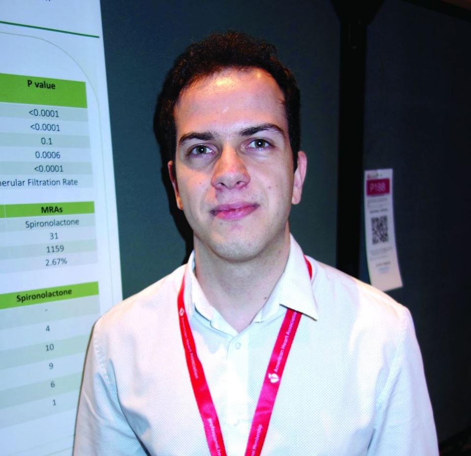
NEW ORLEANS – .
The investigators reviewed hypertension patients whose treatment regimens included one diuretic. Forty on chlorthalidone developed hyponatremia – defined as a serum sodium below 133 mEq/L – across 1,322 prescriptions, for an incidence of 3.03%. There were 31 cases across 1,159 spironolactone prescriptions, an incidence of 2.67%.
Among 14 patients in a substudy who discontinued chlorthalidone after developing hyponatremia at a mean of about 2 weeks, six (43%) subsequently developed hyponatremia on spironolactone, also at an average of about 2 weeks.
The findings suggest that spironolactone is more likely than generally thought to cause hyponatremia, a potentially severe complication of diuretics, and that hyponatremia on chlorthalidone increases the risk, said lead investigator Faris Matanes, MD, a hypertension researcher at the university.
“We used to think” that hyponatremia on spironolactone was “very unlikely, but actually it’s not; the incidence is really close to chlorthalidone,” a well-known cause, and “if a patient develops hyponatremia on chlorthalidone, we should be more careful about giving them spironolactone,” he said.
Almost half the spironolactone cases were on 25 mg/day or less, and over a quarter of the chlorthalidone cases were on 12.5 mg/day. Of the 154 hyponatremia cases across 10,660 hydrochlorothiazide prescriptions (1.44%), over a third were taking 12.5 mg/day or less.
Overall, hyponatremia was diagnosed at a mean of 40.4 days, but sometimes after 2 or more months of treatment.
The findings “mean that even if we start patients on a low dose, we can’t stop checking after one or two normal sodium levels.” Measurements need to be ongoing, Dr. Matanes said at the joint scientific sessions of the American Heart Association Council on Hypertension, AHA Council on Kidney in Cardiovascular Disease, and American Society of Hypertension.
He and his team wanted to get around the limitations of previous diuretic hyponatremia studies, including use of more than one diuretic, markedly poor kidney function, and other confounders. To that end, the study was limited to outpatients on a single diuretic who had normal sodium levels both before and after their hyponatremic episode, and estimated glomerular filtration rates (eGFR) of at least 30 mL/min/1.73 m2. Exclusion criteria included heart failure, cirrhosis, and adrenal insufficiency.
Older white people with lower baseline sodium and eGFR values were most at risk. Contrary to previous reports, hyponatremia wasn’t more likely in men.
The mean sodium level during an episode was 130.2 mEq/L; the majority of patients eventually normalized and continued treatment.
Subjects in the main study were a mean of 66 years old, about two-thirds were white, and about 60% were women. The baseline eGFR was 77.2 mL/min/1.73 m2, and baseline sodium level 135.8 mEq/L.
All but one of the 14 substudy patients were women. Those who became hyponatremic when switched to spironolactone were older (mean 74.2 versus 65.8 years), had lower baseline eGFRs (63.7 versus 69.7 mL/min/1.73 m2), and were more likely to be white, but the differences were not statistically significant.
There was no external funding, and the investigators didn’t have any industry disclosures.
SOURCE: Matanes F et al. Joint Hypertension 2019, Abstracts 187 and 174.
REPORTING FROM JOINT HYPERTENSION 2019
ABPM rarely used for hypertension management in United States
NEW ORLEANS – , according to a University of Florida, Gainesville, analysis of claims data for almost 4 million people.
“With each iteration, evidence-based guidelines have more strongly recommended out-of-office blood pressure measurement, but it’s basically had no impact. If we are going to continue to recommend this aggressively, we need to put some pressure on both payers and providers,” said lead investigator Steven M. Smith, PharmD, of the department of pharmacotherapy & translational research, associate director of the Center for Integrative Cardiovascular and Metabolic Diseases at the university.
“A number of studies show that ambulatory blood pressure monitoring [ABPM] is more strongly predictive of outcomes than office pressure.” It’s “considered the gold standard for hypertension,” he said at the joint scientific sessions of the American Heart Association Council on Hypertension, AHA Council on Kidney in Cardiovascular Disease, and American Society of Hypertension
The reason is that ABPM gives a continual reading of blood pressure over 24 hours, not just an office snapshot, and can do things that office measurements cannot, including ruling out white coat hypertension, identifying masked hypertension, and checking nocturnal dipping and morning surge, both of which are related to cardiovascular risk.
Although common in Canada and Europe, it’s no secret that ABPM hasn’t caught on in the United States. The goal of Dr. Smith’s work was to help quantify the situation.
Using Truven Health Analytics commercial insurance claims data, he and his team identified 3,378,645 adults starting their first hypertension medication and 335,200 starting their fourth from 2008 to 2017. They looked for ABPM claims in the previous 6 months as well as the month after patients started their new medication. The idea was to assess ABPM use in both new and resistant hypertensive patients.
ABPM claims were submitted for 0.15% of patients starting their first drug in 2008, rising to 0.3% in 2017. ABPM was used mostly before treatment initiation.
ABPM use actually declined among resistant patients from about 0.27% in 2008 to about 0.12% in 2017. Use was split about evenly before and after they started their fourth medication.
About 80% of claims – generally for interpreting ABPM results, not the upfront cost of the machine – were paid. Claims submitted tended to come from more high-end plans. Reimbursement rates were similar for more bargain plans, but there were many fewer claims submitted, Dr. Smith said.
He thought plans would at least follow Medicare’s reimbursement policy, which, at the time of the study, covered ABPM to rule out white coat hypertension, “but they didn’t seem to,” he said. Medicare recently added coverage for suspected masked hypertension.
The study doesn’t address why uptake is so low in the United States, but outside the world of hypertension specialists, “physicians don’t see a value in it. They don’t recognize what they would get from ABPM and how that would change what they do,” in part because treatment is currently based on office measurements. There’s also probably uncertainty about how to interpret the results, Dr. Smith said.
Standardization across payers about what they’ll cover and for whom would probably help, he added.
Findings in the study were similar for home blood pressure monitoring, but probably not an accurate gauge of use. Patients mostly buy their own devices and report the results to their physician, without getting insurance involved, he said.
There was no industry funding, and the investigators didn’t have any disclosures.
SOURCE: Smith SM et al. Joint Hypertension 2019, Abstract P2067.
REPORTING FROM JOINT HYPERTENSION 2019
Remember that preeclampsia has a ‘fourth trimester’
NEW ORLEANS – according to Natalie Bello, MD, a cardiologist and assistant professor of medicine at Columbia University in New York.
In medical school, “they told me that you deliver the placenta, and the preeclampsia goes away. Not the case. Postpartum preeclampsia is a real thing. We are seeing a lot of it at our sites, which have a lot of underserved women who hadn’t had great prenatal care” elsewhere, she said.
Headache and visual changes in association with hypertension during what’s been dubbed “the fourth trimester” raise suspicions. Women can progress rapidly to eclampsia and HELLP syndrome (hemolysis, elevated liver enzymes, low platelet count), but sometimes providers don’t recognize what’s going on because they don’t know women have recently given birth. “We can do better; we should be doing better. Please always ask women if they’ve delivered recently,” Dr. Bello said at the joint scientific sessions of the American Heart Association Council on Hypertension, AHA Council on Kidney in Cardiovascular Disease, and American Society of Hypertension.
Hypertension should resolve within 6 weeks of delivery, and blood pressure should be back to baseline by 3 months. To make sure that happens, BP should be checked within a few days of birth and regularly thereafter. It can be tough to get busy and tired new moms back into the office, but “they’ll do whatever it takes” to get their baby to a pediatric appointment, so maybe having pediatricians involved in checking blood pressure would help, she said.
The cutoff point for hypertension in pregnancy is 140/90 mm Hg, and it’s considered severe when values hit 160/110 mm Hg or higher. Evidence is strong for treating severe hypertension to reduce strokes, placental abruptions, and other problems, but the data for treating nonsevere hypertension are less clear, Dr. Bello explained.
The Chronic Hypertension and Pregnancy (CHAP) trial is expected to fill the evidence gap in a few years; women are being randomized to start treatment at either 140/90 mm Hg or 160/105 mm Hg. Meanwhile, the American College of Obstetricians and Gynecologists recently suggested that treatment of nonsevere hypertension might be appropriate in the setting of comorbidities and renal dysfunction (Obstet Gynecol. 2019 Jan;133[1]:e26-e50).
Dr. Bello prefers treating with extended-release calcium channel blocker nifedipine over the beta-blocker labetalol. “We think it is a little more effective,” and the once daily dosing, instead of two or three times a day, helps with compliance. Thiazide diuretics and hydralazine are also in her arsenal, but hydralazine shouldn’t be used in isolation because of its reflex tachycardia risk. The old standby, the antiadrenergic methyldopa, has fallen out of favor because of depression and other concerns. Angiotensin-converting enzyme inhibitors, angiotensin receptor blockers, renin inhibitors, and mineralocorticoid receptor antagonists shouldn’t be used in pregnancy, she said.
Intravenous labetalol and short-acting oral nifedipine are the mainstays for urgent, severe hypertension, along with high-dose intravenous magnesium, especially for seizure control. IV hydralazine or nitroglycerin are other options, the latter particularly for pulmonary edema. “Be careful of synergistic hypotension with magnesium and nifedipine,” Dr. Bello said.
Dr. Bello didn’t have any industry disclosures.
EXPERT ANALYSIS FROM JOINT HYPERTENSION 2019
BP load predicts cardiovascular damage in children
NEW ORLEANS – In children, ambulatory systolic daytime blood pressure load – the amount of time spent above the 95th blood pressure percentile for age and height – predicts cardiovascular target-organ damage, specifically diastolic dysfunction and arterial stiffness, according to an investigation from the American Heart Association Strategically Focused Research Network.
Blood pressure load is considered in the 2017 American Academy of Pediatrics BP guideline, but the new findings add granularity on how to use it in practice. It’s part of an effort “to supply data to guide future guidelines, rather than arbitrarily picking a number – the 95th percentile – out of the sky,” said lead investigator and pediatric cardiologist Elaine Urbina, MD, director of preventative cardiology at the Cincinnati Children’s Hospital Medical Center, and senior author on the 2017 guideline.
In the absence of data linking specific BP levels to hard cardiovascular outcomes, as in adults, “we feel that load is helpful in determining risk categories for kids as we make decisions about who should get lifestyle counseling and who should get medication. It gets a little bit at blood pressure variability” and supplements the arbitrary 95th-percentile threshold, she said.
“If I saw a child with only a mild elevation of mean ambulatory blood pressure but they had increased load, it would prompt me to order an echocardiogram to look for target organ damage, which may then change my therapy from lifestyle to medication,” Dr. Urbina said at the joint scientific sessions of the American Heart Association Council on Hypertension, AHA Council on Kidney in Cardiovascular Disease, and American Society of Hypertension.
These conclusions come from an investigation of 339 healthy adolescents with a mean age of 15.6 years at six sites across the United States. Office BP was averaged over six readings during two visits, and ambulatory pressure was taken every 20 minutes over 26 hours. BP load was correlated with measures of left ventricular mass index (LVMI), systolic and diastolic function (E/e’ ratio), and pulse wave velocity (PWV), a gauge of arterial stiffness.
Overall, 215 subjects spent less than 25% of their time above the 95th percentile and were classified as the low-load group, 62 were above that mark 25%-49% of the time (mid-load group), and 62 were over it at least half of the time (high-load group).
Both load category and load as a continuous variable were significant predictors of arterial stiffness and diastolic dysfunction even after adjustment for age, sex, body mass index, and mean daytime ambulatory systolic blood pressure (P less than 0.0001).
Subjects in the high-load group, for instance, had a PWV above 5.5 m/sec, versus about 5.2 m/sec and less than 5 m/sec in the mid- and low-load groups, respectively. The high-load group had an E/e’ ratio above 7, versus 6 or less in the other groups. There was a trend for higher LVMI and reduced strain as well in the low- versus high-load groups.
Although the findings don’t indicate clinically relevant cardiovascular damage, children with higher loads seem to be “on the road to getting it,” Dr. Urbina said. Greater arterial stiffness means that high pulsatile pressures are transmitted to the microvasculature. Meanwhile, “the strength of the relationship with diastolic dysfunction worries me. It’s a precursor of heart failure with preserved ejection fraction, for which there are no effective therapies. We have to identify the precursors early and treat them so these kids don’t get heart failure later in life.”
Almost two-thirds of the subjects were white, most of the remainder were black, and 58% were boys. There were no statistically significant differences in age, race, sex, or body mass index across the groups, but overall, the children were overweight, and those with high BP load were more insulin resistant and had higher clinic and ambulatory BPs.
The team is assessing cognitive performance as a function of BP load.
Ambulatory pressures were taken by the Spacelabs OnTrak monitor.
The work was funded by the AHA and the National Institutes of Health. The investigators had no commercial disclosures.
SOURCE: Urbina E. Joint Hypertension 2019, Abstract P2056.
REPORTING FROM JOINT HYPERTENSION 2019
Wrong cuff size throws off pediatric BP by 5 mm Hg
NEW ORLEANS – according to investigators from Columbia University in New York.
There are five cuff sizes in pediatrics, depending on a child’s arm circumference. Ideally, it’s measured beforehand so the right cuff size is used, but that doesn’t always happen in everyday practice.
Sometimes, clinicians just estimate arm size before choosing a cuff or opt for the medium-sized cuff in most kids; other times, the correct size has gone missing, said lead investigator Ruchi Gupta Mahajan, MD, a pediatric nephrology fellow at Columbia.
For those situations, she and her colleagues wanted to quantify how much the wrong cuff size throws off blood pressure readings in children, something that’s been done before in adult medicine, but not in pediatrics.
The idea was to give clinicians a decent estimate of true blood pressure even when the cuff isn’t quite right, something that’s particularly important with an increasing emphasis on catching hypertension as early as possible in children, she said.
After her subjects sat quietly for 10 minutes, Dr. Mahajan took automated blood pressure readings on 137 children; once with the right cuff size, once with a cuff one size too small, and once with a cuff one size too big, with a minute apart between readings.
The children were aged 4-12 years old and were in the office for wellness visits. None of them had heart or kidney disease, and none were on steroids or any other medications that affect blood pressure. There were a few more boys than girls, and almost all the children were Hispanic.
Overall, systolic blood pressure was an average of 5 mm Hg less with the larger cuff and 5 mm Hg more with the smaller cuff. The finding was the same in both girls and boys, and it held across age groups and in under, over, and normal weight children.
“I was really surprised there was no difference between ages, 4-12 years of age, its just a single number: 5. [Even] if [you] don’t have the appropriate cuff size,” the finding means that it’s still possible to have a good estimate of blood pressure, Dr. Mahajan said at the joint scientific sessions of the American Heart Association (AHA) Council on Hypertension, AHA Council on Kidney in Cardiovascular Disease, and American Society of Hypertension.
Meanwhile, cuff size didn’t have any statistically significant effect on diastolic pressure.
There was no outside funding for the study and Dr. Mahajan reported having no disclosures.
SOURCE: Mahajan RG et al. Joint Hypertension 2019, Abstract P182.
REPORTING FROM JOINT HYPERTENSION 2019
Visceral adiposity linked to sixfold increase in masked hypertension
NEW ORLEANS – Visceral adiposity, but not body mass index or total body fat, significantly correlated with elevated 24-hour ambulatory systolic blood pressure, greater systolic variability, and masked hypertension in a study from the University of Pennsylvania, Philadelphia.
Subjects in the highest quartile of visceral fat had a 6.3-fold greater odds of masked hypertension – normal in the office, but high at home – compared with those in the lowest quartile (95% confidence interval, 1.2-33.1).
The study findings suggest that central obesity, in particular, should trigger 24-hour ambulatory blood pressure monitoring (ABPM). “Every obese person should get a 24-hour” ABPM, but “we really need to be pushing [it] in people who have central adiposity. These are the patients ... we really need to focus on” because of the risk of masked hypertension, a “ticking time bomb” that greatly increases the risk of cardiovascular events, said lead investigator Jordana B. Cohen, MD, an assistant professor of medicine at the university.
The study also helps explain why body mass index (BMI) hasn’t been consistently linked to masked hypertension in previous studies; some studies likely included subjects with high BMIs but not central obesity.
Waist circumference, a marker of visceral adiposity, also correlated with elevated 24-hour systolic pressure and greater variability, but a trend for masked hypertension was not statistically significant, Dr. Cohen reported at the joint scientific sessions of the American Heart Association (AHA) Council on Hypertension, AHA Council on Kidney in Cardiovascular Disease, and American Society of Hypertension.
It’s long been known that visceral fat – fat around the abdominal organs – is metabolically active and associated with greater cardiovascular risk, but its relationship to blood pressure hadn’t been well described, so Dr. Cohen and her team decided to take a look.
They ran whole body dual x-ray absorptiometry scans on 96 hypertensive adults on a stable dose of one antihypertensive drug for at least 2 months and correlated the findings with ABPM. Subjects were an average of 58 years old, almost 60% were women, almost half were black, and 54% were obese, with BMIs of at least 30 kg/m2.
After adjustment for age, sex, race, and antihypertensive class, the team found a significant, linear correlation between visceral fat and mean 24-hour systolic blood pressure. Patients with a visceral adiposity of about 0.1 kg/m2, for instance, had a mean pressure of around 130 mm Hg, compared with patients with more than 0.6 kg/m2, who had a mean of almost 150 mm Hg. Findings were similar for waist circumference over a range of 70-150 cm.
The correlations were weak (r = 0.3), but Dr. Cohen said they might improve with ongoing enrollment. Both measures also correlated with systolic variability.
Overall, the highest quartiles of waist circumference and visceral adiposity correlated with the highest mean systolic pressures and greatest variability, compared with the lowest quartiles. Visceral adiposity was the only measure significantly linked with masked hypertension. Trends in those directions for increasing BMI and total body fat mass were not statistically significant.
Mean BMI in the study was 31.7 kg/m2, and mean waist circumference was 104 cm. Mean 24-hour systolic blood pressure was 135 mm Hg and mean 24-hour systolic variability was 13 mm Hg. Almost 30% of the subjects had masked hypertension. Drug classes included beta-blockers, calcium channel blockers, diuretics, ACE-inhibitors, and angiotensin receptor blockers.
Dr. Cohen plans to investigate drug response versus visceral adiposity once the recruitment goal of 150 subjects is reached.
There was no external funding, and the investigators reported that they didn’t have any relevant disclosures.
SOURCE: Cohen JB et al. Joint Hypertension 2019, Abstract P2052.
REPORTING FROM JOINT HYPERTENSION 2019

