User login
University of California, San Diego (UCSD): Critical Care Summer Session
Anticoagulation options in trauma are expanding
SAN DIEGO – In the next 5-10 years, reaching for a powdered form of plasma may become the normative first-line treatment for trauma patients who present with severe bleeding. That’s because the current standard of administering fresh frozen plasma is riddled with problems, Dr. Martin Schreiber said at the University of California, San Diego, Critical Care Summer Session.
For one thing, frozen plasma takes 35 minutes to thaw. "That’s a problem, and the clotting factor function of plasma deteriorates as you freeze it and thaw it," said Dr. Schreiber, professor of surgery at Oregon Health & Science University, Portland. "Also, you need it in large volumes and that’s not good for patients with congestive heart failure on Coumadin, and the availability is limited, especially in rural settings."

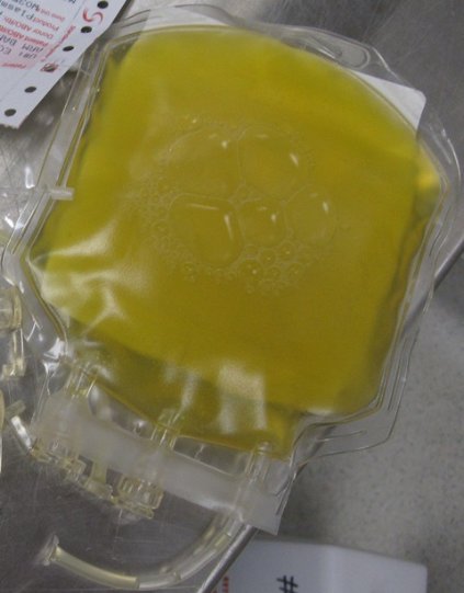
Enter lyophilized plasma (LP), a process developed by HemCon Medical Technologies in which whole blood is sterilely removed, and the plasma component is separated and turned into a powder. The powdered plasma is returned and reconstituted prior to transfusion. "You can put this stuff as a powder on a shelf in nearly any environment," Dr. Schreiber said. "It’s good for at least 3 years, it survives a broad range of temperatures, and you can restore it to plasma within a couple of minutes."
An initial study of LP showed encouraging results with the use of a freeze-dried form of plasma for resuscitation ( J. Trauma 2008;65[5]:975-85). A later study by researchers including Dr. Schreiber evaluated the effects of the lyophilization process on plasma clotting factor levels in swine, by adding the antioxidant ascorbic acid (vitamin C) to the reconstitution solution, and by comparing the efficacy of LP with that of fresh frozen plasma and that of plasma and packed red blood cells in a 1:1 ratio (Arch. Surg. 2009;144[9]:829-34). "What we found was that if we gave LP with packed red blood cells in a 1:1 ratio we had 14% less blood loss than if it’s given as FFP with packed cells, which was significant," Dr. Schreiber said.
"The LP was better in terms of stopping hemorrhage. We also noticed that with the vitamin C, we suppressed inflammation and got reduced IL-6 [interleukin- 6] expression. Now, the Germans, the Dutch, and the French are using LP in their military settings. Our special forces people are also using it. It’s under current development for common use in your hospital in 5-10 years. I think this stuff is good anywhere. With LP we can always maintain a 1:1 ratio, and we don’t have to worry about the thawing process."
Tranexamic acid, a synthetic derivative of lysine, is another anticoagulant therapy that is likely to be used with increasing frequency, he predicted. This agent "binds plasminogen so plasminogen can’t break down fibrin so you can’t get fibrinolysis," said Dr. Schreiber, who has been deployed three times as a combat surgeon.
"This drug has been around forever and is extremely inexpensive. It’s approved by the FDA for use in tooth extraction and the oral form is approved for menorrhagia." Tranexamic acid has also been studied in 53 prospective, randomized studies involving some 3,800 subjects, mostly cardiac patients. "They show that if you use tranexamic acid, you use less blood. It reduces the amount of blood necessary for transfusing people in high-bleeding settings, but no difference in mortality, thrombotic events, myocardial infarction, or stroke. It does not seem to produce a hypercoagulable state."
One study of tranexamic acid use by British surgeons during Afghanistan combat operations found that soldiers who received tranexamic acid were seven times more likely to live, compared with those who did not receive the agent (Arch. Surg. 2012;147:113-9). "There was a survival of 85% in tranexamic acid group, compared with about 70% in those who did not receive it," said Dr. Schreiber. "This has resulted in a change in practice in civilian trauma centers where it is being used widely."
Another anticoagulant being used is the prothrombin complex concentrate known as Kcentra, which contains all four vitamin K–dependent coagulation factors. Distributed by CSL Behring, Kcentra is approved for warfarin reversal in adult patients with acute major bleeding and for those who require emergency surgery. The max dose is 50 units/kg. "That’s about $4,445 for a 70-kg person," Dr. Schreiber said. "Why is it so good? It’s rapidly available, you don’t have to give a lot of fluid, there’s no infectious risk, and you can very rapidly increase coagulation factor function. This is where we’re headed in the future for trauma patients."
Dr. Schreiber said that he had no relevant financial conflicts to disclose.
dbrunk@frontlinemedcom.com
On Twitter @dougbrunk
 |
|
Dr. Frank Podbielski, FCCP, comments: Replacement of blood clotting factors in trauma (and other medical settings) has long been a challenge given the logistics of transfusion and shelf life of blood products. Advances in technology that have yielded powdered forms of blood clotting factors offer clinicians a greater degree of latitude in resuscitation of these patients.
Dr. Francis J. Podbielski, FCCP, is Visiting Clinical Associate Professor of Surgery at the University of Illinois at Chicago - College of Medicine and the Medical Director of the lung cancer program at Jordan Hospital in Plymouth, Massachusetts.
|
|
Dr. Frank Podbielski, FCCP, comments: Replacement of blood clotting factors in trauma (and other medical settings) has long been a challenge given the logistics of transfusion and shelf life of blood products. Advances in technology that have yielded powdered forms of blood clotting factors offer clinicians a greater degree of latitude in resuscitation of these patients.
Dr. Francis J. Podbielski, FCCP, is Visiting Clinical Associate Professor of Surgery at the University of Illinois at Chicago - College of Medicine and the Medical Director of the lung cancer program at Jordan Hospital in Plymouth, Massachusetts.
SAN DIEGO – In the next 5-10 years, reaching for a powdered form of plasma may become the normative first-line treatment for trauma patients who present with severe bleeding. That’s because the current standard of administering fresh frozen plasma is riddled with problems, Dr. Martin Schreiber said at the University of California, San Diego, Critical Care Summer Session.
For one thing, frozen plasma takes 35 minutes to thaw. "That’s a problem, and the clotting factor function of plasma deteriorates as you freeze it and thaw it," said Dr. Schreiber, professor of surgery at Oregon Health & Science University, Portland. "Also, you need it in large volumes and that’s not good for patients with congestive heart failure on Coumadin, and the availability is limited, especially in rural settings."
Enter lyophilized plasma (LP), a process developed by HemCon Medical Technologies in which whole blood is sterilely removed, and the plasma component is separated and turned into a powder. The powdered plasma is returned and reconstituted prior to transfusion. "You can put this stuff as a powder on a shelf in nearly any environment," Dr. Schreiber said. "It’s good for at least 3 years, it survives a broad range of temperatures, and you can restore it to plasma within a couple of minutes."
An initial study of LP showed encouraging results with the use of a freeze-dried form of plasma for resuscitation ( J. Trauma 2008;65[5]:975-85). A later study by researchers including Dr. Schreiber evaluated the effects of the lyophilization process on plasma clotting factor levels in swine, by adding the antioxidant ascorbic acid (vitamin C) to the reconstitution solution, and by comparing the efficacy of LP with that of fresh frozen plasma and that of plasma and packed red blood cells in a 1:1 ratio (Arch. Surg. 2009;144[9]:829-34). "What we found was that if we gave LP with packed red blood cells in a 1:1 ratio we had 14% less blood loss than if it’s given as FFP with packed cells, which was significant," Dr. Schreiber said.
"The LP was better in terms of stopping hemorrhage. We also noticed that with the vitamin C, we suppressed inflammation and got reduced IL-6 [interleukin- 6] expression. Now, the Germans, the Dutch, and the French are using LP in their military settings. Our special forces people are also using it. It’s under current development for common use in your hospital in 5-10 years. I think this stuff is good anywhere. With LP we can always maintain a 1:1 ratio, and we don’t have to worry about the thawing process."
Tranexamic acid, a synthetic derivative of lysine, is another anticoagulant therapy that is likely to be used with increasing frequency, he predicted. This agent "binds plasminogen so plasminogen can’t break down fibrin so you can’t get fibrinolysis," said Dr. Schreiber, who has been deployed three times as a combat surgeon.
"This drug has been around forever and is extremely inexpensive. It’s approved by the FDA for use in tooth extraction and the oral form is approved for menorrhagia." Tranexamic acid has also been studied in 53 prospective, randomized studies involving some 3,800 subjects, mostly cardiac patients. "They show that if you use tranexamic acid, you use less blood. It reduces the amount of blood necessary for transfusing people in high-bleeding settings, but no difference in mortality, thrombotic events, myocardial infarction, or stroke. It does not seem to produce a hypercoagulable state."
One study of tranexamic acid use by British surgeons during Afghanistan combat operations found that soldiers who received tranexamic acid were seven times more likely to live, compared with those who did not receive the agent (Arch. Surg. 2012;147:113-9). "There was a survival of 85% in tranexamic acid group, compared with about 70% in those who did not receive it," said Dr. Schreiber. "This has resulted in a change in practice in civilian trauma centers where it is being used widely."
Another anticoagulant being used is the prothrombin complex concentrate known as Kcentra, which contains all four vitamin K–dependent coagulation factors. Distributed by CSL Behring, Kcentra is approved for warfarin reversal in adult patients with acute major bleeding and for those who require emergency surgery. The max dose is 50 units/kg. "That’s about $4,445 for a 70-kg person," Dr. Schreiber said. "Why is it so good? It’s rapidly available, you don’t have to give a lot of fluid, there’s no infectious risk, and you can very rapidly increase coagulation factor function. This is where we’re headed in the future for trauma patients."
Dr. Schreiber said that he had no relevant financial conflicts to disclose.
dbrunk@frontlinemedcom.com
On Twitter @dougbrunk
Frozen or powdered? Anticoagulation options in trauma are expanding
SAN DIEGO – In the next 5-10 years, reaching for a powdered form of plasma may become the normative first-line treatment for trauma patients who present with severe bleeding. That’s because the current standard of administering fresh frozen plasma is riddled with problems, Dr. Martin Schreiber said at the University of California, San Diego, Critical Care Summer Session.
For one thing, frozen plasma takes 35 minutes to thaw. "That’s a problem, and the clotting factor function of plasma deteriorates as you freeze it and thaw it," said Dr. Schreiber, professor of surgery at Oregon Health & Science University, Portland. "Also, you need it in large volumes and that’s not good for patients with congestive heart failure on Coumadin, and the availability is limited, especially in rural settings."

Enter lyophilized plasma (LP), a process developed by HemCon Medical Technologies in which whole blood is sterilely removed, and the plasma component is separated and turned into a powder. The powdered plasma is returned and reconstituted prior to transfusion. "You can put this stuff as a powder on a shelf in nearly any environment," Dr. Schreiber said. "It’s good for at least 3 years, it survives a broad range of temperatures, and you can restore it to plasma within a couple of minutes."
An initial study of LP showed encouraging results with the use of a freeze-dried form of plasma for resuscitation (J. Trauma 2008;65[5]:975-85). A later study by researchers including Dr. Schreiber evaluated the effects of the lyophilization process on plasma clotting factor levels in swine, by adding the antioxidant ascorbic acid (vitamin C) to the reconstitution solution, and by comparing the efficacy of LP with that of fresh frozen plasma and that of plasma and packed red blood cells in a 1:1 ratio (Arch. Surg. 2009;144[9]:829-34). "What we found was that if we gave LP with packed red blood cells in a 1:1 ratio we had 14% less blood loss than if it’s given as FFP with packed cells, which was significant," Dr. Schreiber said.
"The LP was better in terms of stopping hemorrhage. We also noticed that with the vitamin C, we suppressed inflammation and got reduced IL-6 [interleukin 6] expression. Now, the Germans, the Dutch, and the French are using LP in their military settings. Our special forces people are also using it. It’s under current development for common use in your hospital in 5-10 years. I think this stuff is good anywhere. With LP we can always maintain a 1:1 ratio and we don’t have to worry about the thawing process."
Tranexamic acid, a synthetic derivative of lysine, is another anticoagulant therapy that is likely to be used with increasing frequency, he predicted. This agent "binds plasminogen so plasminogen can’t break down fibrin so you can’t get fibrinolysis," said Dr. Schreiber, who has been deployed three times as a combat surgeon.
"This drug has been around forever and is extremely inexpensive. It’s approved by the FDA for use in tooth extraction and the oral form is approved for menorrhagia." Tranexamic acid has also been studied in 53 prospective, randomized studies involving some 3,800 subjects, mostly cardiac patients. "They show that if you use tranexamic acid, you use less blood. It reduces the amount of blood necessary for transfusing people in high-bleeding settings, but no difference in mortality, thrombotic events, myocardial infarction, or stroke. It does not seem to produce a hypercoagulable state."

One study of tranexamic acid use by British surgeons during Afghanistan combat operations found that soldiers who received tranexamic acid were seven times more likely to live, compared with those who did not receive the agent (Arch. Surg. 2012;147:113-19). "There was a survival of 85% in tranexamic acid group, compared with about 70% in those who did not receive it," said Dr. Schreiber. "This has resulted in a change in practice in civilian trauma centers where it is being used widely."
Another anticoagulant being used is the prothrombin complex concentrate known as Kcentra, which contains all four vitamin K–dependent coagulation factors. Distributed by CSL Behring, Kcentra is approved for warfarin reversal in adult patients with acute major bleeding and for those who require emergency surgery. The max dose is 50 units/kg. "That’s about $4,445 for a 70-kg person," Dr. Schreiber said. "Why is it so good? It’s rapidly available, you don’t have to give a lot of fluid, there’s no infectious risk, and you can very rapidly increase coagulation factor function. This is where we’re headed in the future for trauma patients."
Dr. Schreiber said that he had no relevant financial conflicts to disclose.
On Twitter @dougbrunk
SAN DIEGO – In the next 5-10 years, reaching for a powdered form of plasma may become the normative first-line treatment for trauma patients who present with severe bleeding. That’s because the current standard of administering fresh frozen plasma is riddled with problems, Dr. Martin Schreiber said at the University of California, San Diego, Critical Care Summer Session.
For one thing, frozen plasma takes 35 minutes to thaw. "That’s a problem, and the clotting factor function of plasma deteriorates as you freeze it and thaw it," said Dr. Schreiber, professor of surgery at Oregon Health & Science University, Portland. "Also, you need it in large volumes and that’s not good for patients with congestive heart failure on Coumadin, and the availability is limited, especially in rural settings."
Enter lyophilized plasma (LP), a process developed by HemCon Medical Technologies in which whole blood is sterilely removed, and the plasma component is separated and turned into a powder. The powdered plasma is returned and reconstituted prior to transfusion. "You can put this stuff as a powder on a shelf in nearly any environment," Dr. Schreiber said. "It’s good for at least 3 years, it survives a broad range of temperatures, and you can restore it to plasma within a couple of minutes."
An initial study of LP showed encouraging results with the use of a freeze-dried form of plasma for resuscitation (J. Trauma 2008;65[5]:975-85). A later study by researchers including Dr. Schreiber evaluated the effects of the lyophilization process on plasma clotting factor levels in swine, by adding the antioxidant ascorbic acid (vitamin C) to the reconstitution solution, and by comparing the efficacy of LP with that of fresh frozen plasma and that of plasma and packed red blood cells in a 1:1 ratio (Arch. Surg. 2009;144[9]:829-34). "What we found was that if we gave LP with packed red blood cells in a 1:1 ratio we had 14% less blood loss than if it’s given as FFP with packed cells, which was significant," Dr. Schreiber said.
"The LP was better in terms of stopping hemorrhage. We also noticed that with the vitamin C, we suppressed inflammation and got reduced IL-6 [interleukin 6] expression. Now, the Germans, the Dutch, and the French are using LP in their military settings. Our special forces people are also using it. It’s under current development for common use in your hospital in 5-10 years. I think this stuff is good anywhere. With LP we can always maintain a 1:1 ratio and we don’t have to worry about the thawing process."
Tranexamic acid, a synthetic derivative of lysine, is another anticoagulant therapy that is likely to be used with increasing frequency, he predicted. This agent "binds plasminogen so plasminogen can’t break down fibrin so you can’t get fibrinolysis," said Dr. Schreiber, who has been deployed three times as a combat surgeon.
"This drug has been around forever and is extremely inexpensive. It’s approved by the FDA for use in tooth extraction and the oral form is approved for menorrhagia." Tranexamic acid has also been studied in 53 prospective, randomized studies involving some 3,800 subjects, mostly cardiac patients. "They show that if you use tranexamic acid, you use less blood. It reduces the amount of blood necessary for transfusing people in high-bleeding settings, but no difference in mortality, thrombotic events, myocardial infarction, or stroke. It does not seem to produce a hypercoagulable state."
One study of tranexamic acid use by British surgeons during Afghanistan combat operations found that soldiers who received tranexamic acid were seven times more likely to live, compared with those who did not receive the agent (Arch. Surg. 2012;147:113-19). "There was a survival of 85% in tranexamic acid group, compared with about 70% in those who did not receive it," said Dr. Schreiber. "This has resulted in a change in practice in civilian trauma centers where it is being used widely."
Another anticoagulant being used is the prothrombin complex concentrate known as Kcentra, which contains all four vitamin K–dependent coagulation factors. Distributed by CSL Behring, Kcentra is approved for warfarin reversal in adult patients with acute major bleeding and for those who require emergency surgery. The max dose is 50 units/kg. "That’s about $4,445 for a 70-kg person," Dr. Schreiber said. "Why is it so good? It’s rapidly available, you don’t have to give a lot of fluid, there’s no infectious risk, and you can very rapidly increase coagulation factor function. This is where we’re headed in the future for trauma patients."
Dr. Schreiber said that he had no relevant financial conflicts to disclose.
On Twitter @dougbrunk
EXPERT ANALYSIS AT THE UCSD CRITICAL CARE SUMMER SESSION
Nurse practitioners playing bigger role in critical care
SAN DIEGO – Nurse practitioners are assuming expanded roles and responsibilities in the provision of critical care services, according to Thomas Farley, R.N., N.P.
At the University of California, San Diego, Critical Care Summer Session, Mr. Farley described the expanded role nurse practitioners (NPs) have played in critical care services for about a decade at the University of California, San Francisco (UCSF). In 2004, Dr. Michael Matthay, a pulmonologist, and Dr. Michael Gropper, a critical care anesthesiologist, spearheaded an effort to hire four NPs to work in the university’s medical-surgical intensive care unit (ICU). "At the time, there were increasing limitations on physician trainees, both in the number of trainees and their work-hour restrictions that were becoming more tight," recalled Mr. Farley of the UCSF School of Nursing. "The goal was to provide critical care at the bedside 24 hours per day, which is in line with the Leapfrog Group recommendations for critical care services. At the time, they had 60 adult ICU critical care beds."

Today, 18 NPs work in adult ICUs at UCSF, with expansion to 76 adult critical care beds, including the medical-surgical ICU, cardiothoracic ICU, and neuro ICU. "Initially the NPs were integrated with medical residents," Mr. Farley said. "Now there are teams that don’t have residents, so it’s an NP paired with an attending physician, or sometimes a fellow. There are always two NPs on service. Usually, during the day we have four NPs on, with two on at night." The program was described in 2011 (ICU Director 2011;2:16-19).
Practice trends driving the need for NPs in critical care include the increasing demand for intensivists and a reliance on a team-based approach to care delivery, "understanding that a single provider cannot provide all the needs that any individual critical care patient has," Mr. Farley explained. "I think we’re a little slow to incorporate the use of nurse practitioners on the West Coast. It’s certainly been done for quite some time on the East Coast."
He said that NPs are already incorporated into critical care delivery programs at Memorial Sloan Kettering Cancer Center and Columbia University, both in New York; Emory University, Atlanta; Henry Ford Hospital, Detroit; the Cleveland Clinic; and Vanderbilt University, Nashville, Tenn. Adopters out West include UCSF, UC Davis; UC Los Angeles; UC Fresno; and Oregon Health & Science University, Portland.
At UCSF, critical care NP duties include a consultative role to admitting services, a consultative role to bedside RNs, guidance of house staff, responding to code blue activations, assisting with rapid response consultations, serving on hospital-wide multidisciplinary committees, precepting adult care NP students, as well as attending morning teaching and monthly morbidity and mortality conferences. Procedures performed include insertion of central venous catheters, arterial catheters, chest tubes, and PICC (peripherally inserted central catheter) lines; lumbar puncture; suture and drain removal; and airway intubation.
The use of NPs in critical care "works because we have appropriate conduits for collaboration and supervision that are explained, understood, and taken to heart," Mr. Farley said. "The NPs need to know that they have supportive attending physicians who will assist them when questions arise. We’ve also had buy-in from the ICU nurses."
A growing body of medical literature suggests that this trend is having an effect. A retrospective study of 600 admissions at two ICUs in New York found that patients managed by nurse practitioner/physician assistant teams had no worse outcomes than patients managed by physicians (Chest 2011;139:1347-53). In addition, a recent survey of critical care program directors at academic medical centers found that 86% believe NPs and other advanced practice providers contribute to the continuity of care, 78% believe they save time during rounds and evaluating new patients, and 73% believe they assist in maintaining work flow (J. Crit. Care 2014; 29:112-5).
In the years ahead, Mr. Farley predicted that NPs and other advanced practice providers "are certainly going to be in the ICU, probably even in greater numbers and perhaps in broader roles than they are now."
Mr. Farley said he had no relevant financial conflicts.
On Twitter @dougbrunk
SAN DIEGO – Nurse practitioners are assuming expanded roles and responsibilities in the provision of critical care services, according to Thomas Farley, R.N., N.P.
At the University of California, San Diego, Critical Care Summer Session, Mr. Farley described the expanded role nurse practitioners (NPs) have played in critical care services for about a decade at the University of California, San Francisco (UCSF). In 2004, Dr. Michael Matthay, a pulmonologist, and Dr. Michael Gropper, a critical care anesthesiologist, spearheaded an effort to hire four NPs to work in the university’s medical-surgical intensive care unit (ICU). "At the time, there were increasing limitations on physician trainees, both in the number of trainees and their work-hour restrictions that were becoming more tight," recalled Mr. Farley of the UCSF School of Nursing. "The goal was to provide critical care at the bedside 24 hours per day, which is in line with the Leapfrog Group recommendations for critical care services. At the time, they had 60 adult ICU critical care beds."
Today, 18 NPs work in adult ICUs at UCSF, with expansion to 76 adult critical care beds, including the medical-surgical ICU, cardiothoracic ICU, and neuro ICU. "Initially the NPs were integrated with medical residents," Mr. Farley said. "Now there are teams that don’t have residents, so it’s an NP paired with an attending physician, or sometimes a fellow. There are always two NPs on service. Usually, during the day we have four NPs on, with two on at night." The program was described in 2011 (ICU Director 2011;2:16-19).
Practice trends driving the need for NPs in critical care include the increasing demand for intensivists and a reliance on a team-based approach to care delivery, "understanding that a single provider cannot provide all the needs that any individual critical care patient has," Mr. Farley explained. "I think we’re a little slow to incorporate the use of nurse practitioners on the West Coast. It’s certainly been done for quite some time on the East Coast."
He said that NPs are already incorporated into critical care delivery programs at Memorial Sloan Kettering Cancer Center and Columbia University, both in New York; Emory University, Atlanta; Henry Ford Hospital, Detroit; the Cleveland Clinic; and Vanderbilt University, Nashville, Tenn. Adopters out West include UCSF, UC Davis; UC Los Angeles; UC Fresno; and Oregon Health & Science University, Portland.
At UCSF, critical care NP duties include a consultative role to admitting services, a consultative role to bedside RNs, guidance of house staff, responding to code blue activations, assisting with rapid response consultations, serving on hospital-wide multidisciplinary committees, precepting adult care NP students, as well as attending morning teaching and monthly morbidity and mortality conferences. Procedures performed include insertion of central venous catheters, arterial catheters, chest tubes, and PICC (peripherally inserted central catheter) lines; lumbar puncture; suture and drain removal; and airway intubation.
The use of NPs in critical care "works because we have appropriate conduits for collaboration and supervision that are explained, understood, and taken to heart," Mr. Farley said. "The NPs need to know that they have supportive attending physicians who will assist them when questions arise. We’ve also had buy-in from the ICU nurses."
A growing body of medical literature suggests that this trend is having an effect. A retrospective study of 600 admissions at two ICUs in New York found that patients managed by nurse practitioner/physician assistant teams had no worse outcomes than patients managed by physicians (Chest 2011;139:1347-53). In addition, a recent survey of critical care program directors at academic medical centers found that 86% believe NPs and other advanced practice providers contribute to the continuity of care, 78% believe they save time during rounds and evaluating new patients, and 73% believe they assist in maintaining work flow (J. Crit. Care 2014; 29:112-5).
In the years ahead, Mr. Farley predicted that NPs and other advanced practice providers "are certainly going to be in the ICU, probably even in greater numbers and perhaps in broader roles than they are now."
Mr. Farley said he had no relevant financial conflicts.
On Twitter @dougbrunk
EXPERT ANALYSIS AT THE UCSD CRITICAL CARE SUMMER SESSION
Be alert to less common, but dangerous, thoracic injuries in children
SAN DIEGO – Many thoracic injuries sustained by children and adolescents can be diagnosed with clinical assessment and chest x-ray, and many heal without surgical intervention, according to Dr. Timothy Fairbanks.
"When the pager goes off and says ‘3-year-old involved in a accident,’ that kid may be going home with a physical exam, stickers, and a high five; or he may require a life-saving procedure" in the emergency department. Dr. Fairbanks, of the division of pediatric surgery at Rady Children’s Hospital of San Diego, said at the University of California, San Diego, Critical Care Summer Session. "There’s complete variability in what we see and how serious the injuries can be."

According to data from the National Pediatric Trauma Registry, 86% of injuries are blunt and 14% are penetrating. Chest injuries "are the second leading cause of death to central nervous system injuries," Dr. Fairbanks said. "Mortality rates for blunt and penetrating injuries are similar. In blunt traumas, most kids don’t die from their blunt thoracic injury, but from a CNS injury that they acquired at the same time. Most of the kids who die from penetrating injuries die in the field before reaching the hospital."
Thoracic injuries occur in about 25% of patients who present to a Level I trauma center. While thoracic injuries are less common than abdominal and extremity injuries are, "they are of potentially higher risk and more lethal," he said. "Most can be treated successfully. Male to female injury rate is 2:1 to 3:1 in favor of males having more traumas."
He classifies thoracic trauma injuries by anatomy (rib, pulmonary, bronchial), blunt vs. penetrating, and threat to life (immediate or potentially life threatening). Motor vehicles are the most common mechanism of thoracic injury, and children have an increased risk in all traumas of auto vs. pedestrian, compared with adults. "When children become teenagers, their mechanisms of injuries approach those of the data for adults," Dr. Fairbanks explained. "We see more penetrating knife and gunshot wounds to the chest, although they are rare in our younger patient population. Tracheobronchial lacerations are more common in children, compared with adults. Aortic disruptions are less likely in children."
Common thoracic injuries that he and his associates see at Rady Children’s Hospital are lung contusion, pneumothorax, hemothorax, and fractures to the ribs, sternum, or scapula. Less common "but perhaps more dangerous injuries" are those to the heart, aorta, trachea, bronchi, and diaphragm. "The most common immediately life-threatening thoracic injuries in children are airway obstruction, pneumothorax, hemothorax, and cardiac tamponade," he said. "A large percentage of thoracic injuries have stable vital signs at presentation. Rib fractures are less common in children and are a marker for a significant mechanism of injury or force. Mediastinal structures are more mobile, making tension pneumothorax a bigger problem."
Diagnosis and treatment of thoracic injuries proceeds simultaneously, with an initial goal to rule out life-threatening injuries, which should be identified and treated during the initial resuscitation phase. This includes making sure the airway is clear and secure and providing supplemental oxygen. "If endotracheal intubation is needed, check the position," he advised. "You want to see bilateral chest rise and CO2 waveform on your monitor. It’s very common for a child to have a right mainstem intubation and no breath sounds on the left side." He also makes it a point to assess their ventilation and treat their pneumothorax. "Don’t wait for the chest x-ray if you have a good clinical suspicion," he said.
A chest x-ray can often guide your management decisions in cases of thoracic trauma, while a FAST scan – specifically an ultrasound – is good for seeing a cardiac tamponade.
Sometimes thoracic injuries require an immediate thoracotomy in the emergency department, but its indications "are controversial," Dr. Fairbanks said. "It’s a potentially life-threatening maneuver, especially with penetrating cardiac injuries." The indications for emergency department thoracotomy are post-traumatic arrest or near arrest in all cases of penetrating thoracic injuries; in blunt trauma with loss of vital signs in the ED; or in blunt trauma with loss of vital signs in route to the ED.
"One of the things we struggle with in pediatric trauma is the timing at which they lost the vital signs in the field," he said. "After 20 minutes the chance of survival is almost nil. However, the family is going to want to know that you did everything possible to save their 3- or 4-year-old child."
If the patient is stable, complete the physical exam. "You’re looking for tachypnea, tenderness to the chest wall and abrasions, or signs of thoracic trauma that require further investigation," he said. Important signs include cyanosis, dyspnea, tracheal deviation, hoarseness, jugular engagement, and subcutaneous emphysema.
Chest auscultation is recommended and an anteroposterior (AP) chest x-ray becomes valuable, "because it’s quick, simple, and yields a lot of information," he said. He characterized a thoracic CT scan as "an excellent study" that can help with the diagnosis of aortic and diaphragmatic injuries which can be missed on chest x-ray. "However, there is a lot more radiation with a chest CT," Dr. Fairbanks noted. "Only 1 in 200 chest CTs for trauma yields a new diagnosis." Other studies to consider include EKG, echocardiogram, bronchoscopy, video-assisted thoracic surgery, radionuclide bone scan, and MRI.
A thoracotomy is indicated when the patient is coding or near coding. The other indications are penetrating wound of the heart or great vessels; massive or continuous intrathoracic bleeding; open pneumothorax with major chest wall defect; aortogram indicating injury to the aorta or major branch; massive or continuing air leak, indicating injury to a major airway; cardiac tamponade; esophageal perforation, or diaphragmatic rupture.
Chest wall soft-tissue injuries are usually not clinically significant, "but they’re a marker for a more serious injury under a bruise," he said. "Rib fractures are less common in children. It’s an indicator of significant force. Treatment is pain control and prevention of atelectasis, which is not as big of a problem in kids as it is in adults. They will heal in 6 weeks. Consider child abuse, specifically in cases of multiple rib fractures and those that don’t make sense with the mechanism of reported injury, or rib fractures that are at different stages of healing."
Dr. Fairbanks said that he had no relevant financial conflicts to disclose.
SAN DIEGO – Many thoracic injuries sustained by children and adolescents can be diagnosed with clinical assessment and chest x-ray, and many heal without surgical intervention, according to Dr. Timothy Fairbanks.
"When the pager goes off and says ‘3-year-old involved in a accident,’ that kid may be going home with a physical exam, stickers, and a high five; or he may require a life-saving procedure" in the emergency department. Dr. Fairbanks, of the division of pediatric surgery at Rady Children’s Hospital of San Diego, said at the University of California, San Diego, Critical Care Summer Session. "There’s complete variability in what we see and how serious the injuries can be."
According to data from the National Pediatric Trauma Registry, 86% of injuries are blunt and 14% are penetrating. Chest injuries "are the second leading cause of death to central nervous system injuries," Dr. Fairbanks said. "Mortality rates for blunt and penetrating injuries are similar. In blunt traumas, most kids don’t die from their blunt thoracic injury, but from a CNS injury that they acquired at the same time. Most of the kids who die from penetrating injuries die in the field before reaching the hospital."
Thoracic injuries occur in about 25% of patients who present to a Level I trauma center. While thoracic injuries are less common than abdominal and extremity injuries are, "they are of potentially higher risk and more lethal," he said. "Most can be treated successfully. Male to female injury rate is 2:1 to 3:1 in favor of males having more traumas."
He classifies thoracic trauma injuries by anatomy (rib, pulmonary, bronchial), blunt vs. penetrating, and threat to life (immediate or potentially life threatening). Motor vehicles are the most common mechanism of thoracic injury, and children have an increased risk in all traumas of auto vs. pedestrian, compared with adults. "When children become teenagers, their mechanisms of injuries approach those of the data for adults," Dr. Fairbanks explained. "We see more penetrating knife and gunshot wounds to the chest, although they are rare in our younger patient population. Tracheobronchial lacerations are more common in children, compared with adults. Aortic disruptions are less likely in children."
Common thoracic injuries that he and his associates see at Rady Children’s Hospital are lung contusion, pneumothorax, hemothorax, and fractures to the ribs, sternum, or scapula. Less common "but perhaps more dangerous injuries" are those to the heart, aorta, trachea, bronchi, and diaphragm. "The most common immediately life-threatening thoracic injuries in children are airway obstruction, pneumothorax, hemothorax, and cardiac tamponade," he said. "A large percentage of thoracic injuries have stable vital signs at presentation. Rib fractures are less common in children and are a marker for a significant mechanism of injury or force. Mediastinal structures are more mobile, making tension pneumothorax a bigger problem."
Diagnosis and treatment of thoracic injuries proceeds simultaneously, with an initial goal to rule out life-threatening injuries, which should be identified and treated during the initial resuscitation phase. This includes making sure the airway is clear and secure and providing supplemental oxygen. "If endotracheal intubation is needed, check the position," he advised. "You want to see bilateral chest rise and CO2 waveform on your monitor. It’s very common for a child to have a right mainstem intubation and no breath sounds on the left side." He also makes it a point to assess their ventilation and treat their pneumothorax. "Don’t wait for the chest x-ray if you have a good clinical suspicion," he said.
A chest x-ray can often guide your management decisions in cases of thoracic trauma, while a FAST scan – specifically an ultrasound – is good for seeing a cardiac tamponade.
Sometimes thoracic injuries require an immediate thoracotomy in the emergency department, but its indications "are controversial," Dr. Fairbanks said. "It’s a potentially life-threatening maneuver, especially with penetrating cardiac injuries." The indications for emergency department thoracotomy are post-traumatic arrest or near arrest in all cases of penetrating thoracic injuries; in blunt trauma with loss of vital signs in the ED; or in blunt trauma with loss of vital signs in route to the ED.
"One of the things we struggle with in pediatric trauma is the timing at which they lost the vital signs in the field," he said. "After 20 minutes the chance of survival is almost nil. However, the family is going to want to know that you did everything possible to save their 3- or 4-year-old child."
If the patient is stable, complete the physical exam. "You’re looking for tachypnea, tenderness to the chest wall and abrasions, or signs of thoracic trauma that require further investigation," he said. Important signs include cyanosis, dyspnea, tracheal deviation, hoarseness, jugular engagement, and subcutaneous emphysema.
Chest auscultation is recommended and an anteroposterior (AP) chest x-ray becomes valuable, "because it’s quick, simple, and yields a lot of information," he said. He characterized a thoracic CT scan as "an excellent study" that can help with the diagnosis of aortic and diaphragmatic injuries which can be missed on chest x-ray. "However, there is a lot more radiation with a chest CT," Dr. Fairbanks noted. "Only 1 in 200 chest CTs for trauma yields a new diagnosis." Other studies to consider include EKG, echocardiogram, bronchoscopy, video-assisted thoracic surgery, radionuclide bone scan, and MRI.
A thoracotomy is indicated when the patient is coding or near coding. The other indications are penetrating wound of the heart or great vessels; massive or continuous intrathoracic bleeding; open pneumothorax with major chest wall defect; aortogram indicating injury to the aorta or major branch; massive or continuing air leak, indicating injury to a major airway; cardiac tamponade; esophageal perforation, or diaphragmatic rupture.
Chest wall soft-tissue injuries are usually not clinically significant, "but they’re a marker for a more serious injury under a bruise," he said. "Rib fractures are less common in children. It’s an indicator of significant force. Treatment is pain control and prevention of atelectasis, which is not as big of a problem in kids as it is in adults. They will heal in 6 weeks. Consider child abuse, specifically in cases of multiple rib fractures and those that don’t make sense with the mechanism of reported injury, or rib fractures that are at different stages of healing."
Dr. Fairbanks said that he had no relevant financial conflicts to disclose.
EXPERT ANALYSIS AT THE UCSD CRITICAL CARE SUMMER SESSION
Early ID of placenta accreta key to optimal management
SAN DIEGO – An early and accurate diagnosis of placenta accreta is crucial because maternal mortality can be as high as 7% and perinatal mortality can be as high as 10%.
"Historically, the clinical presentation of placenta accreta was a prolonged third-stage or retained placenta after delivery of the baby and subsequent onset of hemorrhage or the onset of hemorrhage at the time of the termination of pregnancy," Dr. Gladys Ramos said at the University of California, San Diego, Critical Care Summer Session. "Now, with predelivery diagnosis, we have changed the management and the clinical presentation."
Placenta accreta is defined as the abnormal presence of villi attached to the myometrium due to a defect in the decidua basalis. In the 1980s, placenta accreta was believed to be rare. However, a study from 2005 showed an increase in the rate of placenta accreta (Am. J. Obstet. Gynecol. 2005;192:1458-61). The condition now occurs in 1 in every 533 pregnancies.
"Mirroring this rise is a rise in cesarean deliveries," said Dr. Ramos, a perinatologist at the UCSD Medical Center. "We think these two conditions are related. At UCSD, we take care of about 3,200 deliveries per year, and from 1990 to 2008 we have seen a linear increase in the rate of placenta accreta. We take care of about two patients per month with this condition."
Risk factors for the placenta accreta are prior uterine surgery, placenta previa, advanced maternal age, parity, and smoking. The published rate of detection by ultrasound ranges from 80% to 100%, "with a low false-positive rate," Dr. Ramos said. Telltale signs on ultrasound include loss of myometrial interface, a heterogeneous "Swiss cheese–looking" appearance to the placenta, an increase in the vascularity of the placenta, and evidence of bladder invasion.
MRI can be used as an adjunct to ultrasound diagnosis. The published detection rate with MRI ranges from 80% to 88%, "with a very low false-positive rate, which is why we use it as an adjunct to an ultrasound," she said. "We see similar findings on MRI that we do on ultrasound: thickened, dark nodular contour to the placenta; extension of the dark bands within the placenta; and mass effect causing a bulge on T1."
Nurses and sonographers at UCSD have been trained to ask patients upon presentation about risk factors for the condition." Then we look for signs of placenta accreta on ultrasound, including endovaginal ultrasound," Dr. Ramos said. "If we are concerned, we plan ahead with a multidisciplinary approach. If we’re still not sure, we proceed with MRI for diagnosis."
In 1995, clinicians at UCSD developed a multidisciplinary approach to treating patients with placenta accreta, mindful that "it took a cast of thousands to be able to make sure our outcomes were optimal for both mom and baby," said Pat Inzano, R.N., an administrative nurse in labor and delivery at UCSD. "Over the years, we’ve recognized the risk factors and the importance of early and accurate diagnosis."
The approach includes detailed maternal counseling and meticulous planning with colleagues at every conceivable step along the way in the care of the mom and baby, from neonatology and gynecologic surgery to surgical ICU staff and social workers. "When a mother gets the news [of placenta accreta], not only does she probably not understand the pathophysiology of what’s going on, but it takes a long time for her to digest this information," Ms. Inzano explained. "One of the things that’s so important at every level is getting her and her family to understand the diagnosis and reminding her that she’s [receiving] the best possible care."
The multidisciplinary team stages a conference to discuss the patient’s hospital delivery and care; availability of blood products; the need for anesthesia, surgical, and radiological expertise in house; and intensive care capability. Proper consents are also required "because the surgery will involve removing the patient’s uterus and rendering her sterile," she said. "We want to get the patient and the family as comfortable as possible with what’s going to happen. We found that providing tours of every single area that she will be ‘parking in’ is stress-relieving for her and her family, including labor and delivery and the neonatal ICU. We also provide consultations with all of the specialties involved in the case. All of this is education and counseling on a lot of different levels."
The team develops a time-line and schedules a planned delivery, including admission to the hospital, cesarean section/hysterectomy in the main operating room (OR), and unit transfers for epidural, central lines, and femoral balloons. In addition, the team coordinates a hospital tour for the patient and family. Most recommended deliveries are at week 34 because "we don’t want her to get near term and go into labor, which would aggravate a bleed of the placenta accreta," Ms. Inzano said.
The team also crafts a "plan B" for emergent delivery, including a detailed list of whom to contact and their pager numbers. "If we need to emergency deliver this patient in the main OR at 3 in the morning, we have our attending physician call the attending trauma physician to put us on trauma bypass in case we need the blood products," she said. "If we don’t have the type and cross-matched blood available, we activate an [obstetric] hemorrhage protocol in order to obtain O-negative blood in massive quantities until she’s cross-matched."
Ms. Inzano said that the multidisciplinary approach to placenta accreta "has become a smooth operation at our institution, but we never drop our awareness of the severity of what can happen. With the multidisciplinary effort, it brings everyone together; everyone’s on the same page, and everyone knows what to anticipate."
Neither Dr. Ramos nor Ms. Inzano had relevant financial conflicts.
On Twitter @dougbrunk
EXPERT ANALYSIS AT THE UCSD CRITICAL CARE SUMMER SESSION