User login
Skin Cancer Foundation: World Congress on Cancers of the Skin
Fatigue checks assist in preventing wrong-site surgery
EDINBURGH – It’s 4 o’clock in the afternoon on a long Mohs surgery day, and you’ve got another patient with innumerable stages plus reconstructions to do.
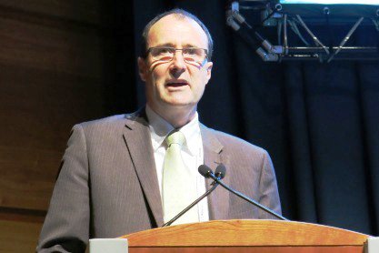
Performing a fatigue check that gives nursing and biomedical staff the opportunity to admit they’re too tired may be the best next step to prevent wrong-site surgery, Dr. Colin Fleming, president of the British Society for Dermatological Surgery (BSDS), posited at the 15th World Congress on Cancers of the Skin.

“When we introduced this, it gave great power to the staff, whom we as doctors were asking to work hard on our behalf,” he said. “It’s been a really valuable tool in increasing safety at the end of a long operating day.”
The fatigue check is separate from the oft-recommended preprocedural surgical pause. Though helpful, research has shown that the surgical pause failed to prevent wrong-site surgery in 10% to a third of cases, said Dr. Fleming, a consultant dermatologist and Mohs surgeon at Ninewells Hospital, Dundee, Scotland.
A study on the usefulness of proposed methods for correct biopsy site identification in cutaneous surgery with the Delphi consensus method was recently published (JAMA Dermatol. 2014;150:550-8), but “I’m not convinced it really tells us anything,” he remarked.
Dr. Fleming suggested coupling the surgical pause with a site check, and he emphasized the value of good quality preoperative photographs of the index lesion. Today’s ubiquitous “selfie” may even have a role, as patients themselves often misidentify the biopsy site.
In addition, dermatology departments should have a protocol incorporating a variety of safety features, he said.
However, only 54% of surgeons said they have a written protocol in place to identify the correct surgical site, based on data from a recent survey of BSDS members conducted by Dr. Fleming and his colleagues.
More than a half of respondents (60/114) acknowledged it was “sometimes” difficult to locate the surgical site, with 47 respondents saying it was difficult to locate the surgical site in 1%-10% of cases and 13 admitting it was difficult to do so in 10%-25% of cases.
The face, scalp, and back were reported as the most challenging areas in which to locate the exact surgical site, Dr. Fleming said at the meeting, sponsored by the Skin Cancer Foundation.
When asked what steps they take in their local written protocols to avoid wrong site surgery, 82 respondents said they check with the patient, 66 use drawings/templates with the site marked, 50 use a mirror or photographs, and 39 respondents double-check with a colleague.
Overall, slightly less than 40% of respondents recalled having no patient in the past 5 years who underwent wrong-site surgery. Approximately 28% had 1 wrong-site surgery patient, 28% had 2-3 patients, 3% had 4-5 patients, and 1% had more than 10 such patients.
“Wrong-site surgery in U.K. dermatology departments is infrequent,” Dr. Fleming concluded, adding that only a small proportion led to formal complaints or a medicolegal case.
The response rate to the web-based survey was 37.5% (115/306 members); 32 respondents were Mohs surgeons, 75 were non-Mohs surgeons, 68 worked at a teaching hospital, and 110 had a regular surgical list of between 500 and 2,000 procedures per year that they were responsible for or did themselves.
EDINBURGH – It’s 4 o’clock in the afternoon on a long Mohs surgery day, and you’ve got another patient with innumerable stages plus reconstructions to do.
Performing a fatigue check that gives nursing and biomedical staff the opportunity to admit they’re too tired may be the best next step to prevent wrong-site surgery, Dr. Colin Fleming, president of the British Society for Dermatological Surgery (BSDS), posited at the 15th World Congress on Cancers of the Skin.
“When we introduced this, it gave great power to the staff, whom we as doctors were asking to work hard on our behalf,” he said. “It’s been a really valuable tool in increasing safety at the end of a long operating day.”
The fatigue check is separate from the oft-recommended preprocedural surgical pause. Though helpful, research has shown that the surgical pause failed to prevent wrong-site surgery in 10% to a third of cases, said Dr. Fleming, a consultant dermatologist and Mohs surgeon at Ninewells Hospital, Dundee, Scotland.
A study on the usefulness of proposed methods for correct biopsy site identification in cutaneous surgery with the Delphi consensus method was recently published (JAMA Dermatol. 2014;150:550-8), but “I’m not convinced it really tells us anything,” he remarked.
Dr. Fleming suggested coupling the surgical pause with a site check, and he emphasized the value of good quality preoperative photographs of the index lesion. Today’s ubiquitous “selfie” may even have a role, as patients themselves often misidentify the biopsy site.
In addition, dermatology departments should have a protocol incorporating a variety of safety features, he said.
However, only 54% of surgeons said they have a written protocol in place to identify the correct surgical site, based on data from a recent survey of BSDS members conducted by Dr. Fleming and his colleagues.
More than a half of respondents (60/114) acknowledged it was “sometimes” difficult to locate the surgical site, with 47 respondents saying it was difficult to locate the surgical site in 1%-10% of cases and 13 admitting it was difficult to do so in 10%-25% of cases.
The face, scalp, and back were reported as the most challenging areas in which to locate the exact surgical site, Dr. Fleming said at the meeting, sponsored by the Skin Cancer Foundation.
When asked what steps they take in their local written protocols to avoid wrong site surgery, 82 respondents said they check with the patient, 66 use drawings/templates with the site marked, 50 use a mirror or photographs, and 39 respondents double-check with a colleague.
Overall, slightly less than 40% of respondents recalled having no patient in the past 5 years who underwent wrong-site surgery. Approximately 28% had 1 wrong-site surgery patient, 28% had 2-3 patients, 3% had 4-5 patients, and 1% had more than 10 such patients.
“Wrong-site surgery in U.K. dermatology departments is infrequent,” Dr. Fleming concluded, adding that only a small proportion led to formal complaints or a medicolegal case.
The response rate to the web-based survey was 37.5% (115/306 members); 32 respondents were Mohs surgeons, 75 were non-Mohs surgeons, 68 worked at a teaching hospital, and 110 had a regular surgical list of between 500 and 2,000 procedures per year that they were responsible for or did themselves.
AT THE WCCS 2014

3-D images aid total body mole mapping
EDINBURGH – The advent of three-dimensional total body photography overcomes many, but not all, of the drawbacks of two-dimensional imaging as an aid to melanoma detection.
“There are challenges to using two-dimensional images, both in terms of acquiring them and reviewing them, but the simplest of which is a 5-mm lesion that’s perfectly perpendicular to the camera will not look like 5 mm if it’s at an angle to the camera, an inherent problem of consistency in looking at size over time on curved surfaces,” Dr. Allan Halpern said at the 15th World Congress on Cancers of the Skin.

Over the last several months, the dermatology service Dr. Halpern heads at Memorial Sloan-Kettering Cancer Center in New York has shifted to 3-D total body photography in patients at high risk of melanoma.
“The new system (Vectra WB360, Canfield Imaging Systems) uses 40 some odd cameras behind a series of panels, and in 3 milliseconds allows the clinician to image the entire patient in 3-D,” he said.
A touch-screen interface allows for additional close-up pictures of areas of particular interest and tags their location to the corresponding body map photographs. The onscreen 3-D images also can be used for consultation and patient education.
In a recent open-ended survey of 199 of his patients who had already undergone 2-D imaging, participants said 3-D mole mapping was faster, easier, less invasive, and required fewer poses, Dr. Halpern said.
Among their concerns, the admittedly motivated patient cohort wanted even better resolution and more images to catch moles in body folds or on the soles of their feet.
Dr. Halpern was quick to acknowledge that, like population-based melanoma screening, total body photography (TBP) has not been shown in a definitive trial to reduce mortality. In fact, clinical practice guidelines issued this year in Germany and Australia strongly recommend training and use of dermoscopy for examination of pigmented lesions, but note that the value of TBP in melanoma-risk patients remains ‘unproven.’
Research also has shown that uptake of TBP in clinical practice has remained relatively flat in the United States since 2000 (J. Am. Acad. Dermatol. 2010:62:794-803). Obstacles include expense, privacy, and security of stored images, and a lack of imaging standards.
The International Society for Digital Imaging of the Skin, however, is working with dermatology specialists, informatics experts, and imaging technology developers through its Melanoma Project to develop international imaging standards and a public-access archive of clinical and dermoscopic images of skin lesions, Dr. Halpern said.
“One of the encouraging things to me is that the CEOs of six of those imaging developers have already signed on. They recognize the importance of having international standards and interchangeability of images,” he said at the meeting sponsored by the Skin Cancer Foundation.
Dr. Halpern has consulted for and shared research grants with Canfield Scientific and consulted for Caliber I.D., Scibase, and DermTech.
EDINBURGH – The advent of three-dimensional total body photography overcomes many, but not all, of the drawbacks of two-dimensional imaging as an aid to melanoma detection.
“There are challenges to using two-dimensional images, both in terms of acquiring them and reviewing them, but the simplest of which is a 5-mm lesion that’s perfectly perpendicular to the camera will not look like 5 mm if it’s at an angle to the camera, an inherent problem of consistency in looking at size over time on curved surfaces,” Dr. Allan Halpern said at the 15th World Congress on Cancers of the Skin.
Over the last several months, the dermatology service Dr. Halpern heads at Memorial Sloan-Kettering Cancer Center in New York has shifted to 3-D total body photography in patients at high risk of melanoma.
“The new system (Vectra WB360, Canfield Imaging Systems) uses 40 some odd cameras behind a series of panels, and in 3 milliseconds allows the clinician to image the entire patient in 3-D,” he said.
A touch-screen interface allows for additional close-up pictures of areas of particular interest and tags their location to the corresponding body map photographs. The onscreen 3-D images also can be used for consultation and patient education.
In a recent open-ended survey of 199 of his patients who had already undergone 2-D imaging, participants said 3-D mole mapping was faster, easier, less invasive, and required fewer poses, Dr. Halpern said.
Among their concerns, the admittedly motivated patient cohort wanted even better resolution and more images to catch moles in body folds or on the soles of their feet.
Dr. Halpern was quick to acknowledge that, like population-based melanoma screening, total body photography (TBP) has not been shown in a definitive trial to reduce mortality. In fact, clinical practice guidelines issued this year in Germany and Australia strongly recommend training and use of dermoscopy for examination of pigmented lesions, but note that the value of TBP in melanoma-risk patients remains ‘unproven.’
Research also has shown that uptake of TBP in clinical practice has remained relatively flat in the United States since 2000 (J. Am. Acad. Dermatol. 2010:62:794-803). Obstacles include expense, privacy, and security of stored images, and a lack of imaging standards.
The International Society for Digital Imaging of the Skin, however, is working with dermatology specialists, informatics experts, and imaging technology developers through its Melanoma Project to develop international imaging standards and a public-access archive of clinical and dermoscopic images of skin lesions, Dr. Halpern said.
“One of the encouraging things to me is that the CEOs of six of those imaging developers have already signed on. They recognize the importance of having international standards and interchangeability of images,” he said at the meeting sponsored by the Skin Cancer Foundation.
Dr. Halpern has consulted for and shared research grants with Canfield Scientific and consulted for Caliber I.D., Scibase, and DermTech.
AT WCCS 2014

More people die from thin melanomas than thick melanomas
EDINBURGH – Contrary to the perception that most melanoma deaths result from thick melanomas, long-term data from Australia show that more people die who initially present with thin melanomas.
Among 4,218 Australians who died from melanoma between 1990 and 2009, thin melanomas (1 mm or less) accounted for 19% of melanoma deaths overall, but increased steadily from 14% of deaths in 1990-1994 to 23% in 2005-2009.
During the most recent time period (2005-2009), more people died from thin melanomas (296 deaths, 23%) than from thick melanomas more than 4 mm in thickness (186 deaths, 14%) or from metastatic presentations (207 deaths, 16%).
The number of deaths in the intermediate thickness categories was also higher for lesions of thickness 1.01-2.0 mm (272 deaths, 21%) than for lesions of thickness 2.01-4.0 mm (267 deaths, 20%).
The patterns of mortality were essentially unchanged when the analyses were restricted to patients with only one primary melanoma, Dr. David Whiteman and Dr. Catherine Olsen of the QIMR Berghofer Medical Research Institute, Brisbane, Australia, reported at the 15th World Congress on Cancers of the Skin.
The melanoma incidence has been rising steadily around the world, mostly because of the greater numbers of thin lesions being diagnosed. The perception that most people who die from melanoma present initially with thick lesions is widespread, but the veracity of this proposition has never been tested because population-based data describing total mortality by the thickness of the primary tumor are scarce, according to the researchers. They used linked histology and death data from the Queensland Cancer Registry, where notification of melanoma has been compulsory since 1982, to calculate age-standardized mortality rates for each year for all melanomas, and by thickness of the first primary lesion. Overall, 67% of patients were male, 68% presented with a single primary lesion, and 68% of all melanomas were thin (1 mm or less).
Deaths from melanoma were most common among those who were in the seventh and eighth decades of life, male, or had a melanoma arising on the trunk. As expected, the intervals from diagnosis to death were significantly shorter for thicker tumors than thinner tumors, Dr. Whiteman and Dr. Olsen noted in the poster presentation.
The average annual rate of change in melanoma mortality increased significantly for men for thin lesions and those of intermediate thickness. Mortality rates from metastatic lesions, however, declined during the observation period.
“From a public health perspective, it can be argued that primary prevention activities aimed at reducing the occurrence of melanoma in the entire population should be accorded the highest priority,” the authors concluded at the meeting, sponsored by the Skin Cancer Foundation.
AT WCCS 2014
Key clinical point: Thin lesions are associated with substantial mortality, so primary prevention of melanoma should remain the principal strategy.
Major finding: From 2005 to 2009, 23% of residents of Queensland, Australia, died from thin melanomas versus 14% with thick melanomas.
Data source: State cancer registry analysis of 4,218 melanoma deaths in Queensland.
Disclosures: The researchers had no relevant financial conflicts to disclose.
Moles quadruple risk for melanoma
EDINBURGH – Patients with moles had more than four times the risk of developing melanoma, compared with those without moles in a large record-linkage study.
The overall rate ratio for melanoma, based on person-years at risk, was 4.68 among patients with moles recorded in their medical record (95% confidence interval, 4.39-4.98), Dr. Eugene Ong reported at the 15th World Congress on Cancers of the Skin.
Rate ratios were also significantly higher for individuals with moles of both sexes and in all age groups, including those aged younger than 25 years (RR, 3.79), 25-59 years (RR, 5.02), and at least 60 years (RR, 4.68).
Prior research has shown that high numbers of melanocytic or dysplastic nevi are strong risk factors for the development of melanoma. The investigators sought to further characterize the risk of melanoma in persons with melanocytic nevus (MN) using linked hospital and mortality records covering the entire population of England from 1999 to 2011.
The analysis included 271,656 patients with a hospital or day-case record of moles and a control cohort of 10,130,417 persons with no moles recorded. Anyone diagnosed with melanoma within 1 year of study entry was excluded.
Patients with a record of moles had a significantly higher risk of developing melanoma both around the site of the mole and elsewhere on their body, and therefore may benefit from increased surveillance, said Dr. Ong of the University of Oxford, England. For patients with a mole on the trunk, the rate ratio for a melanoma on the trunk was 8.99 (95% CI, 7.69-10.46) and 5.66 for a melanoma elsewhere (95% CI, 4.97-6.42).
The investigators were unable to distinguish between different types of moles or to determine the number of moles in each patient. Further, a mole or moles were the principal reason for hospital contact for 91% of patients, so it’s likely they presented with unusual appearing moles in order for them to warrant recording, Dr. Ong acknowledged.
“So while this study does not suggest that everyone with a single mole is far more likely to develop melanoma, it does illustrate the link between moles and skin cancer. This is why it is vital people check their moles regularly and report any changes to their doctor,” he said in a statement released during the meeting, sponsored in part by the Skin Cancer Foundation.
AT THE WCCS 2014
Key clinical point: Individuals with moles are at significantly increased risk for developing melanoma in the same body region as the mole, and in other regions, and could benefit from increased surveillance.
Major finding: The rate ratio for melanoma, based on person-years at risk, was 4.68 times among patients with moles than those without moles (95% CI 4.39-4.98).
Data source: A record-linkage study in 271,656 patients with a diagnosis of moles and 10,130,417 controls without moles.
Disclosures: Dr. Ong had no financial conflicts to disclose.
VIDEO: How red hair and freckles might raise your skin cancer risk
EDINBURGH – Variants in the pigment-associated MC1R gene have been implicated in an increased risk for melanoma and nonmelanoma skin cancers, although the extent of that risk has been inconsistent across studies, according to Dr. Eugene Healy of the University of Southampton (England). In an interview at the 15th World Congress on Cancers of the Skin sponsored by the Skin Cancer Foundation, Dr. Healy discussed how the MC1R gene variants might impact skin cancer risk and the challenges of pinning down genetic data into practical applications for patients.
The video associated with this article is no longer available on this site. Please view all of our videos on the MDedge YouTube channel
EXPERT ANALYSIS FROM WCCS 2014
VIDEO: What to do when cancer patients say they want to die
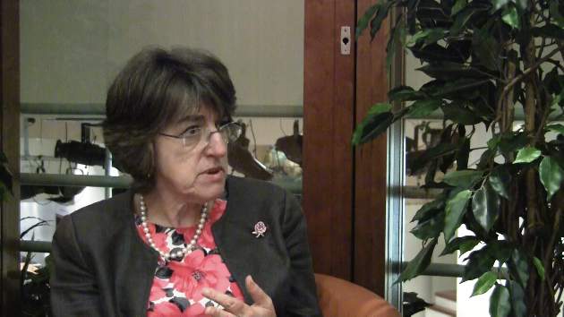
EDINBURGH – When your patient says he or she wants to die, what do you do?
There’s no single answer, but asking the right questions can help patients find peace as well as perspective, according to Dr. Ilora Finlay, who spoke at the 15th World Congress on Cancers of the Skin about the types of conversations and seemingly small actions that can make a big difference for patients coping with advanced cancer.
In an interview at the meeting, Dr. Finlay shared some of her expertise from decades of clinical experience in palliative care.
The congress was sponsored by the Skin Cancer Foundation.
The video associated with this article is no longer available on this site. Please view all of our videos on the MDedge YouTube channel
EXPERT ANALYSIS FROM WCCS 2014
Veterans at Increased Risk for Skin Cancer
EDINBURGH – U.S. military personnel deployed overseas during the past decade have increased risk factors for skin cancer, a new survey suggests.
Researchers reported that 77% of veterans were exposed to 4 or more hours of bright sun during a typical day, but only 27% had ready access to sunscreen while working. Another 32% had no access to sunscreen.
A full 62% of veterans reported getting sunburned while deployed, with 42% having two or more sunburns, 16% four or more sunburns, and 15% at least one blistering sunburn.
Twenty-nine percent of veterans noticed changes in size, shape, or color of moles since deployment, but only 4% reported receiving a skin exam by a physician since returning home.
“This study demonstrates room for improvement for skin cancer prevention in veterans’ populations,” dermatologist Jennifer Powers of Vanderbilt University, Nashville, Tenn., reported at the 15th World Congress on Cancers of the Skin.
The 30-question survey was given to U.S. military workers engaged in Operation Enduring Freedom, Operation Iraqi Freedom, or Operation New Dawn presenting at the Nashville postdeployment clinic, Tennessee Valley Healthcare System, U.S. Department of Veterans. The combat missions occurred at a more equatorial latitude than the mean center of the United States population, increasing the potential for ultraviolet exposure and the development of skin cancer, Dr. Powers noted.
Of the 197 respondents, 64% identified as white, 22% as African American, and 8% as Hispanic. Almost half (48%) were fair-skinned.
Only 22% of veterans reported being made very aware of the risks of skin cancer, compared with 41% who reported not being made aware at all.
Dr. Powers highlighted a prior retrospective review of tumor registries showing that the melanoma incidence increased from 1990-1994 to 2000-2004 among white active-duty military personnel and that rates were significantly higher among those 45 years or older (Cancer Epidemiol. Biomarkers Prev. 2011;20:318-23).
The congress was sponsored by the Skin Cancer Foundation.
AT WCCS 2014

Veterans at increased risk of skin cancer
EDINBURGH – U.S. military personnel deployed overseas during the past decade have increased risk factors for skin cancer, a new survey suggests.
Researchers reported that 77% of veterans were exposed to 4 or more hours of bright sun during a typical day, but only 27% had ready access to sunscreen while working. Another 32% had no access to sunscreen.

A full 62% of veterans reported getting sunburned while deployed, with 42% having two or more sunburns, 16% four or more sunburns, and 15% at least one blistering sunburn.
Twenty-nine percent of veterans noticed changes in size, shape, or color of moles since deployment, but only 4% reported receiving a skin exam by a physician since returning home.
“This study demonstrates room for improvement for skin cancer prevention in veterans’ populations,” dermatologist Jennifer Powers of Vanderbilt University, Nashville, Tenn., reported at the 15th World Congress on Cancers of the Skin.
The 30-question survey was given to U.S. military workers engaged in Operation Enduring Freedom, Operation Iraqi Freedom, or Operation New Dawn presenting at the Nashville postdeployment clinic, Tennessee Valley Healthcare System, U.S. Department of Veterans. The combat missions occurred at a more equatorial latitude than the mean center of the United States population, increasing the potential for ultraviolet exposure and the development of skin cancer, Dr. Powers noted.
Of the 197 respondents, 64% identified as white, 22% as African American, and 8% as Hispanic. Almost half (48%) were fair-skinned.
Only 22% of veterans reported being made very aware of the risks of skin cancer, compared with 41% who reported not being made aware at all.
Dr. Powers highlighted a prior retrospective review of tumor registries showing that the melanoma incidence increased from 1990-1994 to 2000-2004 among white active-duty military personnel and that rates were significantly higher among those 45 years or older (Cancer Epidemiol. Biomarkers Prev. 2011;20:318-23).
The congress was sponsored by the Skin Cancer Foundation.
EDINBURGH – U.S. military personnel deployed overseas during the past decade have increased risk factors for skin cancer, a new survey suggests.
Researchers reported that 77% of veterans were exposed to 4 or more hours of bright sun during a typical day, but only 27% had ready access to sunscreen while working. Another 32% had no access to sunscreen.
A full 62% of veterans reported getting sunburned while deployed, with 42% having two or more sunburns, 16% four or more sunburns, and 15% at least one blistering sunburn.
Twenty-nine percent of veterans noticed changes in size, shape, or color of moles since deployment, but only 4% reported receiving a skin exam by a physician since returning home.
“This study demonstrates room for improvement for skin cancer prevention in veterans’ populations,” dermatologist Jennifer Powers of Vanderbilt University, Nashville, Tenn., reported at the 15th World Congress on Cancers of the Skin.
The 30-question survey was given to U.S. military workers engaged in Operation Enduring Freedom, Operation Iraqi Freedom, or Operation New Dawn presenting at the Nashville postdeployment clinic, Tennessee Valley Healthcare System, U.S. Department of Veterans. The combat missions occurred at a more equatorial latitude than the mean center of the United States population, increasing the potential for ultraviolet exposure and the development of skin cancer, Dr. Powers noted.
Of the 197 respondents, 64% identified as white, 22% as African American, and 8% as Hispanic. Almost half (48%) were fair-skinned.
Only 22% of veterans reported being made very aware of the risks of skin cancer, compared with 41% who reported not being made aware at all.
Dr. Powers highlighted a prior retrospective review of tumor registries showing that the melanoma incidence increased from 1990-1994 to 2000-2004 among white active-duty military personnel and that rates were significantly higher among those 45 years or older (Cancer Epidemiol. Biomarkers Prev. 2011;20:318-23).
The congress was sponsored by the Skin Cancer Foundation.
AT WCCS 2014
Key clinical point: Skin cancer prevention can be improved among U.S. military personnel.
Major finding: Seventy-seven percent of veterans were exposed to 4 or more hours of bright sun during a typical day, but 27% had ready access to sunscreen.
Data source: Survey of 197 recent veterans.
Disclosures: Dr. Powers reported no conflicts of interest. The study was supported by the Nashville Tennessee Valley Healthcare System, which employs some of the coauthors.

Closing large dermal defects much like a Victorian corset
EDINBURGH – Barbed absorbable sutures are a useful new tool to facilitate dermal closure of facial and nonfacial defects following tumor resection.
“These are not the bad old sutures that you might of heard about before, that were nonabsorbable sutures and attempted for use in cosmetic procedures,” Dr. John Strasswimmer said at the 15th World Congress on Cancers of the Skin.
Last year, Dr. Strasswimmer, medical director of melanoma and cutaneous oncology at the Lynn Cancer Institute in Boca Raton, Fla., reported his initial experience using a procedure he calls “Corseta” to close a large Mohs defect on the trunk of an 83-year-old man (JAMA Dermatol. 2013;149:853-4).
The procedure employs a barbed, bioabsorbable suture (Ethicon’s Stratafix and Covidien’s V-Loc) that is run in a continuous vertical looping manner in the subcutaneous layer, with minimal to no undermining of the wound. Undermining is typically used in cutaneous surgery to relieve tension or provide structure around anatomical landmarks, but it can increase the risk of bleeding, swelling, and patient discomfort, he said.
Instead, the first suture pass is placed in the deepest portion of the subcutaneous tissue and brought out within the more superficial subcutaneous layer. Each bite of the barbed suture extends peripherally at least 2.0 cm from the edge of the wound, so the point of tension is lateral to the wound margins. At every two passes, tension is placed evenly across the sutures to close the deepest layer of tissue and to engage the barbs, much like closing of a Victorian corset, Dr. Strasswimmer said.
The second arm of the suture is passed in a similar manner in the subcutaneous plane, superficial to the first pass.
“This is a lacing, not a suturing technique,” he said. “You get tissue approximation, but more importantly, because we’re bringing in all that deep tissue, you automatically get beautiful wound-edge eversion and very nice cosmetic results.”
Because the sutures have barbs cut into them, however, a 0-0 weight polydioxane or other absorbable material suture can have a breaking strength of a #2-0 suture. “You have to look very carefully at the manufacturer’s sizing and strength requirements,” Dr. Strasswimmer cautioned.
Since their initial case report, Dr. Strasswimmer and his colleagues have expanded use of the Corseta technique to more than 600 facial and nonfacial reconstructions. The Corseta procedure is not as helpful for curved topography such as the central face or scalp, he said in an interview. Still, of the 600 or so cases, none required conversion to another closure technique.
“The traditional closure technique would not have worked in those challenging cases,” Dr. Strasswimmer said. “In the most difficult situations, such as older patients with severely atrophic skin, even the best suturing won’t work. In that case, the Corseta at least produces a partial closure, thereby reducing the wound and accelerating healing.” The Corseta procedure is often coupled with tumescent anesthesia to decrease the risk of bleeding, particularly in patients on anticoagulation, he noted.
The conference was sponsored by the Skin Cancer Foundation.
EXPERT ANALYSIS FROM WCCS 2014
Mobile Apps Get Skin in the Sun Protection Game
EDINBURGH – Mobile phone apps that provide real-time, personalized sun risk and protection information are gaining ground in the fight against skin cancer.
The SunSmart app uses geolocation to alert iPhone, Android, and Samsung phones users in Australia when the UV Index reaches 3 or more and protection is required.
"The great advantage of a smartphone app for communicating the UV Index is that it can really communicate very directly to individuals based on their location," Craig Sinclair said at the 15th World Congress on Cancers of the Skin.
The SunSmart app also provides precise information related to the UV index and behavior prompts associated with it. It can, for example, calculate how much sunscreen should be applied based on the user’s height, weight, and choice of clothing; provide reminders to reapply sunscreen every 2 hours; and track whether a user is getting enough vitamin D based on their skin type, time spent outdoors, and clothing choices, said Mr. Sinclair of the Cancer Council Australia, Victoria, developer of the app.
The World UV app, created by the British Association of Dermatologists (BAD) and the United Kingdom’s Met Office (national weather service), takes sun protection one step further by providing a guide to the Fitzpatrick skin classification scale so users can determine the effects of the current UV rating on their specific skin type.
The app uses simple icons to relay the current UV index in real time and whether skin protection is required. Separate tabs allow access to more information on their Fitzpatrick skin type, what type of protection is required, and sun protection tips.
The app uses geolocation to pinpoint users but also allows them to select from more than 10,000 locations worldwide – making it a truly global app, Nina Goad, head of communications at BAD, said at the meeting.
Since its informal launch in 2012, more than 150,000 people in 200 countries have downloaded the free World UV app. The app has been most popular in the United States, United Kingdom, Belgium, Spain, and Australia, she said.
The launch of the SunSmart app in late 2010 has prompted roughly 130,000 Australians to download the free app. An online survey of 78 users in February 2011 showed that 85% felt it was easy to use and 87% said it met or exceeded expectations, Mr. Sinclair said at the meeting, which was sponsored by the Skin Cancer Foundation.
A follow-up survey of 380 persons revealed that 72% "agreed" or "strongly agreed" that using the app improved their sun protection that summer.
Another 86% said that it made them more aware of the times of day when sun protection is required and 81% said they were able to recognize the difference between UV level and temperature, which is "always a difficult issue to communicate," Mr. Sinclair said.
"These are self-reports, so there is undoubtedly some bias associated with them, but nonetheless, showing reasonably promising results," he added.
Both Mr. Sinclair and Ms. Goad said the apps will continue to be refined to increase downloads and return visits but noted that their respective nonprofits do not have the funding for large-scale advertising campaigns.
EXPERT ANALYSIS FROM THE WCCS 2014