SAN FRANCISCO – The (ccRCC), according to findings from an analysis of nearly 5,000 tumor samples.
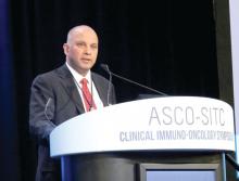
Similar associations may exist in other solid tumors, Shridar Ganesan, MD, reported at the ASCO-SITC Clinical Immuno-Oncology Symposium.
There is a known correlation between mutation burden and response to immune checkpoint blockade in some, but not all, cancer types. Increasing mutation burden is generally associated with increasing response rate, but there are exceptions, said Dr. Ganesan, chief of molecular oncology at the Rutgers Cancer Institute of New Jersey, New Brunswick.Merkel cell carcinoma, for example, has a high response rate despite its relatively low mutation burden. This is explained by the presence of infection with the Merkel cell polyomavirus in a low mutation burden subset, he said, noting that expression of the virus likely leads to expression of antigens that makes the disease both highly immunogenic and responsive to immune checkpoint therapy.
Expression of exogenous viruses is associated with response to immune checkpoint therapy in other cancers with low mutation burden, such as Epstein-Barr virus-positive gastric cancer, natural killer lymphoma, and Hodgkin disease.
“Intriguingly, renal cell carcinoma also has a relatively higher response rate to immune checkpoint therapy than would be anticipated by its relatively low mutation burden. However, this has no evidence of exogenous virus infection. Therefore, we turned our attention to endogenous retroviruses, which are an abundant potential stimulant of innate immunity in cancer,” he said.
Endogenous retroviruses (ERVs) now comprise about 5-8% of the human genome, and almost all of them have mutations that disable key coding genes of the retrovirus, meaning most cannot produce all the proteins necessary for viral replication.
“They’re essentially genomic fossils,” Dr. Ganesan said, adding that they all are silenced epigenetically in most normal adult tissues, both by DNA methylation and histone methylation. “However, inappropriate expression of endogenous retroviruses has been reported by multiple groups in some cancers and has been associated with evidence of immune activation. One can imagine that endogenous retroviral expression can be immunogenic both by expression of some [open reading frames] that are not disabled ... or just the viral RNA itself, which can be detected by cytoplasmic sensors ... and activate innate immunity,” he said.
To assess whether ERV expression corresponds with evidence of immune checkpoint activation in cancer, he and his colleagues conducted a pan-cancer analysis of more than 4,900 tumors across 21 cancer types using data from the Cancer Genome Atlas (TCGA).
“When we did this for about 66 annotated endogenous retroviruses in the TCGA database, we could see that different cancers had different amounts of retrovirus associated with immune activation ... in fact the strongest signal seen in this analysis was in clear cell renal cell carcinoma,” he said.
Signals were also seen in estrogen receptor–positive/human epidermal growth factor receptor 2-negative breast cancer, head and neck squamous cell carcinoma, and colon cancer.
A variety of ERVs were upregulated in RCC; different panels were associated with different cancer types. But two – ERVK.2 and ERV3.2 – were consistently upregulated across all the tumor types.
A closer look at ERV expression showed three clear clusters: extremely-high, intermediate, and low expression of ERV, and the very-high expression cluster had increased expression of numerous checkpoint genes, including CD8A, PD1, CTLA4, and LAG3 among others, compared with the low expression cluster.
This leads to the question of why a subset of RCCs have ERV expression.
In an attempt to answer that question, a transcript analysis was conducted to determine which transcripts are differentially regulated between the high and low ERV-expressing groups, and gene ontology analysis was performed.
“The results were quite striking. If you look at the modules that are differentially expressed between the high-ERV and the low-ERV groups, what pops up is really a lot of histone methyltransferase modules and chromatin regulation modules, ” he said, noting that this makes sense because of the silencing of ERV expression by histone methylation and DNA methylation, and suggests that “there is some deep abnormality in chromatin modulation in this subset of RCCs.
“This is intriguing, because RCCs are known to have mutations in chromatin modifying genes,” he said.
A look into whether the high-ERV–expressing group was enriched in any of these mutations showed some enrichment of BAP1 in the high- vs. low-expressing group, and that is currently being looked at further, he noted.
The next question was whether ERV expression correlated with response to immune checkpoint blockade in ccRCC, and this was looked at in a nonrandomized group of 15 patients with metastatic ccRCC who were treated with single-agent immune checkpoint therapy who had either clearly documented partial response or progressive disease. In 13 patient samples for which RNA expression of ERV3.2 was successfully measured by quantitative real-time polymerase chain reaction using two primer sets, ERV3.2 expression was significantly higher in responders vs. nonresponders in both primer sets (P less than .05 and .005).
“In summary, we have shown that expression of ERVs correlates with immune activation and increased expression of immune checkpoint genes in a subset of ccRCC, and perhaps several other solid tumor classes, and the expression of ERV3.2 is perhaps associated with response to PD1 blockade in this small preliminary cohort of ccRCC patients,” he said, noting that abnormal expression of ERV may be a biomarker of immune checkpoint therapy response in some cancers with a low mutation burden. “Mechanisms underlying ERV expression need to be investigated and may reflect underlying chromatin alterations or epigenetic abnormalities.”
Dr. Ganesan reported that his spouse is employed by Merck and that he is a consultant and/or advisory board member for Novartis, Roche, and Inspirata. He also holds patents with Inspirata.
sworcester@frontlinemedcom.com
SOURCE: Panda A et al., ASCO-SITC, Abstract #104.


