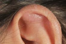
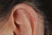
A 53-year-old white female presented for routine evaluation of psoriasis vulgaris. She was scheduled to see an ENT for evaluation of a problem with soreness and swelling involving her left ear. She denied any ear trauma, but did note sleeping on her left side because of some right hip pain. She noted gradual swelling of her ear with some tenderness, beginning 3 weeks before presentation. On physical examination, there was a 1.5- by 2.0-cm cystic mass involving the scaphoid fossa of her left ear. Aspiration using a 21-gauge needle and a 3-cc syringe yielded yellow serous fluid.
Diagnosis: Auricular Pseudocyst. The patient presented 4 days later with recurrence. Another drainage was performed following the same process as described. She presented 7 days after her second treatment with a third recurrence.
Auricular pseudocysts are uncommon, noninflammatory, fluctuant swellings of the ear believed to be caused by accumulation of glycosaminoglycans and subsequent ischemic necrosis of the cartilage, or from repeated minor trauma to the ear. They can be a therapeutic challenge.
Many treatment options have been used with variable success, including simple aspiration, plaster of paris casts, injectable agents (steroids, trichloroacetic acid, and minocycline), and surgical intervention incorporating incision, drainage, and curettage. Some clinicians apply sclerosing agents, such as fibrin glue, as part of their surgical intervention to eliminate the space. More complicated surgery involves excision of the anterior cartilage of the ear and compression buttoning of the pseudocyst.
Surgical intervention is not always successful and can lead to cauliflower and floppy ear deformities. Surgical bolsters have been reported in numerous publications and are a proven alternative to surgery, but can be bulky and uncomfortable.
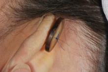
In this patient’s case, a single button was selected to fit in the inside of the patient’s ear. The intention was to use a button with smooth edges large enough to fit comfortably over the superior crura of the antihelix and the scapha, directly over the region of the pseudocyst. A larger button was selected for behind the ear, again with rounded and smooth edges. Both of the buttons were placed in a sterilization pack and autoclaved.
The pseudocyst was drained, and the anterior and posterior portion of the ear where the suture was placed was anesthetized using 2% lidocaine with epinephrine. Using 5-0 Prolene suture, both buttons were secured into place. The suture was passed through the skin and cartilage, allowing the secured buttons to apply direct pressure against the skin and perichondrium of the ear. The buttons were kept in place for a week.
Using needle aspiration of auricular pseudocysts followed by placement of button bolsters has been successful in two patients. No other surgical or sclerosing intervention was used. Button bolsters are a safe, comfortable, and effective form of treatment for auricular pseudocysts. The buttons should be kept in place for 7-14 days.
Dr. Samlaska is in private practice in Nevada.


