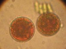
Certain plant constituents have been established as exerting photoprotective effects, such as antioxidants, which include carotenoids, tocopherols, vitamin C, and flavonoids and other polyphenols. Researchers have shown that carotenoids are superior scavengers of singlet oxygen and sufficient scavengers of other reactive oxygen species (Nutrition 2001;17:818-22). As dietary antioxidants, carotenoids have been shown to confer photoprotective benefits to human skin, efficiently scavenging peroxyl radicals and hampering lipid peroxidation (Photochem. Photobiol. 2002;75:503-6).
Carotenoid pigments, which consist of more than 600 compounds, are secondary metabolic plant products that confer yellow, green, red, or orange color to fruits and vegetables. They are precursors of vitamin A (retinol). Astaxanthin, a member of the xanthophyll class of carotenoids and closely related to beta-carotene, lutein, and zeaxanthin (J. Nat. Prod. 2006;69:443-9), is a red-orange carotenoid present in plants and algae that is also known to contribute to the color of seafood such as salmon, shrimp, and lobster. It is synthesized by only a few bacteria, including Brevibacterium, Mycobacterium lacticola, Agrobacterium auratim, and Haematococcus pluvialis, and the yeast Phaffia rhodozyma (Bioresour. Technol. 2004;92:209-14). Astaxanthin is also found in bird species, such as flamingos and quails, but cannot be synthesized by them (J. Nat. Prod. 2006;69:443-9).
Sources. The richest source of astaxanthin is the green microalga H. pluvialis (J. Nat. Prod. 2006;69:443-9), which contains more than 80% astaxanthin in its cells (Bioresour. Technol. 2004;92:209-14). Because of its rapid growth and high astaxanthin content, H. pluvialis is now produced at industrial scale, and its commercial production has grown significantly worldwide during the last decade (Trends Biotechnol. 2003;21:210-6). Astaxanthin also can be derived from the fermentation of the pink yeast Xanthophyllomyces dendrorhous, extracted from crustacea (e.g., Antarctic krill, Euphausia superba), which have a limited ability to synthesize astaxanthin from dietary carotenoids, and produced synthetically. Mammals can neither produce astaxanthin nor convert dietary astaxanthin into vitamin A (Trends Biotechnol. 2003;21:210-6).
Although this potent carotenoid is expensive to produce, isolate, and purify, investigators recently demonstrated a method of fermenting P. rhodozyma (X. dendrorhous) yeast cells with brewer malt waste, yielding a significant amount of astaxanthin with demonstrated antiproliferative effectiveness in two tested breast cancer cell lines (Int. J. Mol. Med. 2005;16:931-6). Indeed, astaxanthin is known to exhibit antiproliferative properties against skin and breast cancer (Int. J. Mol. Med. 2005;16:931-6).
Research Findings. In in vitro studies performed during the last two decades, astaxanthin has been shown to exhibit significantly greater antioxidant capacity than beta-carotene (Nutr. Res. 1995;15:1695-1704; Arch. Biochem. Biophys. 1992;297:291-5; Lipids 1989;24:659-61). In fact, this xanthophyllic carotenoid has demonstrated up to several-fold stronger antioxidant activity than vitamin E and beta-carotene (Trends Biotechnol. 2003;21:210-6; Physiol. Chem. Phys. Med. NMR 1990;22:27-38), as well as other carotenoids (J. Nat. Prod. 2006;69:443-9; Trends Biotechnol. 2003;21:210-6; Crit. Rev. Food Sci. Nutr. 2006;46:185-96).
Astaxanthin also appears to have the potential to be significantly more effective than beta-carotene and lutein at preventing UV-induced lipid photo-oxidation (J. Dermatol. Sci. 1998;16:226-30; Trends Biotechnol. 2003;21:210-6).
In a study conducted in 1998, investigators found that beta-carotene, lutein, and astaxanthin exhibited protective activity against UVA-induced oxidative stress in cultured rat kidney fibroblasts. Of the three carotenoids, astaxanthin demonstrated the most potent protective capacity (J. Dermatol. Sci. 1998;16:226-30).
Ten years ago, investigators tested a proprietary algal extract containing a significant amount of astaxanthin to determine its protective capacity against UVA-induced DNA damage in human skin fibroblasts, human melanocytes, and human intestinal CaCo-2 cells. They also compared the effects with those of a synthetic astaxanthin. The results showed that the synthetic astaxanthin prevented UVA-induced damage in all cell types at all tested concentrations (10 nmol, 100 nmol, and 10 micromol), whereas the algal extract conferred protection in all cell types only at the highest concentration. In addition, 2-hour exposure to UVA led to significant cellular superoxide dismutase activity in fibroblast cells, along with a substantial decrease in cellular glutathione content. Eighteen-hour preincubation with 10 micromol of either form of astaxanthin, however, prevented both of these UVA-induced changes in fibroblast cells. Simultaneous incubation with either form of astaxanthin also prevented UVA-induced depletion of glutathione content in CaCo-2 cells, but did not affect superoxide dismutase activity. Overall, the researchers concluded that the algal extract containing astaxanthin performed comparably to synthetic astaxanthin, and warrants attention as an antioxidant that may deliver health benefits (J. Dermatol. Sci. 2002;30:73-84).
Recently, investigators studied in vitro the effects of the carotenoids astaxanthin and canthaxanthin on gap junctional intercellular communication, which is integral in homeostasis, growth control, and cell development, and is known to be impaired in cancer cells. They exposed primary human skin fibroblasts to 0.001 to 10 micromol/L of the carotenoids, and used a dye transfer assay to measure gap junctional communication. Intercellular communication increased after 24 and 72 hours of incubation with canthaxanthin, but decreased significantly with astaxanthin, with a reverse seen upon withdrawal of astaxanthin. Using Western blot analysis, the investigators found that astaxanthin influences channel function by altering the phosphorylation pattern of the protein connexin, lowering the phosphorylation state (J. Nutr. 2005;135:2507-11).


