(A) Paratubal cyst CORRECT
Paratubal, or paraovarian, cysts typically are round or oval avascular hypoechoic cysts (long arrow) separate from the adjacent ovary (short arrow). Since they are congenital remnants of the Wolffian duct, they arise from the mesosalpinx, specifically the broad ligament or fallopian tube.1,2 They usually are seen in close proximity to but separate from the ovary without distorting the ovary’s architecture.1,2
B) Hydrosalpinx INCORRECT
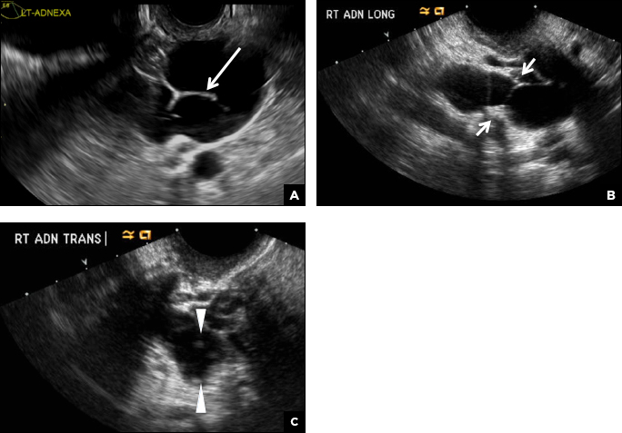
A hydrosalpinx appears as an elongated C- or S-shaped, thin-walled tubular serpiginous cystic lesion separate from the ovary. It often has incomplete septations that are infolding of the tube on itself (long arrow).3 Other findings include diametrically opposed indentations (short arrows) of the wall (Waist sign) and short linear mucosal or submucosal folds (arrowhead) that when viewed in cross section appear similar to the spokes of a cogwheel (Cogwheel sign).1–3 Prior tubal infection or gynecologic surgery represent risk factors for hydrosalpinx.

Hydrosalpinx. (A) Transvaginal pelvic ultrasound of the left adnexa demonstrates an elongated C- or S-shaped, thin-walled tubular serpiginous cystic lesion with incomplete septations (long arrow). (B) Longitudinal image of the right adnexa shows the dilated fallopian tube with diametrically opposed indentations of the wall consistent with the Waist sign (short arrows). (C) Transverse image of the dilated fallopian tube viewed in cross section has the appearance of several short mural nodules similar to the spokes of a cogwheel (arrowheads).
C) Peritoneal inclusion cyst INCORRECT
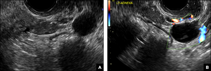
A peritoneal inclusion cyst appears as an anechoic cystic mass that conforms passively to the shape of the peritoneal cavity/pelvic sidewall (long arrow) and may contain entrapped ovaries (short arrow) within or along the periphery of the fluid collection.1,2 Septations within it are likely from peritoneal adhesions (arrowhead) and may show vascularity.2 Prior (often multiple) gynecologic surgeries represent a risk factor for peritoneal inclusion cysts.

Peritoneal inclusion cyst. (A) Longitudinal transvaginal pelvic ultrasound of the left adnexa demonstrating an anechoic cystic lesion that conforms passively to the shape of the peritoneal cavity/pelvic sidewall (long arrow) with a thick septation (arrowhead). (B) Transverse image demonstrates the left ovary entrapped within the fluid collection (short arrow).
D) Dilated pelvic veins INCORRECT
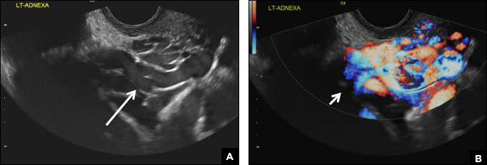
Dilated pelvic veins appear on sonography as a cluster of elongated and tubular cystic lesions in the adnexa along the broad ligament and demonstrate low level echoes due to sluggish flow (long arrow) and visible red blood cell rouleaux formation. This can be confirmed on color Doppler images (short arrow) and help differentiate it from hydrosalpinx.

Dilated pelvic veins. (A) Transvaginal pelvic ultrasound of the left adnexa reveals a cluster of elongated and tubular cystic lesions that demonstrate low level echoes due to sluggish flow (long arrow). (B) Color Doppler ultrasound confirms vascularity within these dilated pelvic veins (short arrow).


