GYN Image Quiz

Adenomyosis in the spotlight, but which sign is featured?

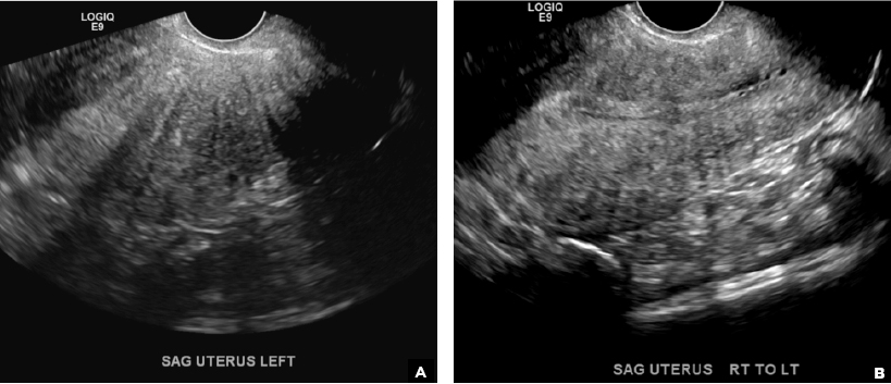
Can you identify which feature of adenomyosis is found on these ultrasound images?

Author and Disclosure Information

A 37-year-old woman with heavy menstrual bleeding and dysmenorrhea presents to her gynecologist. Physical exam suggests an enlarged uterus; the gynecologist orders pelvic ultrasonography.

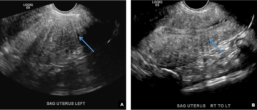
Recognizing that, in a given patient, multiple features of adenomyosis may be imaged, which imaging feature of adenomyosis is highlighted above?

Globular enlarged uterus

Cystic myometrial spaces

Asymmetric myometrial thickening

Indistinct endomyometrial interface

Myometrial linear striations

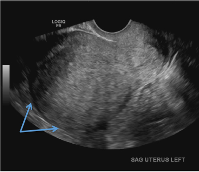
A) Globular enlarged uterus INCORRECT
A homogeneously enlarged uterus in the absence of fibroids is characteristic of adenomyosis.1

Transvaginal pelvic ultrasound demonstrates a homogenously enlarged uterus (arrows) without evidence of a focal fibroid.

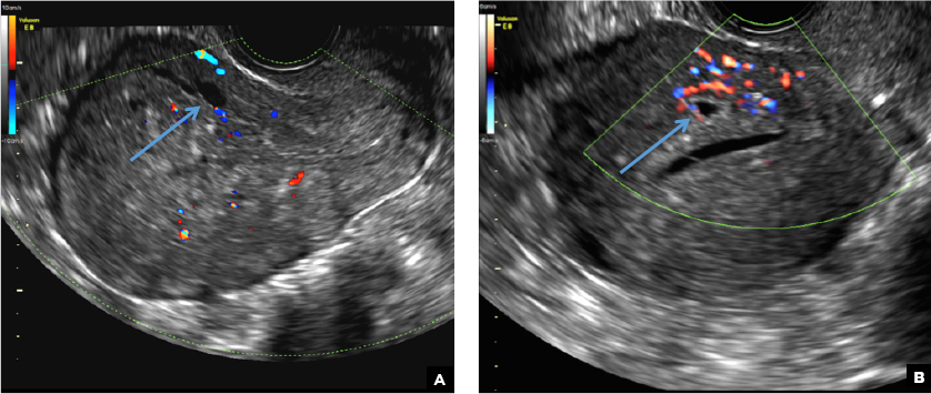
B) Cystic myometrial spaces INCORRECT
Myometrial cysts are dilated cystic glands or foci of hemorrhage within heterotopic endometrial tissue.2 They are often less than 5 mm in size but can be extensive and of variable sizes and can be differentiated from arcuate veins seen in the outer myometrium by the use of color Doppler.1,2

Transvaginal pelvic ultrasound on 2 different patients demonstrates well-defined anechoic cysts in the myometrium without vascular flow on color Doppler (long arrows).

C) Asymmetric myometrial thickening INCORRECT
The finding of asymmetric uterine wall thickening in adenomyosis is usually seen when there is focal disease and presents with wall thickening that demonstrates anteroposterior asymmetry.1

(A) Transvaginal pelvic ultrasound demonstrates asymmetric wall thickening of the posterior myometrium (long arrow). (B) Transvaginal pelvic ultrasound of a different patient demonstrates asymmetric wall thickening of the anterior myometrium (long arrow).

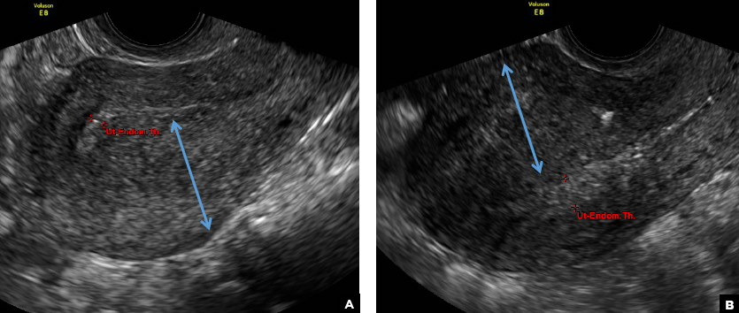
D) Indistinct endomyometrial interface INCORRECT
The heterotopic endometrial tissue invading the myometrium obscures the normal endometrial myometrial border with pseudo-widening of the endometrial echo resulting in poor definition of the endomyometrial junction.1,2

Transvaginal pelvic ultrasounds from 2 different patients demonstrate poor definition of the endomyometrial interface (short arrows) in addition to the globular appearance of the uterus (long arrows).

E) Myometrial linear striations CORRECT
A hyperplastic reaction to the infiltration of the heterotrophic endometrial glands into the myometrium results in radiating linear echogenic striations (sometimes referred to as the "venetian blinds" sign).1,2

Transvaginal pelvic ultrasound images demonstrate myometrial linear echogenic striations (long arrows) radiating from the anterior uterine wall.

Recommended Reading

Optimizing HPV vaccination
MDedge ObGyn
Novel device aims to make cervical cancer screening more accessible
MDedge ObGyn
Ob.gyns. split on cosmetic genital surgery
MDedge ObGyn
Hormonal IUD is most cost-effective menorrhagia management
MDedge ObGyn
Oral prophylaxis and vaginal ring effective in adolescent HIV prevention
MDedge ObGyn
FDA extends Liletta IUD duration of use to 4 years
MDedge ObGyn
HPV vaccination rose after ACA implementation
MDedge ObGyn
C-section raises hysterectomy complication risk later
MDedge ObGyn
Metronidazole clears PID anaerobes with no drop in antibiotic compliance
MDedge ObGyn
Copper IUDs increase bacterial vaginosis risk
MDedge ObGyn