CHICAGO – Autologous engineered skin substitutes (EES) reduce mortality and donor-site harvesting in children with extensive, deep burns, a randomized, paired-site comparison showed.
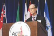
“Autologous ESS offer an alternative therapy for closure of extensive, excised, full-thickness burns,” Steven Boyce, Ph.D., said at the annual meeting of the American Burn Association.
The skin substitutes were engineered using split-thickness skin biopsies from 16 children with full-thickness burns covering an average total body surface area (TBSA) of 78% and a high of 95%.
The ESS were composed of autologous cultured keratinocytes and dermal fibroblasts attached to a collagen-based, biopolymer scaffold.
Clinicians are increasingly turning to tissue engineering as a way to address the shortfalls associated with the prevailing standard of care, autologous skin grafting, including lack of donor sites in larger burns, increased morbidity from autograft harvesting and skin grafts, and scarring.
At the 2014 ABA meeting, Canadian researchers reported on an autologous skin substitute that had both dermal and epidermal components. It took 2 months to prepare, whereas the Cincinnati model requires about 1 month to prepare and offers greater availability and stability of the closed wounds, Dr. Boyce said in an interview.
For the current study, Dr. Boyce and his associates compared mortality after treatment with ESS in 16 children with data from 1,008 patients from the National Burn Repository who were of similar ages, had 77% full-thickness TBSA burns, and were treated with meshed and expanded split-thickness skin autografts (AG). One patient died before ESS were prepared, leaving 15 patients in whom 2,056 ESS grafts were applied in 59 operations. Their average age was 6.3 years, 13 were male, and the average time to first ESS was 32 days.
Mortality was 6.25% (1/16) with the skin substitutes and 30.3% (305/1,008) with the autografts (P value < .05), said Dr. Boyce, a professor with the University of Cincinnati and director of the Skin Substitute Laboratories, Shriners Hospitals for Children, both in Cincinnati.
Rates of engraftment at postoperative day 14, however, significantly favored the AG group at 96.5 vs. 83.5% for ESS (P < .05).
One major reason for reduced rates of engraftment is lack of blood vessels in current models of engineered skin, he explained.
At day 28, the percentage of TBSA closed was approximately 30% for ESS and 47% for AG, while the ratio of closed-to-donor areas was significantly greater for ESS (108.7 vs. 4.0; P < .001).
The correlation between the percentage TBSA closed with ESS and TBSA burned generated an R-squared value of 0.65 (P < .001), Dr. Boyce said.
Regraftment rates were similar between graft types. Antibody formation to the biopolymer scaffold at postoperative day 28 was similar between patients receiving 1 or multiple ESS.
“Stable wound closure [with ESS] is interpreted to result from preservation of epidermal progenitor cells, formation in vitro of basement membrane between fibroblasts and keratinocytes, and generation of fibrovascular tissue by the biopolymer scaffold,” he said.
Tissue engineering offers patients the opportunity to survive with otherwise statistically lethal burns and “the data are very clear that happens with this product,” session comoderator and past ABA president Dr. David Ahrenholz said in an interview.
That said, “the product is very expensive, so I would not use it for patients in whom I had adequate donor sites, but it’s fairly easy to identify those patients on admission,” he added.
The study was supported by grants from the U.S. Department of Defense, National Institute of General Medical Sciences, and Shriners Hospitals for Children. The authors reported no current relevant financial disclosures.
On Twitter @pwendl


