User login
There’s More Where That Came From
ANSWER
The correct answer is pityriasis rosea (choice “d”).
Virtually all patients with this condition believe that they have a terrible case of “ringworm” (ie, fungal infection; choice “a”)—an opinion all too often corroborated by the medical provider unacquainted with pityriasis rosea. The two can be difficult to distinguish, but the “herald” patch, oval shape, odd color, and fine scale all serve to confirm the diagnosis. When necessary, a KOH prep or biopsy can be done.
Psoriasis (choice “b”) can manifest acutely, but it is distinctly salmon-pink under coarse, white scale that affects the palms, nails (with pits), and scalp. Psoriasis lesions are round (rather than oval), with coarser scale and without a herald patch.
The lack of antecedent sores and denial of sexual contact ruled out secondary syphilis (choice “c”). Patients with secondary syphilis often present with low-grade fever, malaise, and palmar scaly papules—all of which were missing in this case. Syphilis serology (rapid plasma reagin) can be easily obtained if doubt persists.
DISCUSSION
Pityriasis rosea (PR) is a papulosquamous eruption that is common in younger populations. About 40% of affected patients present with a large scaly lesion, which is followed by the appearance of multiple smaller oval lesions within days. In most cases, PR is fairly easy to diagnose: the lesion’s oval shape, pinkish-brown color, centripetal fine scale, herald patch, and adherence to skin tension lines are all characteristic findings.
Though the exact organism has not been identified, PR is almost certainly viral in origin. Like many viral exanthems, occurrence tends to peak in the spring and fall. There are also indications that the body builds immunity to the infection, since recurrence outside the acute phase is rare.
Treatment options are unsatisfactory, though exposure to UV sources appears to help. Patients must be informed that their condition will, unfortunately, last for at least six to nine weeks, during which crops of lesions will come and go.

About 10 days ago, an asymptomatic, scaly lesion arose overnight on this 21-year-old man’s upper left chest, near the anterior deltoid area. Within the past week, multiple scaly lesions—much smaller than the original—have appeared on his trunk and arms.
The patient claims to be in good health otherwise, denying fever, malaise, or myalgia. He denies sexual contact of any kind in the past two months. There is no personal or family history of skin disease or atopy.
On exam, the patient looks his stated age, is afebrile, and is in no acute distress. On his chest is an oval, pinkish brown, papulosquamous lesion with very fine, sparse scaling in the center. The long axis of the lesion parallels local skin tension lines.
About 20 additional lesions are observed elsewhere, primarily on the truncal skin, sparing the upper neck, face, and palms. All are oval, with the same odd color and scale, and follow skin tension lines. The patient’s elbows, knees, scalp, and nails are unaffected, as are the areas around and below the waist.

Slow and Steady: A Worrisome Pace
ANSWER
The false statement is that most melanomas are raised (choice “a”), since most melanomas are essentially macular (flat).
DISCUSSION
Misconceptions about melanomas often delay diagnosis, costing lives. In truth, the majority of melanomas are difficult, if not impossible, to feel on palpation. This is because they arise in the skin, rather than on it.
Malignant melanomas are cancers of melanocytes (the cells that line the basal cell layer). Overexposure to UV sources damages the nuclei of these cells, compromising their ability to repair the damage. This can lead to focally unregulated cell growth.
Initially, this uncontrolled cell replication spreads horizontally. Over time, though, it can grow vertically and penetrate deep enough to invade the vasculature (roughly 1 mm deep), and spread to the liver, lung, or brain. This is why ascertaining the depth of a melanoma is critical for predicting prognosis and determining the extent of additional surgery and search for evidence of metastatic disease.
Melanomas do not typically itch or bleed until they are advanced (if at all). And most arise de novo (as new lesions, rather than pre-existing). So, contrary to popular belief, moles rarely turn into melanomas.
It is also true that approximately 80% of all melanomas arise on skin normally covered by clothing, despite the role of sunlight in the development of melanoma. For reasons not totally understood, the combination of fair, su

For more than two years, a 43-year-old man has had an asymptomatic lesion on his right deltoid area. His primary care provider has repeatedly assured him of its benignancy “because it is flat.” But its slow, steady, horizontal growth concerns the patient, who is otherwise healthy.
He does admit to a significant history of sun exposure, explaining that he burns easily but acquires a tan by summer’s end each year. Several members of his immediate family have had skin cancers removed.
Examination reveals a round, dark macule, measuring 1.1 cm, on the right lateral deltoid area. The lesion has poorly defined borders and a variety of colors—mostly black and brown, with flecks of red and pink. There is no palpable component, and no nodes can be detected in the area.
The rest of his type II skin has abundant evidence of UV overexposure, including solar lentigines and weathering, especially around the neck.
The lesion is anesthetized, removed by deep shave technique, and submitted to pathology. The report shows a superficial, spreading melanoma with a Breslow depth of 0.75 mm. Malignant cells extend to the dermoepidermal junction (Clark level II). Only a few mitotic cells can be seen, with no intravascular invasion or signs of ulceration.

What’s Her Dry-agnosis?
ANSWER
The correct answer is eczema/atopic dermatitis (choice “c”).
Patients with eczema have a low threshold for itching, so they scratch, often making the condition appear far worse than it really is. In such cases, it’s typical for the problem to be mistaken for impetigo (choice “a”) or yeast infection (choice “d”). The latter, much like psoriasis (choice “b”), is quite rare in the perioral area. Furthermore, the patient had no indicative signs of psoriasis.
DISCUSSION
Eczema is one of several manifestations of atopic dermatitis, a syndrome that affects more than 20% of newborns in this country. These children have extraordinarily dry, thin skin that overreacts to wetting and drying, as well as scratching. Certain areas are especially prone to these changes and consequently develop scaling and itching.
The perioral area is one, in large part because it is kept moist by food, drink, nasal secretions, and saliva (due to habitual lip licking). The scaly rash becomes inflamed; on people with skin of color, this frequently manifests with hyperpigmentation.
When picked enough, this type of rash can become impetiginized—that is, superficially infected with staph or strep. But in this patient’s case, antibiotics were of no practical use.
A topical steroid ointment (hydrocortisone 2.5%) was used, and the patient was urged to stop picking (or licking!) and to use moisturizers (eg, petroleum jelly). The family was educated about the problem and its origins, and the parents were reassured of the self-limiting nature of postinflammatory hyperpigmentation (a major source of concern).
In this case, the key to making the correct diagnosis was the significance of the personal and family history of atopy—and an appre
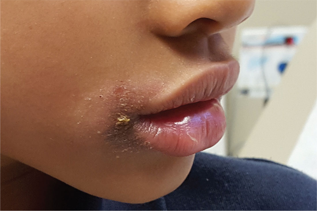
The persistent rash around this 5-year-old African-American girl’s mouth is causing a great deal of concern for her parents, who request referral to dermatology after four months of attempted treatment. Oral and topical antibiotics, as well as antifungal products (nystatin and fluconazole), have been used to no good effect.
The patient is often seen licking her lips, which dries and irritates them. Fine crusting surrounds her mouth, particularly the left lateral oral commissure. The skin in the affected areas is darker than the rest.
Elsewhere, her type IV skin is quite dry, with focal areas of scaling on the arms and antecubital region. No rash is seen on extensor areas, nor are there any changes in her nails.
The child is markedly atopic, as are her siblings, who are present for the exam. The patient and her siblings are all congested, breathing through their mouths (“allergies,” according to their parents).

All Is Not Swell
ANSWER
The correct answer is elephantiasis nostras verrucosa (ENV; choice “d”). Cellulitis (choice “a”), venous insufficiency (choice “b”), and lymphedema (choice “c”) are all factors in the broader diagnosis of ENV.
DISCUSSION
ENV is an unusual condition that represents hypertrophic fibrosis secondary to repeated episodes of lymphangitis. This begins with venous insufficiency, which is made worse by increasing obesity (which impedes venous return) and repeated bouts of cellulitis. With ENV, fibroblasts are increased due to extravasation of high-molecular-weight protein (lymphorrhea), which leads to a buildup of keratinocytes, ultimately expressing as extreme hyperkeratosis.
In this patient’s case, his sedentary lifestyle and constant seated position contribute to the problem. Many of his past treatments were reasonable, but—as in many ENV cases—his condition is beyond the point of treatment.
Typically, in-home treatment includes compression and elevation of the legs. Topical application of urea creams is often used to soften the rough skin, but in this patient’s case, the cream burned so badly that it was of no use. Alas, the very things he needs to do are those he cannot: walk, burn calories, and avoid long periods of inactivity.
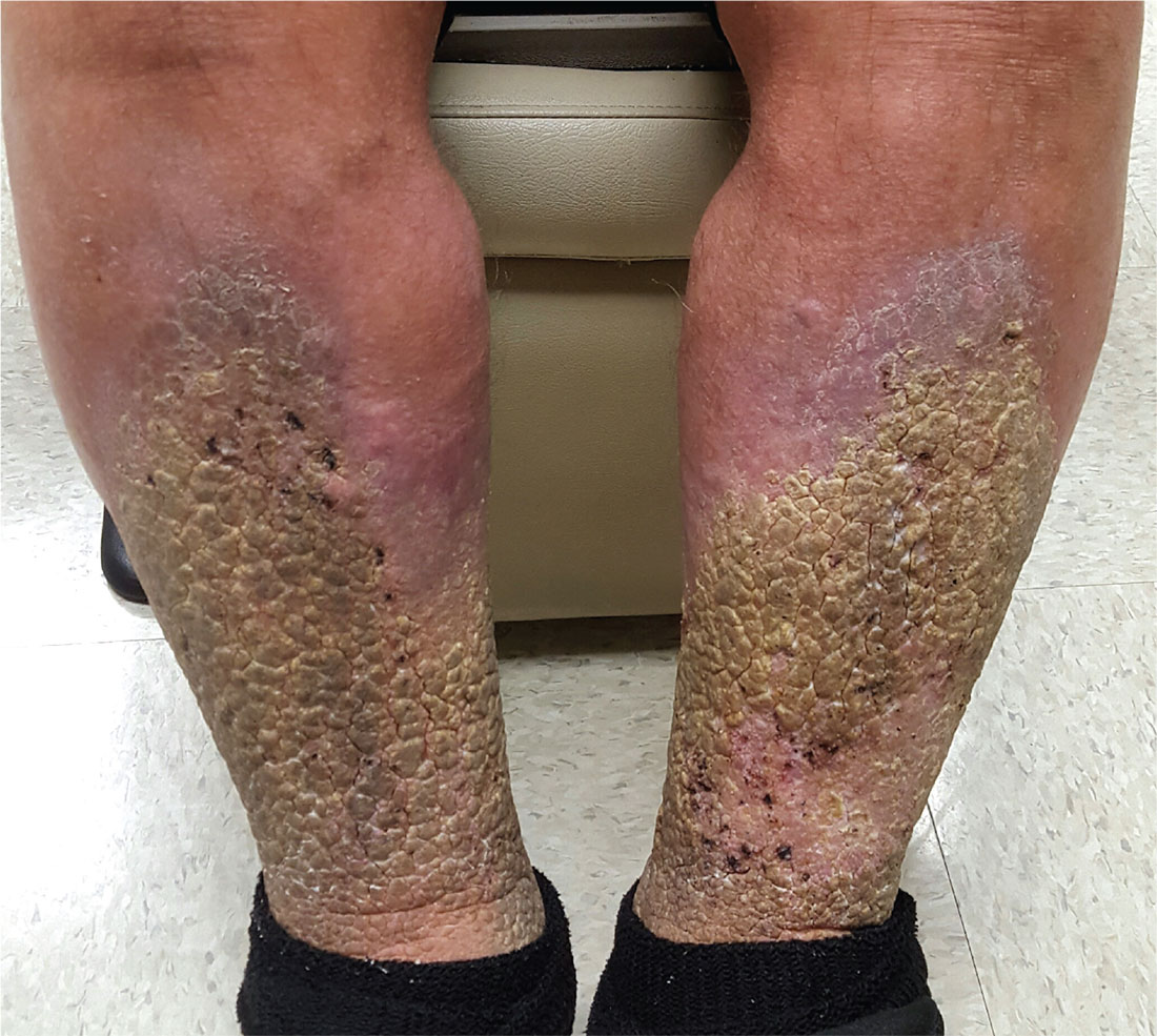
A 70-year-old man is referred to dermatology after trying “everything else” for problems he has had for at least 15 years. In that time, he has been hospitalized repeatedly for swelling and pain in his legs, with odoriferous drainage.
Despite extensive treatment attempts—multiple antibiotics, oral and topical steroids, and OTC creams—the condition is worsening. In ho
The patient denies having cancer or deep vein thrombosis (both of which he has been thoroughly checked for), as well as congestive heart failure. He states that almost every morning, upon rising, the swelling in his legs is considerably lessened.
Both legs are swollen, red, and edematous from just below the knees down. Advanced, pebbly, hyperkeratotic plaques cover the lower two-thirds of both legs, favoring the anterior over the posterior portions. Pitting edema is elicited with minimal digital pressure but does not cause any pain.The patient is in no acute distress but is clearly uncomfortable. He has been confined to a wheelchair for years due to back problems; he can barely stand when asked to do so. He is extremely obese.

Boy’s Dark Side Comes Out
ANSWER
The correct answer is Becker nevus (BN; choice “c”).
Because the lesion was not congenital, a number of possibilities, including congenital melanocytic nevus (choice “a”), were ruled out. Another differential item that was eliminated was nevus spilus (choice “b”), a tan congenital nevus covered with darker, punctate macules that usually manifest on extremities. These have little, if any, malignant potential.
And while BN is not associated with malignancy, it is possible for it to co-exist with melanoma (choice “d”). Therefore, atypical BN cases may need to be followed or serially biopsied.
DISCUSSION
BN is unusual but not rare. Also referred to as Becker melanosis, the condition affects approximately 0.5% of the population. It primarily manifests in young men during early puberty, although women can develop BN.
Histologic signs of BN include epidermal thickening, elongation of rete ridges, and increased bundles of smooth muscle in the dermis.
Androgen causation is strongly indicated by the condition’s predominance in males and onset at puberty, as well as its associated hypertrichosis and increased density of androgen receptors in affected areas. Variations of the condition can involve the legs, arms, or face. It is possible for BN to manifest without additional hair growth. Hypoplasia of ipsilateral pectoral structures (or the ipsilateral breast in women) has also been reported.
Several types of lasers can be used to lighten the hyperpigmentation and remove hairs. This treatment modality yields variable results.

This 14-year-old boy’s family is alarmed by a darkening patch of skin on the right side of his chest and shoulder, which first appeared two years ago. Over the span of a few months, dark hairs grew on the hyperpigmented area.
The family’s primary care provider assured them that it was likely benign, but the lack of a specific diagnosis left them concerned. They requested referral to dermatology.
A large portion of the patient’s right anterior chest and shoulder is completely covered by uniformly hyperpigmented, hypertrichotic skin, which feels a bit thicker than the unaffected skin. The lesion’s borders are quite irregular and uneven; beyond them, no hairs can be seen. The problem is confined to the chest and shoulder; although the medial border extends to the sternal area, the lower edge stops short of the pectoral area.
The patient denies any symptoms and claims to be healthy in all other respects. There is no family history of similar problems.

When You Can’t Make a Rash Diagnosis
ANSWER
The false statement—and therefore the correct choice—is that MF is almost always fatal (choice “a”). MF can often be controlled, if not completely cured.
DISCUSSION
In its early stages, cutaneous T-cell lymphoma (CTCL) can manifest with an innocuous-appearing rash, notable for its chronicity and resistance to treatment. One example is poikiloderma vasculare atrophicans (PVA), which is identified by nonblanchable atrophic patches with fine surface vascularity that often manifest around the waistline or groin. Left undiagnosed and untreated, PVA can slowly progress to a more advanced stage, as was the case with this patient.
Several cancers can present with rash, including extramammary Paget disease, superficial squamous cell carcinoma (Bowen disease), and various types of metastatic cancer (eg, breast, colon, lung).
The confirmation of CTCL/MF may require serial biopsies over time, as the diagnostic signs can take years to become detectable. These specimens must be accompanied by pertinent clinical information to suggest a differential that includes this lymphoma.
Early-stage CTCL can be controlled with topical steroids, but as the condition advances, specialized treatment is needed. The tumor stage observed in
For years, this 64-year-old man has complained of itching and a rash around his head and neck. He has consulted several primary care providers—and even a dermatologist. A punch biopsy performed by that provider yielded no clear diagnosis. The patient was advised to return for follow-up but never did so.
Treatment was attempted with a succession of medications; none resulted in any improvement. The list includes antifungal creams (econazole, clotrimazole, and miconazole), an oral antifungal medication (a one-month course of terbinafine 250 mg/d), and a corticosteroid (a one-month course of prednisone 20 mg/d).
In addition to the rash and pruritus, the patient feels “lumps” in the affected areas. He also reports feeling more tired than usual. Prior to the onset of these symptoms, his only complaint was lifelong eczema.
Large infiltrative plaques are seen on both sides of his neck, extending into his ears and onto his scalp. A few exceed 8 cm in diameter, and all have smooth surfaces with no epidermal disturbance. Several discrete, 2- to 4-cm, fixed nodules are also seen and felt on his neck below these plaques.
A 5-mm punch biopsy is performed on one of the plaques on his occipital scalp; the pathology report shows only chronic changes consistent with eczema. The decision is made to perform another biopsy. A deeper, wider, 5-cm wedge from the left preauricular plaque is taken and submitted. The report shows changes consistent with tumor-stage mycosis fungoides (MF).

Seeing Redness and Ear-itation
ANSWER
The correct diagnosis is relapsing polychondritis (RP; choice “a”). The lack of surface changes in the affected skin rules out contact dermatitis, while the lack of a positive response to antibiotics and absence of an entrance wound eliminate the possibility of an infectious etiology.
DISCUSSION
There are no tests to confirm the diagnosis of RP. It is a rare autoimmune condition that usually manifests in the later decades of life and equally affects men and women.
RP’s ability to appear in cartilage anywhere in the body and in a variety of forms makes timely diagnosis almost impossible. But this case illustrates some diagnostically useful signs to watch for.
The unexplained erythema in the ear, which very obviously spared the cartilage-free lobe, prompted a biopsy of the cartilage; this showed changes consistent with RP. A subsequent review of the patient’s ophthalmology records indicated a chronic episcleritis, most likely due to inflammation of eyelid cartilage.
Further testing was performed to rule out other explanations, such as gout, or autoimmune diseases, such as lupus. Results were negative.
The patient was then referred to a pulmonologist, who found no respiratory involvement, and a rheumatologist, for further evaluation (including blood work) to rule out other conditions and end-organ (eg, renal) involvement.
On follow-up, the patient was responding well to prednisone prescribed by her rheumatologist. Given her limited disease, her prognosis is fairly good.
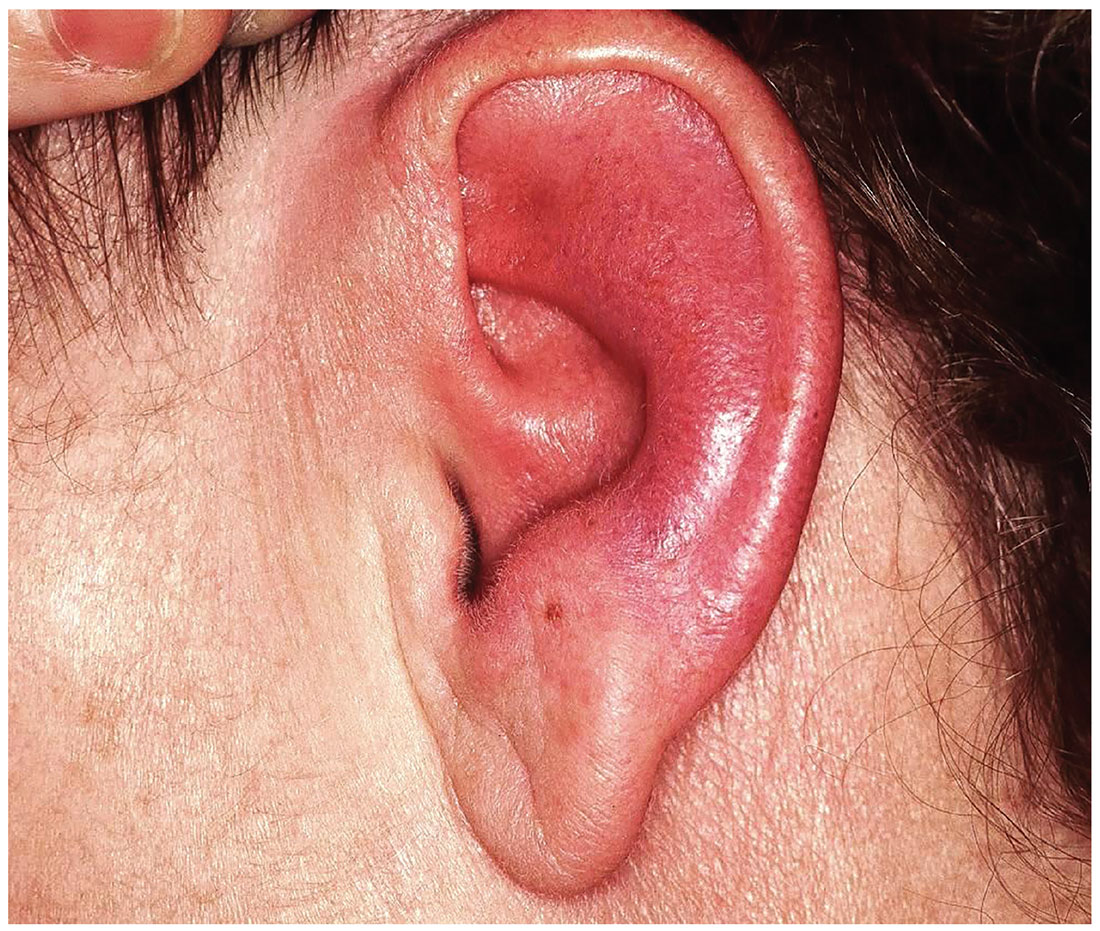
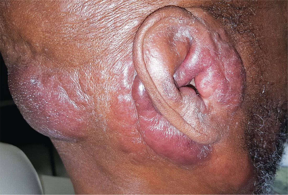
Several months ago, family members pointed out that this 60-year-old woman’s left ear was red. She consulted her primary care provider, who prescribed antibiotics. But when these failed to clear the problem, she was referred to dermatology.
Today, the patient complains of some discomfort in the ear but denies actual pain; she is, for example, able to sleep despite the problem. She reports that the redness manifested slowly but has spread over time to encompass most of her ear.
Uniformly distributed, bright red erythema on the left ear spares only the lobe. No wound or epidermal component (eg, scaling or blistering) is noted. However, there is increased warmth and tenderness on palpation of the erythematous portion. No nodes can be felt in the vicinity, nor are any abnormalities observed in the other ear.
The patient denies other skin problems, joint pain, and breathing difficulty. But she does have an ongoing history of irritation in both eyes. She has been seeing an ophthalmologist for months without relief. On examination, both eyes appear injected, with slightly swollen eyelids. Inspection and palpation of the nose reveal no abnormalities.

More Than a Spot of Bother
ANSWER
The correct answer is vitiligo (choice “b”).
DISCUSSION
Vitiligo develops when pigment cells (melanocytes) fail or die. Although there appears to be a hereditary component in some cases, as well as a connection to autoimmune disease, environmental factors (eg, intense sun exposure, stress) may also play a role.
This patient has nonsegmental vitiligo (NSV), the most common form. It is usually symmetrically distributed on high-friction areas, such as hands, knees, and elbows, as well as around the eyes and mouth. Segmental vitiligo, which affects only 10% of all vitiligo patients, tends to manifest during adolescence and typically remains confined to one area.
Unfortunately, for the majority of those with NSV (including the case patient), the condition tends to be progressive—and it responds poorly to treatment with topical steroids, calcineurin inhibitors, or phototherapy. As seen in this case, it can cause pigment loss in or around lesions; in fact, if left alone, the lesion may completely lose color. And NSV can encompass wider areas of involvement—to the extent that some patients lose all the pigment in their bodies. The resulting psychiatric fallout is considerable, especially in darker-skinned patients.
This patient's lesion will likely double in size by adulthood, which will not only subject him to ridicule but also increase the risk for malignant transformation. For this reason, he was advised to have the lesion excised under general anesthesia. He was also started on a regimen of a topical steroid cream and a calcineurin inhibitor on alternating days, but his prognosis is, in all honesty, poor.
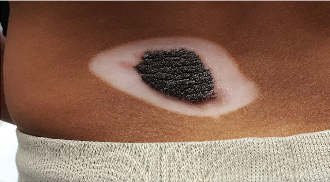
A 5-year-old boy is referred to dermatology for evaluation of recent color changes to the skin around a congenital lesion. Located on his mid low back, the polygonal lesion measures 8 x 5 cm and is uniformly dark brown with a mammillated, hair-bearing surface. The plaque has grown proportionately with the child but otherwise remained stable.
A year ago, however, the patient’s family noticed that the normal brown skin around the lesion was turning white. This “halo” became noticeably larger over the span of the year—effectively doubling the size of the lesion.
The child’s type V skin is in sharp contrast to the porcelain white band that parallels the margins of his lesion. The surface of the depigmented skin is completely smooth, with no epidermal changes. Faint but definite depigmentation is noted on the periocular skin of both eyes, in addition to well-defined depigmentation on his fingertips and the perionychial areas of all 10 fingers.
The family asserts that the boy is otherwise healthy and that there is no family history of similar phenomena. The rest of the examination is unremarkable.

Thinking Pimple? That’s Too Simple
ANSWER
The correct answer is to perform a punch biopsy (choice “b”). This will help establish the exact nature of the problem, which will dictate rational treatment.
The patient doesn’t have acne, so the suggested treatment options (choices “a,” “c,” and “d”) would be of no use. With cryotherapy, furthermore, there is a risk of leaving a permanent blemish on her skin.
DISCUSSION
A sample of one lesion was obtained via 3-mm punch biopsy and the resulting defect closed with a single suture. Pathologic examination showed the specimen to be a vellus hair cyst (VHC). In this case, it was one of many, making the diagnosis eruptive vellus hair cysts.
VHC, which can be acquired or inherited, typically manifests in the first two decades of life. In this developmental abnormality, a gradual disruption occurs between the proximal and distal portions of the vellus hair follicle, usually at the level of the infundibulum. As a result, the characteristic papule (which holds the retained hair) forms and the hair bulb atrophies.
The lesions may be solitary or appear in clusters on the body; they are easily mistaken for acne, milia, or even molluscum. As this case demonstrates, biopsy is often necessary to establish the correct diagnosis.
One final note about biopsy: It is best to incise each lesion with an 18-gauge needle tip or #11 blade and express the contents. This tedious process causes some discomfort for the patient, but it is quite effective and, if done correctly, should not leave a permanent mark on the skin.
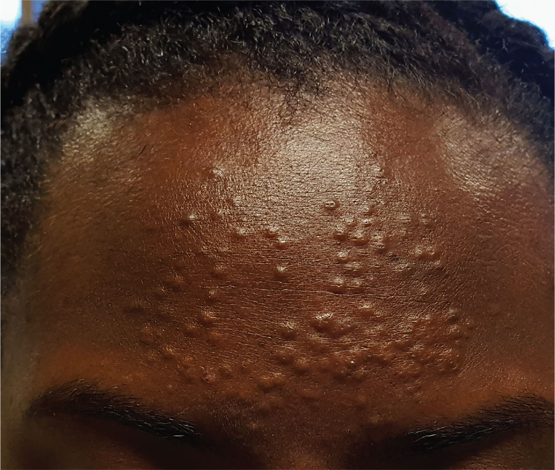
A 13-year-old African-American girl is brought in by her mother for evaluation of lesions that manifested on her forehead several years ago. Over time, the lesions have multiplied from just a few papules to a current total of about 30. Attempted treatment with topical benzoyl peroxide and two retinoids (tazarotene and adapalene)—for a presumptive diagnosis of acne—has yielded no improvement.
The lesions are quite obvious but asymptomatic; they are not tender, inflamed, or pustulant. The soft, 2- to 3-mm intradermal papules are grouped in a fairly round 7-cm area of the patient’s forehead. No punctum is seen with any of the lesions.
The child’s type V skin is otherwise clear, with no sign of acne. Her hair, teeth, and nails appear normal. According to her mother, the patient is healthy apart from this eruption.

15 and Going Gray
ANSWER
The correct answer is seborrheic dermatitis (choice “d”).
Psoriasis (choice “a”) can manifest in a similar fashion but would also appear elsewhere (eg, the elbows, knees, or nails). The scaling seen with psoriasis is usually white on a salmon-colored base and much coarser.
Vitiligo (choice “b”) presents with total, porcelain-white depigmentation and is not scaly at all; this patient’s loss of pigment was only partial.
Rashes like this are often thought to be fungal, but a fungal infection (choice “c”) is rarely seen on the face and would not demonstrate this exact pattern.
DISCUSSION
Seborrheic dermatitis (SD) is an extremely common papulosquamous skin condition that manifests primarily in oil-rich areas, such as the central face, scalp (dandruff), and in and behind the ears. On white skin, it yields a pinkish-brown scaly rash, but on darker skin, SD causes partial depigmentation—sometimes to an alarming extent. The distribution in this case is typical, with nasolabial, glabellar, ear, and brow involvement.
SD might be caused by a number of factors, some related to climate or genetics. One prevailing theory is that it involves an inflammatory reaction resulting from commensal yeast organisms (Malassezia furfur) feeding on sebum. In this case, the contribution of stress was clear.
TREATMENT/PROGNOSIS
Treatment comprised daily use of ketoconazole shampoo on the scalp and face and a topical ketoconazole cream. The condition cleared within two weeks. Although the chances of a recurrence are quite high, the patient is now armed with information and products to avoid the worst effects.
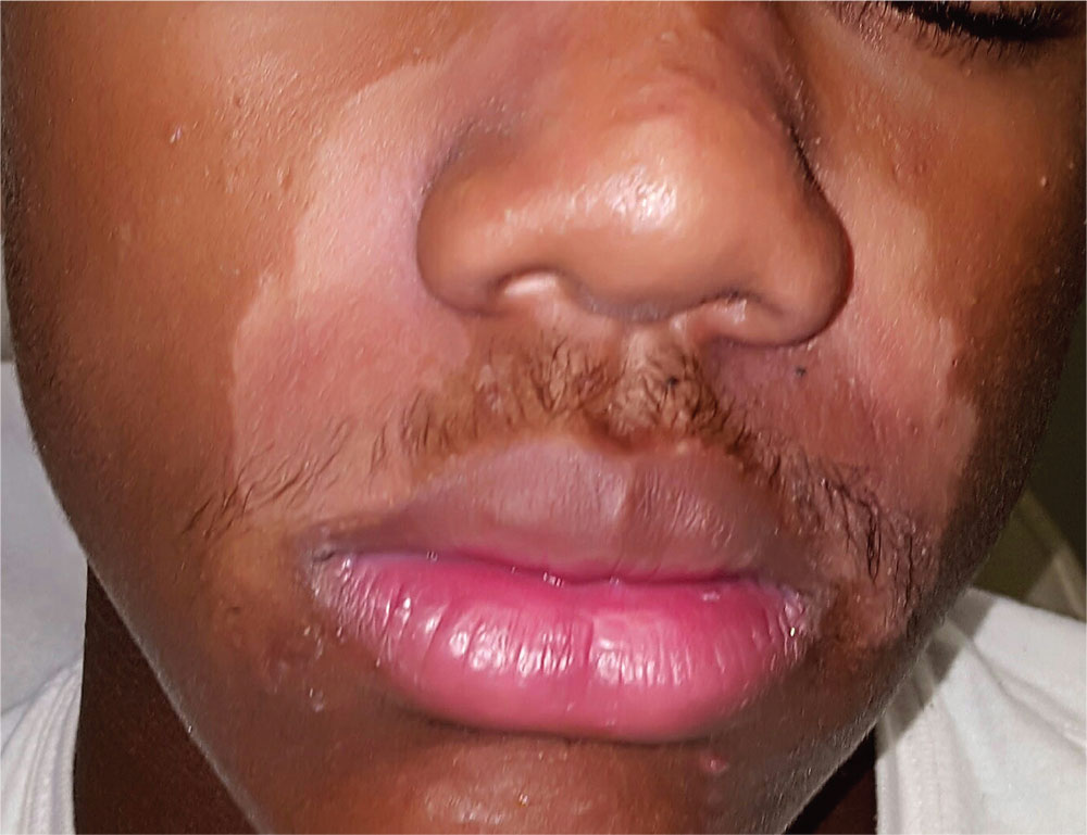
A 15-year-old African-American boy is brought in by his family for evaluation of a condition that began several months ago. Despite multiple attempts at treatment, the uniform but partial depigmentation of the skin around his nose persists. It is made obvious by his type V skin, extending up into the glabellar area and into both eyebrows.
Fine but definite scaling is confined to the depigmented areas. There is heavier white to gray scaling on his frontal scalp, and light scaling in and behind both ears. His elbows, knees, and nails appear normal. He is asymptomatic and in excellent health otherwise, but he considers his condition “unsightly and embarrassing.”
More history-taking reveals that around the time this problem manifested, he was facing the threat of a one-year suspension from school for disciplinary reasons.
