User login
Woman Loses Weight, Gains Skin Problem
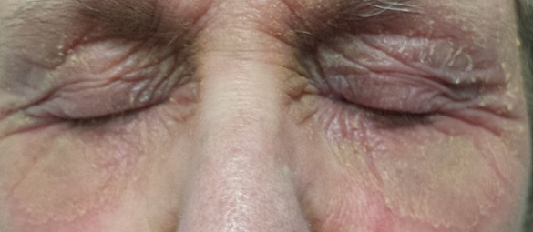
Since early summer, a 51-year-old woman has had an itchy rash on her trunk. It manifested after she intentionally lost more than 200 lb, which resulted in deeper and more pronounced skinfolds. The same skinfolds are repeatedly involved in flares of the rash, which is worse on particularly hot days. She was diagnosed with “yeast infection” but the problem failed to respond to oral fluconazole and topical nystatin.
The patient has multiple health problems, including diabetes, chronic anxiety, and hypertension.

EXAMINATION
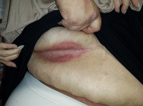
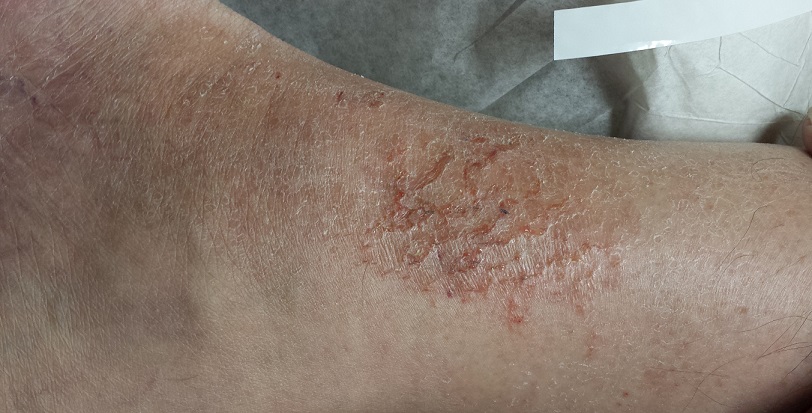
There is a deep, linear, concave fold in the skin of the right flank, the long axis of which is transversely oriented. Within this fold, a fiery red rash is seen; its margins exactly match the outline of skin-on-skin contact. The surface of the affected skin is macerated and wet looking. There is neither tenderness nor increased warmth on palpation.
What is the diagnosis?
DISCUSSION
This phenomenon is called intertrigo, and it’s a subject of much confusion. This is especially true in primary care, where almost any intertriginous rash is labeled “yeast infection” and treated with anti-yeast medications that fail as monotherapy. In fact, true yeast infections in or on the skin are quite unusual.
Here is what happens when skin is held on skin: Moisture can accumulate, temperature rises, and skin becomes macerated and starts to break down. Organisms already present on the skin begin to multiply and can contribute to the inflammatory burden. Any pre-existing skin disease (eg, seborrhea, eczema or psoriasis) can flare under these conditions, further breaking down the epidermal barrier. With the right mix of these and other factors (severe obesity, diabetes, immunosuppression, hot weather), yeast can play a bigger role in the problem—but it is usually a minor factor.
Intertrigo is common under the breasts, in the axillae, and in abdominal panniculi. It can also be seen, as in this case, in large folds on the trunk, or even on the legs. Most cases of diaper rash involve at least a component of intertrigo.
The factors that predispose to intertrigo tend to be chronic, making the problem chronic as well. The patient and family (if appropriate) need to understand this, as well as the seasonal effects (the condition will be worse in warm months but improve in winter).
Treatment should be directed at reducing moisture in the area. This can be achieved by opening it up to air as much as possible, using aluminum acetate solution soaks to dry out the skin, placing strips of cotton or linen cloth to separate skinfolds, or applying antiperspirants or talcum-based powder.
For acute treatment, class 4 or 5 steroid creams can be used for short periods. An OTC topical anti-yeast cream, such as clotrimazole or miconazole, can be added.
The onset of such a rash, without obvious predisposing factors, should prompt consideration of other items in the differential. These include cutaneous T-cell lymphoma, Paget’s disease, or zinc deficiency–related conditions. More common lookalikes include contact or irritant dermatitis.
TAKE-HOME LEARNING POINTS
• As its name suggests, intertrigo is a condition affecting the intertriginous areas (eg, skin folds); common locations are under breasts or in the axillae, groin, or panniculi.
• Though yeast organisms can play a role in the development of intertrigo, treatment with anti-yeast products as monotherapy rarely work.
• Instead, management of intertrigo must be directed at the causative factors: heat, sweat, and friction.
• Intertrigo is difficult to treat, in large part because the major causative factors (weight, heat, sweat) are hard to change.
Since early summer, a 51-year-old woman has had an itchy rash on her trunk. It manifested after she intentionally lost more than 200 lb, which resulted in deeper and more pronounced skinfolds. The same skinfolds are repeatedly involved in flares of the rash, which is worse on particularly hot days. She was diagnosed with “yeast infection” but the problem failed to respond to oral fluconazole and topical nystatin.
The patient has multiple health problems, including diabetes, chronic anxiety, and hypertension.
EXAMINATION
There is a deep, linear, concave fold in the skin of the right flank, the long axis of which is transversely oriented. Within this fold, a fiery red rash is seen; its margins exactly match the outline of skin-on-skin contact. The surface of the affected skin is macerated and wet looking. There is neither tenderness nor increased warmth on palpation.
What is the diagnosis?
DISCUSSION
This phenomenon is called intertrigo, and it’s a subject of much confusion. This is especially true in primary care, where almost any intertriginous rash is labeled “yeast infection” and treated with anti-yeast medications that fail as monotherapy. In fact, true yeast infections in or on the skin are quite unusual.
Here is what happens when skin is held on skin: Moisture can accumulate, temperature rises, and skin becomes macerated and starts to break down. Organisms already present on the skin begin to multiply and can contribute to the inflammatory burden. Any pre-existing skin disease (eg, seborrhea, eczema or psoriasis) can flare under these conditions, further breaking down the epidermal barrier. With the right mix of these and other factors (severe obesity, diabetes, immunosuppression, hot weather), yeast can play a bigger role in the problem—but it is usually a minor factor.
Intertrigo is common under the breasts, in the axillae, and in abdominal panniculi. It can also be seen, as in this case, in large folds on the trunk, or even on the legs. Most cases of diaper rash involve at least a component of intertrigo.
The factors that predispose to intertrigo tend to be chronic, making the problem chronic as well. The patient and family (if appropriate) need to understand this, as well as the seasonal effects (the condition will be worse in warm months but improve in winter).
Treatment should be directed at reducing moisture in the area. This can be achieved by opening it up to air as much as possible, using aluminum acetate solution soaks to dry out the skin, placing strips of cotton or linen cloth to separate skinfolds, or applying antiperspirants or talcum-based powder.
For acute treatment, class 4 or 5 steroid creams can be used for short periods. An OTC topical anti-yeast cream, such as clotrimazole or miconazole, can be added.
The onset of such a rash, without obvious predisposing factors, should prompt consideration of other items in the differential. These include cutaneous T-cell lymphoma, Paget’s disease, or zinc deficiency–related conditions. More common lookalikes include contact or irritant dermatitis.
TAKE-HOME LEARNING POINTS
• As its name suggests, intertrigo is a condition affecting the intertriginous areas (eg, skin folds); common locations are under breasts or in the axillae, groin, or panniculi.
• Though yeast organisms can play a role in the development of intertrigo, treatment with anti-yeast products as monotherapy rarely work.
• Instead, management of intertrigo must be directed at the causative factors: heat, sweat, and friction.
• Intertrigo is difficult to treat, in large part because the major causative factors (weight, heat, sweat) are hard to change.
Why Are You Still Prescribing a 66-Year-Old Drug?
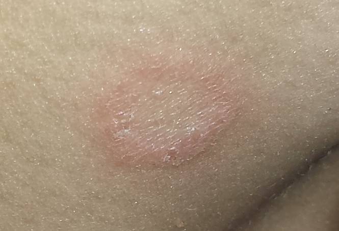
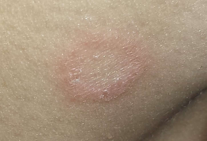
Several weeks ago, this 14-year-old boy developed an itchy spot on his neck. Concerned that the problem was “ringworm,” the boy’s grandfather took him to the primary care provider, who prescribed nystatin cream. This produced modest improvement in the appearance of the lesion but had no effect on the associated itching. At that point, they were referred to dermatology for further evaluation.
The patient denies any other skin problems. The only animal he has been exposed to is his own dog, who has been part of the household for years. The boy is not involved in contact sports (eg, football, wrestling), and he reports that none of his siblings or friends have any skin complaints.
He is otherwise healthy and does not take any prescription medications.
EXAMINATION
The “rash” consists of a single, 2-cm lesion on the patient’s anterolateral neck. It is perfectly round and slightly erythematous, with a cleared center and scaly advancing margin. Palpable adenopathy is evident just above the lesion. There are no other lesions elsewhere, and the patient’s skin is otherwise unremarkable.
What is the diagnosis?
DISCUSSION
A KOH prep revealed abundant fungal elements, confirming the diagnosis of tinea corporis. This conclusion had already been reached empirically by the primary care provider, who chose nystatin cream for treatment. When that drug faltered, diagnostic doubt reared its head. The KOH settled the question once and for all—a crucial step, since nystatin is often prescribed for conditions that have no likelihood of responding to it (eg, eczema, psoriasis, granuloma annulare).
Nystatin, a polyene antifungal, was discovered and brought to market in 1950—that’s nearly 66 years ago! At that time, very few antifungal agents were available, so nystatin gained instant acceptance practically overnight. Since then, although dozens of newer and better antifungals have come on the market, nystatin continues to be prescribed out of sheer habit. A lot of water has gone down the river since 1950!
Nystatin was first isolated and developed by two scientists (Brown and Fuller) who worked in the lab at the New York State Health Department in the ’40s and ’50s. A popular practice at the time was to collect soil samples from local farmers to see what bacteria could be isolated. Brown and Fuller found a unique species of Streptomyces that they named S noursei after the dairy farmer whose soil they sampled.
They noted that this organism exuded a substance that inhibited yeast, mold, and fungi in vitro; when this substance was isolated and purified, it worked topically as well. They named this substance nystatin in honor of their employer, the New York State Health Department.
As charming as this history is, in the intervening 60+ years, better antifungals have been introduced and organisms have become less responsive to nystatin. In my opinion, except for unusual selective instances, there is no reason to prescribe nystatin instead of imidazole and allylamine (eg, miconazole, terbinafine, or naftifine).
For this patient, I prescribed oxiconazole lotion for twice-daily application to this small and limited lesion. With more extensive disease, I might add an oral antifungal, such as terbinafine. The patient’s lesion should resolve in two weeks or less.
One final note: In dermatology, we discourage the use of the term ringworm because it contributes to the “ick” factor associated with “worms”—which are not even involved in tinea corporis.
TAKE-HOME LEARNING POINTS
• Nystatin is a 66-year-old relic that has no place in treating ordinary dermatophytosis.
• “Newer” antifungals are more effective and kill a wider range of organisms, including yeast and fungi.
• In my experience, most nystatin is prescribed for conditions that have no chance of responding to it (eg, eczema, psoriasis, or granuloma annulare), because a differential was not considered.
• Primary care offices that are comfortable doing wet preps for clue cells and trichomonas feel no such need to perform KOHs to confirm fungal infection, and so diagnostic confusion results.
• There is no medical entity called ringworm. This is lay terminology based on the misbelief that worms are somehow involved in tinea.
It Started With a Bug Bite (He Thinks)
An 81-year-old man is brought in by his wife for evaluation of a very itchy rash on his bilateral lower tibial areas. He says the problem started about six months ago, after a spate of summer yardwork during which he sustained what he assumed was a bug bite. It was itchy, so he scratched it.
Of course, in the way of most itches, the scratching offered temporary relief, after which the itching resumed. The patient tried any number of OTC products, including rubbing alcohol, hydrogen peroxide, tea tree oil, several different essential oils, and triple-antibiotic cream and ointment. The worse the itching became, the more products he applied—all to no avail.
The patient describes his health as otherwise decent. He does have type 2 diabetes, which he says is in good control.

EXAMINATION
The lower anterior tibial areas of both legs are covered by a scaly red rash. The left leg is more heavily affected, and obvious edema can be seen distal to the rash on that leg. The surface scales of the rash have a polygonal look, resembling a dried lake bed or finely cracked porcelain. The edges of the cracks turn upward, resulting in a rough feel on palpation.
The patient’s skin is quite dry in general but otherwise within normal limits.
What is the diagnosis?
DISCUSSION
Leg skin is unique in many respects. For one thing, it’s down there, where gravity takes and often holds blood and other fluids that might not accumulate elsewhere. It’s also a long trip for blood to get out to the extremities and often a longer return trip.
Leg skin is also remarkable because it has far fewer sebaceous glands than the scalp, face, and chest, which means it tends to be quite dry. This is especially true in older patients already prone to dry skin and in those who seldom moisturize to counteract this problem (in other words, men!).
This is why most patients with asteatotic eczema (AE) are men. Also known as eczema craquele and xerotic eczema, AE is especially common in the dry winter months, when long, hot showers are so appealing, as are wearing warmer clothes and sleeping under heavy covers.
Patients with AE, including this one, often make matters worse by applying a multiplicity of contactants. The edema noted in the exam, although due to the AE, also served to worsen the condition by making the skin tighter and drier still.
At this point, the problem often starts to take on aspects of lichen simplex chronicus, in which more scratching leads to more itching (and then more scratching, and so on). Clearly, what this patient needed (and got) was a definitive diagnosis and a treatment plan dictated by that diagnosis.
AE can be a challenge to treat, but the first step is to help the patient understand the nature of the problem and his role in the solution. The patient also needs to stop applying nonprescribed/recommended contactants, which don’t help and may exacerbate the problem.
To achieve relief, the patient can soak the leg with wet compresses for 10 minutes, remove excess water, and then apply a medium-strength steroid ointment (eg, triamcinolone 0.5%) in a thin but thorough coat. The area can then be covered with an occlusive covering, such as saran wrap, taped in place. This should be left on all night. During the day, the patient should apply only petroleum jelly to the affected area.
This approach will take 90% of patients out of the crisis stage. After a week or two, attention must shift to preventing recurrence, with generous use of emollients such as petroleum jelly. The patient should also be instructed to avoid using harsh (colored, scented, high pH) soaps, switch to shorter, relatively cool showers, and stop using anything but his hand to wash with (ie, no washcloths or loofahs).
For nondiabetic patients with severe AE that persists despite these measures, an intramuscular injection of a glucocorticoid (eg, triamcinolone 40 - 60 mg) can work wonders.
TAKE-HOME LEARNING POINTS
• Asteatotic eczema (AE), also called xerotic eczema or eczema craquele, is quite common, especially on the lower legs of older men.
• The particular rash of AE is said to resemble the cracked surface of a porcelain vessel.
• AE is often accompanied by edema distal to the rash.
• A topical steroid ointment applied to water-soaked skin, held in place overnight with an occlusive dressing, usually takes the patient out of the crisis phase.
• Prevention is then directed at avoiding drying of the affected areas.
An 81-year-old man is brought in by his wife for evaluation of a very itchy rash on his bilateral lower tibial areas. He says the problem started about six months ago, after a spate of summer yardwork during which he sustained what he assumed was a bug bite. It was itchy, so he scratched it.
Of course, in the way of most itches, the scratching offered temporary relief, after which the itching resumed. The patient tried any number of OTC products, including rubbing alcohol, hydrogen peroxide, tea tree oil, several different essential oils, and triple-antibiotic cream and ointment. The worse the itching became, the more products he applied—all to no avail.
The patient describes his health as otherwise decent. He does have type 2 diabetes, which he says is in good control.
EXAMINATION
The lower anterior tibial areas of both legs are covered by a scaly red rash. The left leg is more heavily affected, and obvious edema can be seen distal to the rash on that leg. The surface scales of the rash have a polygonal look, resembling a dried lake bed or finely cracked porcelain. The edges of the cracks turn upward, resulting in a rough feel on palpation.
The patient’s skin is quite dry in general but otherwise within normal limits.
What is the diagnosis?
DISCUSSION
Leg skin is unique in many respects. For one thing, it’s down there, where gravity takes and often holds blood and other fluids that might not accumulate elsewhere. It’s also a long trip for blood to get out to the extremities and often a longer return trip.
Leg skin is also remarkable because it has far fewer sebaceous glands than the scalp, face, and chest, which means it tends to be quite dry. This is especially true in older patients already prone to dry skin and in those who seldom moisturize to counteract this problem (in other words, men!).
This is why most patients with asteatotic eczema (AE) are men. Also known as eczema craquele and xerotic eczema, AE is especially common in the dry winter months, when long, hot showers are so appealing, as are wearing warmer clothes and sleeping under heavy covers.
Patients with AE, including this one, often make matters worse by applying a multiplicity of contactants. The edema noted in the exam, although due to the AE, also served to worsen the condition by making the skin tighter and drier still.
At this point, the problem often starts to take on aspects of lichen simplex chronicus, in which more scratching leads to more itching (and then more scratching, and so on). Clearly, what this patient needed (and got) was a definitive diagnosis and a treatment plan dictated by that diagnosis.
AE can be a challenge to treat, but the first step is to help the patient understand the nature of the problem and his role in the solution. The patient also needs to stop applying nonprescribed/recommended contactants, which don’t help and may exacerbate the problem.
To achieve relief, the patient can soak the leg with wet compresses for 10 minutes, remove excess water, and then apply a medium-strength steroid ointment (eg, triamcinolone 0.5%) in a thin but thorough coat. The area can then be covered with an occlusive covering, such as saran wrap, taped in place. This should be left on all night. During the day, the patient should apply only petroleum jelly to the affected area.
This approach will take 90% of patients out of the crisis stage. After a week or two, attention must shift to preventing recurrence, with generous use of emollients such as petroleum jelly. The patient should also be instructed to avoid using harsh (colored, scented, high pH) soaps, switch to shorter, relatively cool showers, and stop using anything but his hand to wash with (ie, no washcloths or loofahs).
For nondiabetic patients with severe AE that persists despite these measures, an intramuscular injection of a glucocorticoid (eg, triamcinolone 40 - 60 mg) can work wonders.
TAKE-HOME LEARNING POINTS
• Asteatotic eczema (AE), also called xerotic eczema or eczema craquele, is quite common, especially on the lower legs of older men.
• The particular rash of AE is said to resemble the cracked surface of a porcelain vessel.
• AE is often accompanied by edema distal to the rash.
• A topical steroid ointment applied to water-soaked skin, held in place overnight with an occlusive dressing, usually takes the patient out of the crisis phase.
• Prevention is then directed at avoiding drying of the affected areas.
The Diagnosis Either You Know or You Don’t
A 54-year-old woman self-refers to dermatology for evaluation of asymptomatic lesions on her legs. Over the past several years, they have slowly grown larger, redder, and shinier. She denies any other skin problems. Medical history is significant for type 2 diabetes, which, according to the patient, is under good control.
The patient has sought care from a variety of professionals, including her primary care provider, her endocrinologist, and her gynecologist. Various diagnoses, including “fungal infection,” have been posited; treatment with antifungal cream and oral medications produced no effect.
She also consulted the proprietor of her local health food store, who actually examined her before diagnosing “yeast infection” and advising her to change her diet.
EXAMINATION
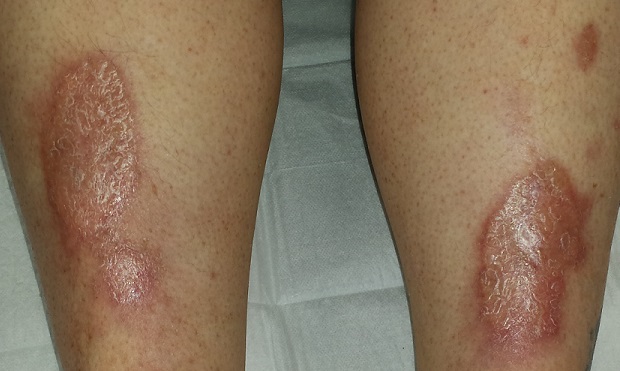
There are large, oval, reddish brown shiny plaques, measuring about 8 x 4 cm, on both anterior tibial areas. On closer inspection, the affected skin is yellowish pink and appears quite atrophic. This effect is more pronounced toward the centers of the lesions, which also display distinct telangiectasias. The borders of the plaques are slightly raised.
Examination of the rest of the patient’s skin reveals no other abnormalities.
What is the diagnosis?
DISCUSSION
Necrobiosis lipoidica (NL), though uncommon, is far from rare; it is seen somewhat regularly in dermatology practices. As this case demonstrates, it is one of hundreds of dermatologic diagnoses that you either know about or you don’t. Providers in the latter group invariably call it “fungal infection” because of its rounded borders—and because they simply have nothing else to call it.
True fungal infections are usually very superficial, involving only the outer layer of skin. This means they will manifest with scaling, a feature notably absent in this case. But more to the point, what was needed was a specific diagnosis, rather than another blind attempt at empiric treatment. Correct diagnosis dictates correct treatment.
NL was first described by Oppenhein in 1929, when he saw it in a diabetic patient. But it got its modern name in 1932 from Urbach, who thought it was invariably connected to diabetes. Now we know nondiabetic persons can develop it as well.
A number of theories exist as to the origins of this condition. One pinpoints microangiopathy of the same sort seen in the kidneys and eyes of diabetic patients. Another, proposed to account for the fact that NL is not exclusively seen in diabetes, holds that an autoimmune process might be involved—an opinion bolstered by the finding of increased TNF-alpha in the sera and skin of NL patients.
In any case, these atrophic plaques can grow quite large, eventually breaking down and ulcerating focally. Although the condition is usually asymptomatic, 25% of NL patients with advanced disease report pain.
Definitive diagnosis is made by punch biopsy, though visual diagnosis is quite adequate for those who can recognize the condition. NL lesions are most commonly seen on bilateral anterior tibial areas but can occasionally develop on the arm, face, or even genitals. The lesions can also koebnerize (ie, form along lines of trauma), occasionally being seen in surgical scars or even insulin injection sites.
Treatment, though largely unsatisfactory, includes topical and intralesional steroids. Particular care is taken to avoid or at least limit the formation of ulcers. In advanced cases, patients are referred to a wound care specialist. Oddly enough, neither the severity nor the prognosis of NL seem to have anything to do with how well or poorly the patient’s diabetes is controlled.
This patient was started on topical clobetasol cream, to be applied mostly to the actively advancing peripheral margin. Her prognosis, as with all NL patients, is guarded.
TAKE-HOME LEARNING POINTS
• Necrobiosis lipoidica (NL) is a disease of collagen degeneration that induces a granulomatous response that manifests microscopically with microangiopathy.
• NL usually appears on bilateral anterior tibial areas (but can affect the arms, face, or genitalia), as pinkish brown plaques with atrophic centers that tend to be yellow and telangiectatic.
• Though NL is usually associated with diabetes, it can develop in nondiabetic persons as well.
• The most feared complication of NL is eventual ulceration, which is why treatment is directed at taking care to avoid wounds to the plaques.
A 54-year-old woman self-refers to dermatology for evaluation of asymptomatic lesions on her legs. Over the past several years, they have slowly grown larger, redder, and shinier. She denies any other skin problems. Medical history is significant for type 2 diabetes, which, according to the patient, is under good control.
The patient has sought care from a variety of professionals, including her primary care provider, her endocrinologist, and her gynecologist. Various diagnoses, including “fungal infection,” have been posited; treatment with antifungal cream and oral medications produced no effect.
She also consulted the proprietor of her local health food store, who actually examined her before diagnosing “yeast infection” and advising her to change her diet.
EXAMINATION
There are large, oval, reddish brown shiny plaques, measuring about 8 x 4 cm, on both anterior tibial areas. On closer inspection, the affected skin is yellowish pink and appears quite atrophic. This effect is more pronounced toward the centers of the lesions, which also display distinct telangiectasias. The borders of the plaques are slightly raised.
Examination of the rest of the patient’s skin reveals no other abnormalities.
What is the diagnosis?
DISCUSSION
Necrobiosis lipoidica (NL), though uncommon, is far from rare; it is seen somewhat regularly in dermatology practices. As this case demonstrates, it is one of hundreds of dermatologic diagnoses that you either know about or you don’t. Providers in the latter group invariably call it “fungal infection” because of its rounded borders—and because they simply have nothing else to call it.
True fungal infections are usually very superficial, involving only the outer layer of skin. This means they will manifest with scaling, a feature notably absent in this case. But more to the point, what was needed was a specific diagnosis, rather than another blind attempt at empiric treatment. Correct diagnosis dictates correct treatment.
NL was first described by Oppenhein in 1929, when he saw it in a diabetic patient. But it got its modern name in 1932 from Urbach, who thought it was invariably connected to diabetes. Now we know nondiabetic persons can develop it as well.
A number of theories exist as to the origins of this condition. One pinpoints microangiopathy of the same sort seen in the kidneys and eyes of diabetic patients. Another, proposed to account for the fact that NL is not exclusively seen in diabetes, holds that an autoimmune process might be involved—an opinion bolstered by the finding of increased TNF-alpha in the sera and skin of NL patients.
In any case, these atrophic plaques can grow quite large, eventually breaking down and ulcerating focally. Although the condition is usually asymptomatic, 25% of NL patients with advanced disease report pain.
Definitive diagnosis is made by punch biopsy, though visual diagnosis is quite adequate for those who can recognize the condition. NL lesions are most commonly seen on bilateral anterior tibial areas but can occasionally develop on the arm, face, or even genitals. The lesions can also koebnerize (ie, form along lines of trauma), occasionally being seen in surgical scars or even insulin injection sites.
Treatment, though largely unsatisfactory, includes topical and intralesional steroids. Particular care is taken to avoid or at least limit the formation of ulcers. In advanced cases, patients are referred to a wound care specialist. Oddly enough, neither the severity nor the prognosis of NL seem to have anything to do with how well or poorly the patient’s diabetes is controlled.
This patient was started on topical clobetasol cream, to be applied mostly to the actively advancing peripheral margin. Her prognosis, as with all NL patients, is guarded.
TAKE-HOME LEARNING POINTS
• Necrobiosis lipoidica (NL) is a disease of collagen degeneration that induces a granulomatous response that manifests microscopically with microangiopathy.
• NL usually appears on bilateral anterior tibial areas (but can affect the arms, face, or genitalia), as pinkish brown plaques with atrophic centers that tend to be yellow and telangiectatic.
• Though NL is usually associated with diabetes, it can develop in nondiabetic persons as well.
• The most feared complication of NL is eventual ulceration, which is why treatment is directed at taking care to avoid wounds to the plaques.

The Patient Who Didn’t Complain Enough?
A 70-year-old woman presents to dermatology with asymptomatic lesions on her legs that, she reports, have been there for decades but are now becoming larger and more unsightly. She’s tried several courses of antifungal creams and pills with no success.
In all this time, she hasn’t seen a dermatology provider, because “no one ever felt the need” to refer her since her diagnosis was so “obvious.” When the changes began, however, her children finally convinced her to consult a specialist.
Her medical history is otherwise unremarkable, with no problems directly related to her skin issues. She denies both personal and family history of diabetes.
EXAMINATION
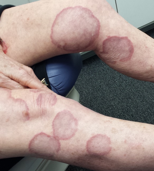
There are numerous impressive, reddish brown, annular plaques with slightly raised borders and clearing centers on the patient’s legs. There are also a few on her arms and one or two on her trunk. There is no epidermal component (scale or other surface disruption) on any of the lesions, which range in size from 2 cm to 15 cm.
What is the diagnosis?
DISCUSSION
It’s hard to believe that any patient could go this long with such a florid condition and never be seen by dermatology. Patients have an obligation to speak up—to complain, as it were—but some are too compliant. This woman’s primary care providers should have attached some significance to the nonresponse to treatment, one explanation for which might have been erroneous diagnosis.
Everybody knows that big round things are fungal, except when they aren’t. That’s where dermatology providers shine. Our training is all about development of differentials, as in: What are the various potential explanations for big round lesions? What would those things look like, and how could we distinguish them from fungal infection?
Here’s one huge clue: Fungal infection commonly refers to a relatively trivial, superficial infection of the outermost layer of the skin, the stratum corneum. By its very nature, it almost always involves scaling; if those scales are examined microscopically, the fungal elements can be seen, thus confirming the diagnosis.
When there is no scale, and the condition fails to respond to antifungal medications, alternative diagnoses must be considered. One major item that fits the bill is granuloma annulare (GA), an asymptomatic condition that presents with reddish brown, distinctly annular papules and plaques with raised (papular) borders and cleared centers. Though GA is extremely common, it is almost always misdiagnosed initially as fungal infection.
Besides the lack of scaling, GA lesions have some other characteristics that aid diagnosis. They’re commonly seen on extremities, especially the dorsa of feet and hands, and are far more common on women (especially young ones) than on men.
Although usually reddish brown, the lesions will be darker on patients with darker skin. It should be mentioned that this patient’s lesions were unusually large and extensive. Most cases are far more modest in size and distribution.
When necessary, punch biopsy shows what is called a palisaded granuloma. This, taken in context with the clinical picture, nails down the diagnosis.
One other piece of information is good to know: GA is quite common. You will see it, regularly.
Since the cause of GA is unknown, treatment can be problematic. Fortunately, the problem almost always resolves, with or without treatment. A few of this patient’s lesions were treated (for cosmetic reasons) with intralesional steroid injection.
TAKE-HOME LEARNING POINTS
• Granuloma annulare (GA) is a benign asymptomatic condition that presents with annular, reddish brown intradermal lesions.
• GA is commonly seen on the dorsa of feet, hands, and arms, especially of young women.
• GA lesions are intradermal, not epidermal, so there is no scaling or other disturbance of the skin surface.
• GA treatment is problematic, but the problem usually resolves on its own.
A 70-year-old woman presents to dermatology with asymptomatic lesions on her legs that, she reports, have been there for decades but are now becoming larger and more unsightly. She’s tried several courses of antifungal creams and pills with no success.
In all this time, she hasn’t seen a dermatology provider, because “no one ever felt the need” to refer her since her diagnosis was so “obvious.” When the changes began, however, her children finally convinced her to consult a specialist.
Her medical history is otherwise unremarkable, with no problems directly related to her skin issues. She denies both personal and family history of diabetes.
EXAMINATION
There are numerous impressive, reddish brown, annular plaques with slightly raised borders and clearing centers on the patient’s legs. There are also a few on her arms and one or two on her trunk. There is no epidermal component (scale or other surface disruption) on any of the lesions, which range in size from 2 cm to 15 cm.
What is the diagnosis?
DISCUSSION
It’s hard to believe that any patient could go this long with such a florid condition and never be seen by dermatology. Patients have an obligation to speak up—to complain, as it were—but some are too compliant. This woman’s primary care providers should have attached some significance to the nonresponse to treatment, one explanation for which might have been erroneous diagnosis.
Everybody knows that big round things are fungal, except when they aren’t. That’s where dermatology providers shine. Our training is all about development of differentials, as in: What are the various potential explanations for big round lesions? What would those things look like, and how could we distinguish them from fungal infection?
Here’s one huge clue: Fungal infection commonly refers to a relatively trivial, superficial infection of the outermost layer of the skin, the stratum corneum. By its very nature, it almost always involves scaling; if those scales are examined microscopically, the fungal elements can be seen, thus confirming the diagnosis.
When there is no scale, and the condition fails to respond to antifungal medications, alternative diagnoses must be considered. One major item that fits the bill is granuloma annulare (GA), an asymptomatic condition that presents with reddish brown, distinctly annular papules and plaques with raised (papular) borders and cleared centers. Though GA is extremely common, it is almost always misdiagnosed initially as fungal infection.
Besides the lack of scaling, GA lesions have some other characteristics that aid diagnosis. They’re commonly seen on extremities, especially the dorsa of feet and hands, and are far more common on women (especially young ones) than on men.
Although usually reddish brown, the lesions will be darker on patients with darker skin. It should be mentioned that this patient’s lesions were unusually large and extensive. Most cases are far more modest in size and distribution.
When necessary, punch biopsy shows what is called a palisaded granuloma. This, taken in context with the clinical picture, nails down the diagnosis.
One other piece of information is good to know: GA is quite common. You will see it, regularly.
Since the cause of GA is unknown, treatment can be problematic. Fortunately, the problem almost always resolves, with or without treatment. A few of this patient’s lesions were treated (for cosmetic reasons) with intralesional steroid injection.
TAKE-HOME LEARNING POINTS
• Granuloma annulare (GA) is a benign asymptomatic condition that presents with annular, reddish brown intradermal lesions.
• GA is commonly seen on the dorsa of feet, hands, and arms, especially of young women.
• GA lesions are intradermal, not epidermal, so there is no scaling or other disturbance of the skin surface.
• GA treatment is problematic, but the problem usually resolves on its own.

The Lesion That Grew Unbearable
A 49-year-old woman presents to dermatology with a lesion on her back. It’s been there for at least 20 years, slowly growing to its present size; it is now so prominent that it shows through her clothes and is subject to traumatization. The visibility of the lesion, particularly when the patient wears a swimsuit, is a source of considerable embarrassment.
Notable medical history includes polycystic ovarian syndrome and related diabetes and dyslipidemia. Family history reveals that the patient’s mother died of heart disease in her 40s.

EXAMINATION
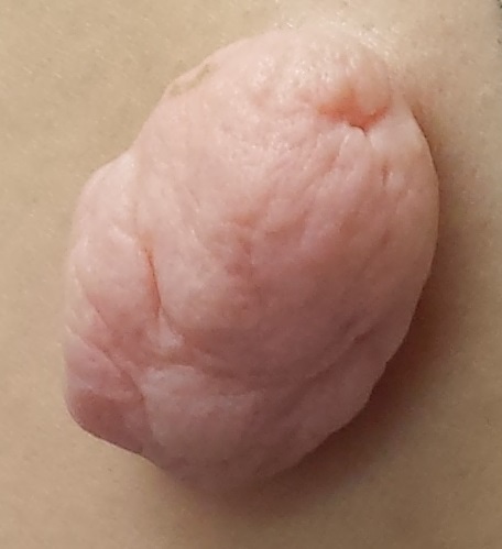
The lesion, which measures 3 cm x 3 cm, is an impressive, pedunculated, doughy, rubbery, skin-colored mass protruding from her left mid back. No redness or edema is seen on or around the lesion. On palpation, the lesion is found to be uniformly soft and rubbery.
The patient is quite obese and has numerous skin tags in skin folds, under the arms, and around the neck.
What is the diagnosis?
DISCUSSION
The lesion was excised with deep margins and submitted to pathology. The report showed benign histology, consistent with a fibroepitheliomatous polyp.
In one sense, it’s hard to believe this huge lesion was a “skin tag,” and there are other things it might have been. But the microscopic examination of the tissue proved it was simply a large version of the same skin tags we see around necks and in axillae. These lesions are known by various names, including fibroepitheliomatous polyps (FEP) and acrochordons. A more typical FEP would be the size of a grain of rice, but they can take on various forms and sizes.
There is a differential for lesions that look like FEPs; it includes seborrheic keratoses, warts, molluscum contagiosum, neurofibromas, and fibrolipomas. Even melanoma and squamous cell carcinoma can assume a tag-like morphology. So it pays to take a close look at these lesions, checking to see if they’re soft and pliable, as well as skin colored. With any odd feature, including increased size, they need to be removed and sent for pathologic examination.
Heredity appears to play a role in the development of FEPs, as do age and obesity. There are studies showing an association between FEPs and insulin resistance and others identifying FEPs as a marker for increased risk for atherosclerotic vessel disease. In any case, 59% of the population has them by age 70, with men and women affected equally. Of the general population, 46% has some sort of skin tag somewhere.
Treatment may entail cryotherapy, excision, or electrodessication. With lesions as large as the case patient’s, excision is the only reasonable option. Deep margins are required, since a “shave” would leave a gaping, open wound. For the case patient, there was no postoperative sequelae aside from some scarring.
TAKE-HOME LEARNING POINTS
• Skin tags, also known as fibroepitheliomas, fibroepitheliomatous polyps, or acrochordons, are typically the size of a grain of rice, and tend to appear in skin folds (eg, axillae, neck, groin).
• Studies have shown that there may be an association between having skin tags and developing CAD or insulin resistance.
• Obesity, age, and heredity also appear to be factors in developing skin tags.
• Large or odd tags need to be captured and sent for pathologic examination.
• Skin tags are extremely common, affecting 59% of the population older than 70.
A 49-year-old woman presents to dermatology with a lesion on her back. It’s been there for at least 20 years, slowly growing to its present size; it is now so prominent that it shows through her clothes and is subject to traumatization. The visibility of the lesion, particularly when the patient wears a swimsuit, is a source of considerable embarrassment.
Notable medical history includes polycystic ovarian syndrome and related diabetes and dyslipidemia. Family history reveals that the patient’s mother died of heart disease in her 40s.
EXAMINATION
The lesion, which measures 3 cm x 3 cm, is an impressive, pedunculated, doughy, rubbery, skin-colored mass protruding from her left mid back. No redness or edema is seen on or around the lesion. On palpation, the lesion is found to be uniformly soft and rubbery.
The patient is quite obese and has numerous skin tags in skin folds, under the arms, and around the neck.
What is the diagnosis?
DISCUSSION
The lesion was excised with deep margins and submitted to pathology. The report showed benign histology, consistent with a fibroepitheliomatous polyp.
In one sense, it’s hard to believe this huge lesion was a “skin tag,” and there are other things it might have been. But the microscopic examination of the tissue proved it was simply a large version of the same skin tags we see around necks and in axillae. These lesions are known by various names, including fibroepitheliomatous polyps (FEP) and acrochordons. A more typical FEP would be the size of a grain of rice, but they can take on various forms and sizes.
There is a differential for lesions that look like FEPs; it includes seborrheic keratoses, warts, molluscum contagiosum, neurofibromas, and fibrolipomas. Even melanoma and squamous cell carcinoma can assume a tag-like morphology. So it pays to take a close look at these lesions, checking to see if they’re soft and pliable, as well as skin colored. With any odd feature, including increased size, they need to be removed and sent for pathologic examination.
Heredity appears to play a role in the development of FEPs, as do age and obesity. There are studies showing an association between FEPs and insulin resistance and others identifying FEPs as a marker for increased risk for atherosclerotic vessel disease. In any case, 59% of the population has them by age 70, with men and women affected equally. Of the general population, 46% has some sort of skin tag somewhere.
Treatment may entail cryotherapy, excision, or electrodessication. With lesions as large as the case patient’s, excision is the only reasonable option. Deep margins are required, since a “shave” would leave a gaping, open wound. For the case patient, there was no postoperative sequelae aside from some scarring.
TAKE-HOME LEARNING POINTS
• Skin tags, also known as fibroepitheliomas, fibroepitheliomatous polyps, or acrochordons, are typically the size of a grain of rice, and tend to appear in skin folds (eg, axillae, neck, groin).
• Studies have shown that there may be an association between having skin tags and developing CAD or insulin resistance.
• Obesity, age, and heredity also appear to be factors in developing skin tags.
• Large or odd tags need to be captured and sent for pathologic examination.
• Skin tags are extremely common, affecting 59% of the population older than 70.
A Streak of Trouble in Fingernail?
A 14-year-old girl is brought to dermatology by her mother, following referral from her pediatrician for evaluation of fingernail changes. Initially, a faint brown streak was observed in the nail of her left fourth finger. Over time, the spot darkened and widened, and the adjacent nail plate flattened.
A few weeks before presentation to dermatology, the adjacent cuticle darkened. The patient denies any sensitivity in the area. Her mother denies any significant or relevant medical history.
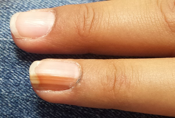
EXAMINATION
The dark brown streak measures 4.5 mm in width and runs longitudinally the entire length of the nail plate. The discolored area coincides exactly with flattening of the nail plate, which, when viewed on edge, also exhibits darkening. Significantly, the adjacent cuticle is similarly affected.
No changes are observed on other digits. The child is of Native American ancestry, with type IV skin.
What is the diagnosis?
DISCUSSION
Generically, this problem is termed melanonychia; this case represents the most common type, longitudinal melanonychia (LM). Typically, it originates in the nail matrix with increased melanin production; this causes darkening of the onchocytes, which migrate distally as the nail plate grows.
This benign version is an extremely common problem, especially in darker-skinned patients. Almost 100% of African-Americans older than 50 have it, often in several nails, as do 77% of African Americans older than 20 and 10% to 20% of Japanese persons. Comparatively, only 1% to 2% of white individuals are affected.
The main significance of LM, of course, is the fact that it can represent melanoma, which, for a variety of reasons, is often belatedly identified in subungual areas and is therefore associated with relatively poor survival. Five-year survival rates for subungual melanoma are around 30%, while 10-year survival is only 13%—much lower than for melanomas elsewhere on the body. For example, patients with melanomas removed from other areas of the hand demonstrate 100% five-year survival rates.
Subungual melanoma presenting in this manner is considered one of the so-called acrolentiginous melanomas, which present in peripheral “acral” locations (eg, scalp, soles, mucosal surfaces). While these are not inherently more dangerous in terms of biologic behavior, they tend to escape detection because of their location and (often) atypical appearance, thus having more time in which to become invasive.
Perhaps the most useful and puzzling conundrum associated with acrolentiginous melanomas is this: Darker-skinned patients are, in general, at low risk for melanoma compared to fair-skinned redheads and blondes. However, when dark-skinned patients do develop melanoma, it tends to manifest in an acral area (one reason why melanoma in these patients has a relatively poor prognosis).
Darkening under or in the nails can have other causes; it has been associated with Cushing disease, Addison disease, and alkaptonuria, to name just a few. It can also be associated with skin diseases, including psoriasis, Darier disease, lichen planus, and lichen striatus. A number of drugs, among them minocycline, can produce focal discoloration in or under the nails.
This subungual lesion was biopsied at its proximal origin, under digital block and using a tourniquet. The 3-mm punch specimen was sent for pathologic examination, which showed benign nevoid tissue—effectively ruling out melanoma. Even if the nail is permanently deformed, this was considered a small price to pay for the patient’s peace of mind.
TAKE-HOME LEARNING POINTS
• Dark streaks under fingernails are common in those with darker skin, in whom they often appear in multiple fingers. In the absence of change, these are usually safe.
• The related conundrum is that when those with darker skin develop melanoma, the subungual areas, or other light-skinned areas (sole, mouth, palms), are often where it manifests.
• New and/or changing areas of subungual pigmentation need to be referred to dermatology for evaluation and possible biopsy.
• When this discoloration also involves the adjacent cuticle or other perionychial skin, even more urgency is added to the referral.
A 14-year-old girl is brought to dermatology by her mother, following referral from her pediatrician for evaluation of fingernail changes. Initially, a faint brown streak was observed in the nail of her left fourth finger. Over time, the spot darkened and widened, and the adjacent nail plate flattened.
A few weeks before presentation to dermatology, the adjacent cuticle darkened. The patient denies any sensitivity in the area. Her mother denies any significant or relevant medical history.
EXAMINATION
The dark brown streak measures 4.5 mm in width and runs longitudinally the entire length of the nail plate. The discolored area coincides exactly with flattening of the nail plate, which, when viewed on edge, also exhibits darkening. Significantly, the adjacent cuticle is similarly affected.
No changes are observed on other digits. The child is of Native American ancestry, with type IV skin.
What is the diagnosis?
DISCUSSION
Generically, this problem is termed melanonychia; this case represents the most common type, longitudinal melanonychia (LM). Typically, it originates in the nail matrix with increased melanin production; this causes darkening of the onchocytes, which migrate distally as the nail plate grows.
This benign version is an extremely common problem, especially in darker-skinned patients. Almost 100% of African-Americans older than 50 have it, often in several nails, as do 77% of African Americans older than 20 and 10% to 20% of Japanese persons. Comparatively, only 1% to 2% of white individuals are affected.
The main significance of LM, of course, is the fact that it can represent melanoma, which, for a variety of reasons, is often belatedly identified in subungual areas and is therefore associated with relatively poor survival. Five-year survival rates for subungual melanoma are around 30%, while 10-year survival is only 13%—much lower than for melanomas elsewhere on the body. For example, patients with melanomas removed from other areas of the hand demonstrate 100% five-year survival rates.
Subungual melanoma presenting in this manner is considered one of the so-called acrolentiginous melanomas, which present in peripheral “acral” locations (eg, scalp, soles, mucosal surfaces). While these are not inherently more dangerous in terms of biologic behavior, they tend to escape detection because of their location and (often) atypical appearance, thus having more time in which to become invasive.
Perhaps the most useful and puzzling conundrum associated with acrolentiginous melanomas is this: Darker-skinned patients are, in general, at low risk for melanoma compared to fair-skinned redheads and blondes. However, when dark-skinned patients do develop melanoma, it tends to manifest in an acral area (one reason why melanoma in these patients has a relatively poor prognosis).
Darkening under or in the nails can have other causes; it has been associated with Cushing disease, Addison disease, and alkaptonuria, to name just a few. It can also be associated with skin diseases, including psoriasis, Darier disease, lichen planus, and lichen striatus. A number of drugs, among them minocycline, can produce focal discoloration in or under the nails.
This subungual lesion was biopsied at its proximal origin, under digital block and using a tourniquet. The 3-mm punch specimen was sent for pathologic examination, which showed benign nevoid tissue—effectively ruling out melanoma. Even if the nail is permanently deformed, this was considered a small price to pay for the patient’s peace of mind.
TAKE-HOME LEARNING POINTS
• Dark streaks under fingernails are common in those with darker skin, in whom they often appear in multiple fingers. In the absence of change, these are usually safe.
• The related conundrum is that when those with darker skin develop melanoma, the subungual areas, or other light-skinned areas (sole, mouth, palms), are often where it manifests.
• New and/or changing areas of subungual pigmentation need to be referred to dermatology for evaluation and possible biopsy.
• When this discoloration also involves the adjacent cuticle or other perionychial skin, even more urgency is added to the referral.

The 15-Year Itch
A 58-year-old woman presents to dermatology with a 15-year history of an itchy rash on her left leg. A native of India, she has been in this country for more than 20 years and enjoys generally good health.
But about 15 years ago, she experienced some personal problems that caused great stress. About that same time, she developed a small rash on her left leg, which she began to scratch.
Over time, the lesion has become larger and more pruritic, prompting her to scratch and rub it more. Lately, she has started to use a hairbrush to scratch it. The itching has taken on a whole new level of intensity.
Her primary care provider referred her to a wound care clinic, where her lesion was treated with twice-weekly whirlpool therapy, followed by debridement and dressing with a zinc oxide–based paste. Although this calmed the affected site a bit, when treatment ceased, the rash flared again.

EXAMINATION
The patient has type IV skin, consistent with her origins. The lesion is an impressive, elongated oval plaque measuring about 20 x 10 cm. It covers most of the anterolateral portion of her left leg and calf.
An underlying area of brown macular hyperpigmentation extends an additional 2 to 3 cm around the periphery of the plaque. The central area is slightly edematous, quite pink, and shiny. No focal breaks in the skin are observed. There is no increased warmth or tenderness on palpation.
What is the diagnosis?
DISCUSSION
This complaint and its location are typical of an extremely common dermatologic entity called lichen simplex chronicus (LSC), formerly known as neurodermatitis. All cases of LSC start with a relatively minor, itchy trigger—such as dry skin, eczema, or a bug bite—that the patient begins to scratch or rub. This has the effect of lowering the threshold for itching by making the nerves more numerous and sensitive; the patient then reacts by scratching or rubbing even more. As a result, the epidermal layer thickens and, particularly in patients with darker skin, postinflammatory hyperpigmentation develops.
The patient will persistently respond to the itching by scratching, a reaction that becomes habitual (and in some cases, even pleasurable) and perpetuates the cycle. Although the original insult has long since resolved, the problem has taken on a life of its own.
In the 15-year history of this lesion, the patient had seen a number of clinicians but, incredibly, never a dermatology provider. She had taken several courses of oral antibiotics, used triple-antibiotic ointment, and tried tea tree oil, emu oil, antifungal creams, and most recently, topical triamcinolone cream—the last of which helped a bit.
The actual solution to the problem is utterly simple, at least in concept: Stop scratching. But there’s the rub—the impulse to scratch is quite powerful, and habits are difficult to break.
That’s where we intervened, with the use of a stronger topical steroid ointment (clobetasol 0.05% bid) on an occlusive dressing (eg, an elastic bandage wrap), which potentiates the steroid and serves as a barrier to the patient’s scratching. A soft cast (eg, an Unna boot) would accomplish the same thing but is more troublesome to apply.
Given this patient’s skin type, this area of her leg will always be discolored. She should, however, be able to control the problem from now on. If this treatment attempt were to fail, a biopsy would be needed to rule out other diagnostic possibilities, such as psoriasis or lichen planus.
TAKE-HOME LEARNING POINTS
• Lichen simplex chronicus (LSC) is quite common, especially on the anterolateral leg.
• LSC is always secondary to an original trigger, such as xerosis, eczema, bug bite, or even psoriasis.
• The chronicity of the problem, sometimes extreme (as in this case), is not only common but often diagnostic.
• Postinflammatory color changes, especially on darker skinned individuals, are common with LSC.
• Other common areas for LSC include the scrotum/vulvae and nuchal scalp.
A 58-year-old woman presents to dermatology with a 15-year history of an itchy rash on her left leg. A native of India, she has been in this country for more than 20 years and enjoys generally good health.
But about 15 years ago, she experienced some personal problems that caused great stress. About that same time, she developed a small rash on her left leg, which she began to scratch.
Over time, the lesion has become larger and more pruritic, prompting her to scratch and rub it more. Lately, she has started to use a hairbrush to scratch it. The itching has taken on a whole new level of intensity.
Her primary care provider referred her to a wound care clinic, where her lesion was treated with twice-weekly whirlpool therapy, followed by debridement and dressing with a zinc oxide–based paste. Although this calmed the affected site a bit, when treatment ceased, the rash flared again.
EXAMINATION
The patient has type IV skin, consistent with her origins. The lesion is an impressive, elongated oval plaque measuring about 20 x 10 cm. It covers most of the anterolateral portion of her left leg and calf.
An underlying area of brown macular hyperpigmentation extends an additional 2 to 3 cm around the periphery of the plaque. The central area is slightly edematous, quite pink, and shiny. No focal breaks in the skin are observed. There is no increased warmth or tenderness on palpation.
What is the diagnosis?
DISCUSSION
This complaint and its location are typical of an extremely common dermatologic entity called lichen simplex chronicus (LSC), formerly known as neurodermatitis. All cases of LSC start with a relatively minor, itchy trigger—such as dry skin, eczema, or a bug bite—that the patient begins to scratch or rub. This has the effect of lowering the threshold for itching by making the nerves more numerous and sensitive; the patient then reacts by scratching or rubbing even more. As a result, the epidermal layer thickens and, particularly in patients with darker skin, postinflammatory hyperpigmentation develops.
The patient will persistently respond to the itching by scratching, a reaction that becomes habitual (and in some cases, even pleasurable) and perpetuates the cycle. Although the original insult has long since resolved, the problem has taken on a life of its own.
In the 15-year history of this lesion, the patient had seen a number of clinicians but, incredibly, never a dermatology provider. She had taken several courses of oral antibiotics, used triple-antibiotic ointment, and tried tea tree oil, emu oil, antifungal creams, and most recently, topical triamcinolone cream—the last of which helped a bit.
The actual solution to the problem is utterly simple, at least in concept: Stop scratching. But there’s the rub—the impulse to scratch is quite powerful, and habits are difficult to break.
That’s where we intervened, with the use of a stronger topical steroid ointment (clobetasol 0.05% bid) on an occlusive dressing (eg, an elastic bandage wrap), which potentiates the steroid and serves as a barrier to the patient’s scratching. A soft cast (eg, an Unna boot) would accomplish the same thing but is more troublesome to apply.
Given this patient’s skin type, this area of her leg will always be discolored. She should, however, be able to control the problem from now on. If this treatment attempt were to fail, a biopsy would be needed to rule out other diagnostic possibilities, such as psoriasis or lichen planus.
TAKE-HOME LEARNING POINTS
• Lichen simplex chronicus (LSC) is quite common, especially on the anterolateral leg.
• LSC is always secondary to an original trigger, such as xerosis, eczema, bug bite, or even psoriasis.
• The chronicity of the problem, sometimes extreme (as in this case), is not only common but often diagnostic.
• Postinflammatory color changes, especially on darker skinned individuals, are common with LSC.
• Other common areas for LSC include the scrotum/vulvae and nuchal scalp.
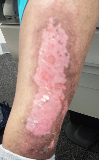
Lesion Sprang Up Under His Nose (Well, to One Side, Actually …)
An 80-year-old man is brought in by family for evaluation of a lesion on his nose. It manifested several years ago, at a smaller size, but has recently and abruptly grown. Although asymptomatic, the lesion is disturbing to the patient, who can now see it out of the corner of his eye.
The patient worked all of his adult life in the outdoors, as a farm and ranch hand. He has an extensive history of nonmelanoma skin cancer; several lesions have been removed from his face and arm.
Since the patient lives alone and rarely has visitors, it has been months since anyone has seen him. But as soon as his son-in-law saw the patient, he was sufficiently alarmed by the lesion to insist that care be sought.

EXAMINATION
The patient’s facial skin shows abundant evidence of chronic, severe sun damage: a whitish, spongy look to the skin on his forehead and upper cheeks and a great deal of discoloration and scaling.
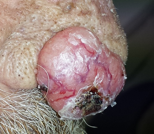
The lesion in question is a 3 x 1.5–cm, round, bulbous, smooth mass covering the right alar bulb. The surface is glassy-looking, with multiple telangiectasias. It is very firm but nontender on palpation. Shave biopsy is performed.
What is the diagnosis?
DISCUSSION
The biopsy results confirmed the suspicion of basal cell carcinoma (BCC). BCCs typically grow very slowly, often taking years to become noticeable, although not every BCC follows the rules. Some are more aggressive than others, both in terms of growth and clinical behavior.
It’s quite likely that in this case, the patient’s social isolation created the impression that his lesion grew abruptly and dramatically. (A subsequent eye exam revealed a number of problems, including severe presbyopia and advanced cataracts, so the patient himself might not have noticed the lesion for a while.) However, due to the large size and aggressive nature of the lesion—and the fact that the patient lives more than two hours from the nearest city, rendering his other treatment option, radiation, impractical—he was referred for Mohs surgery.
This process will establish clear surgical margins and provide acceptable closure. The latter may require reconstruction of the nose, depending on the depth of the cancer. Mohs surgeons often co-manage such cases with their counterparts in ENT or plastic surgery.
The differential for this lesion included keratoacanthoma , squamous cell carcinoma, and cyst.
TAKE-HOME LEARNING POINTS
• Basal cell carcinoma (BCC) is typically very slow growing, but there are exceptions.
• Social isolation can allow lesions and conditions to advance before they’re detected.
• Shave biopsy is indicated only for possible nonmelanoma skin cancers. Possible melanomas require excision, multiple punches, or deep shave to establish depth (a key prognostic factor).
• Rapid growth of BCCs suggests more aggressive clinical behavior, which in turn suggests the need for controlled margins to ensure complete removal.
An 80-year-old man is brought in by family for evaluation of a lesion on his nose. It manifested several years ago, at a smaller size, but has recently and abruptly grown. Although asymptomatic, the lesion is disturbing to the patient, who can now see it out of the corner of his eye.
The patient worked all of his adult life in the outdoors, as a farm and ranch hand. He has an extensive history of nonmelanoma skin cancer; several lesions have been removed from his face and arm.
Since the patient lives alone and rarely has visitors, it has been months since anyone has seen him. But as soon as his son-in-law saw the patient, he was sufficiently alarmed by the lesion to insist that care be sought.
EXAMINATION
The patient’s facial skin shows abundant evidence of chronic, severe sun damage: a whitish, spongy look to the skin on his forehead and upper cheeks and a great deal of discoloration and scaling.
The lesion in question is a 3 x 1.5–cm, round, bulbous, smooth mass covering the right alar bulb. The surface is glassy-looking, with multiple telangiectasias. It is very firm but nontender on palpation. Shave biopsy is performed.
What is the diagnosis?
DISCUSSION
The biopsy results confirmed the suspicion of basal cell carcinoma (BCC). BCCs typically grow very slowly, often taking years to become noticeable, although not every BCC follows the rules. Some are more aggressive than others, both in terms of growth and clinical behavior.
It’s quite likely that in this case, the patient’s social isolation created the impression that his lesion grew abruptly and dramatically. (A subsequent eye exam revealed a number of problems, including severe presbyopia and advanced cataracts, so the patient himself might not have noticed the lesion for a while.) However, due to the large size and aggressive nature of the lesion—and the fact that the patient lives more than two hours from the nearest city, rendering his other treatment option, radiation, impractical—he was referred for Mohs surgery.
This process will establish clear surgical margins and provide acceptable closure. The latter may require reconstruction of the nose, depending on the depth of the cancer. Mohs surgeons often co-manage such cases with their counterparts in ENT or plastic surgery.
The differential for this lesion included keratoacanthoma , squamous cell carcinoma, and cyst.
TAKE-HOME LEARNING POINTS
• Basal cell carcinoma (BCC) is typically very slow growing, but there are exceptions.
• Social isolation can allow lesions and conditions to advance before they’re detected.
• Shave biopsy is indicated only for possible nonmelanoma skin cancers. Possible melanomas require excision, multiple punches, or deep shave to establish depth (a key prognostic factor).
• Rapid growth of BCCs suggests more aggressive clinical behavior, which in turn suggests the need for controlled margins to ensure complete removal.
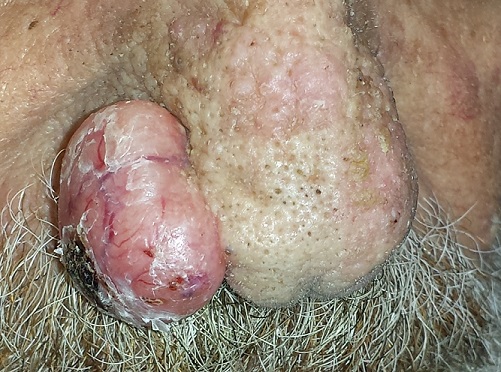
The Eyes Have It, and It Itches Like Crazy
For several months, a 69-year-old woman has had a rash around her eyes. It is terribly symptomatic, burning and itching with or without treatment (attempts at which have encompassed moisturizers, petroleum jelly, topical vitamin E oil, and most recently, application of triple-antibiotic cream three times a day). She finally requests referral to dermatology from her primary care provider.
When the rash manifested, she reports, she made some alterations to her routine, eliminating or changing the type of makeup, soap, cleanser, and laundry detergent she used. None of these changes helped.
Even before the distressing symptoms started, a friend had suggested the patient might have an eye problem. She consulted an ophthalmologist, who prescribed eye drops (the patient doesn’t recall any details); these only produced more burning and itching around her eyes.
The patient’s history is significant for atopy, with a childhood history of seasonal allergies, asthma, and eczema.

EXAMINATION
There is marked erythema and scaling in the bilateral periocular areas that spills onto both upper and lower lids. Very little edema is seen. The eyes themselves are free of changes.
What is the diagnosis?
DISCUSSION
For many patients, any problem that manifests close to the eye is deemed an “eye problem,” even when the eye itself is uninvolved. Eyelid dermatitis is an extremely common complaint, and this patient’s history is quite typical: The worse the problem gets, the more attempts the patient makes to relieve symptoms.
When this patient presented to dermatology, she was applying six different products (all OTC) to the affected areas. None helped, and in fact, most seemed to worsen the problem. Even if one had helped, she would never have known which. But desperation drives patients to do irrational things, especially when the problem is out in the open for the whole world to see.
Virtually every patient I’ve seen with eyelid dermatitis has (like this patient) already stopped using makeup and changed or discontinued use of laundry detergent and other products. These are almost never the problem; if any of them were, the effects would not be so sharply limited to the periocular area.
Rather, the sharp margins of this condition suggested irritant contact dermatitis. It often appears in this context: The periocular skin is unique in that it’s the thinnest in the female body. This means it is easily traumatized by rubbing and scratching and can be quite permeable to various contactants. This is especially true in atopic patients, whose skin is not only thin and dry but also overreactive to insult.
Though we may never know the full story, I suspect this patient had a more modest case of eczema on her eyelids until she began to apply product after product. One of them—triple-antibiotic cream—is a notorious topical sensitizer. In one sense, this patient is a victim of overattention to the problem.
I advised her to stop use of all the contactants and prescribed hydrocortisone 2.5% ointment for twice-daily application. I also gave her a prescription for a two-week taper of prednisone. When she returned for follow-up three weeks later, the rash was completely resolved.
Other conditions that can cause eyelid dermatitis include seborrhea and psoriasis.
TAKE-HOME LEARNING POINTS
• Eyelid dermatitis, an extremely common complaint, is rarely seen in men and is almost never caused by makeup, soap, or shampoo.
• Eyelid dermatitis is not an eye problem—rather, it is a skin problem that happens to occur near the eye.
• The differential for eyelid dermatitis includes atopic dermatitis, seborrhea and psoriasis.
• Stopping use of all contactant products (except prescription medications) is necessary.
• Class 5 or 6 steroids, especially hydrocortisone 2.5% in ointment form, are useful. In severe cases, a tapering course of oral glucocorticoids (prednisone) is extremely helpful.
For several months, a 69-year-old woman has had a rash around her eyes. It is terribly symptomatic, burning and itching with or without treatment (attempts at which have encompassed moisturizers, petroleum jelly, topical vitamin E oil, and most recently, application of triple-antibiotic cream three times a day). She finally requests referral to dermatology from her primary care provider.
When the rash manifested, she reports, she made some alterations to her routine, eliminating or changing the type of makeup, soap, cleanser, and laundry detergent she used. None of these changes helped.
Even before the distressing symptoms started, a friend had suggested the patient might have an eye problem. She consulted an ophthalmologist, who prescribed eye drops (the patient doesn’t recall any details); these only produced more burning and itching around her eyes.
The patient’s history is significant for atopy, with a childhood history of seasonal allergies, asthma, and eczema.
EXAMINATION
There is marked erythema and scaling in the bilateral periocular areas that spills onto both upper and lower lids. Very little edema is seen. The eyes themselves are free of changes.
What is the diagnosis?
DISCUSSION
For many patients, any problem that manifests close to the eye is deemed an “eye problem,” even when the eye itself is uninvolved. Eyelid dermatitis is an extremely common complaint, and this patient’s history is quite typical: The worse the problem gets, the more attempts the patient makes to relieve symptoms.
When this patient presented to dermatology, she was applying six different products (all OTC) to the affected areas. None helped, and in fact, most seemed to worsen the problem. Even if one had helped, she would never have known which. But desperation drives patients to do irrational things, especially when the problem is out in the open for the whole world to see.
Virtually every patient I’ve seen with eyelid dermatitis has (like this patient) already stopped using makeup and changed or discontinued use of laundry detergent and other products. These are almost never the problem; if any of them were, the effects would not be so sharply limited to the periocular area.
Rather, the sharp margins of this condition suggested irritant contact dermatitis. It often appears in this context: The periocular skin is unique in that it’s the thinnest in the female body. This means it is easily traumatized by rubbing and scratching and can be quite permeable to various contactants. This is especially true in atopic patients, whose skin is not only thin and dry but also overreactive to insult.
Though we may never know the full story, I suspect this patient had a more modest case of eczema on her eyelids until she began to apply product after product. One of them—triple-antibiotic cream—is a notorious topical sensitizer. In one sense, this patient is a victim of overattention to the problem.
I advised her to stop use of all the contactants and prescribed hydrocortisone 2.5% ointment for twice-daily application. I also gave her a prescription for a two-week taper of prednisone. When she returned for follow-up three weeks later, the rash was completely resolved.
Other conditions that can cause eyelid dermatitis include seborrhea and psoriasis.
TAKE-HOME LEARNING POINTS
• Eyelid dermatitis, an extremely common complaint, is rarely seen in men and is almost never caused by makeup, soap, or shampoo.
• Eyelid dermatitis is not an eye problem—rather, it is a skin problem that happens to occur near the eye.
• The differential for eyelid dermatitis includes atopic dermatitis, seborrhea and psoriasis.
• Stopping use of all contactant products (except prescription medications) is necessary.
• Class 5 or 6 steroids, especially hydrocortisone 2.5% in ointment form, are useful. In severe cases, a tapering course of oral glucocorticoids (prednisone) is extremely helpful.