User login
A Lifetime Of Sunshine
A 68-year-old man is seen for evaluation of a facial sore, which has been present for “at least a month.” It concerns him because, while the lesion is not painful, it won’t heal. The patient denies any preceding trauma to the area but does admit to having several skin cancers removed in his lifetime.
His lesion has been previously diagnosed as “pyoderma” and treated unsuccessfully with topical mupirocin and oral cephalexin.
He has held a number of jobs throughout his life, all of which involved being out in the sun all day—often seven days a week. And it wasn’t until he turned 45 that he finally started wearing a hat.
EXAMINATION
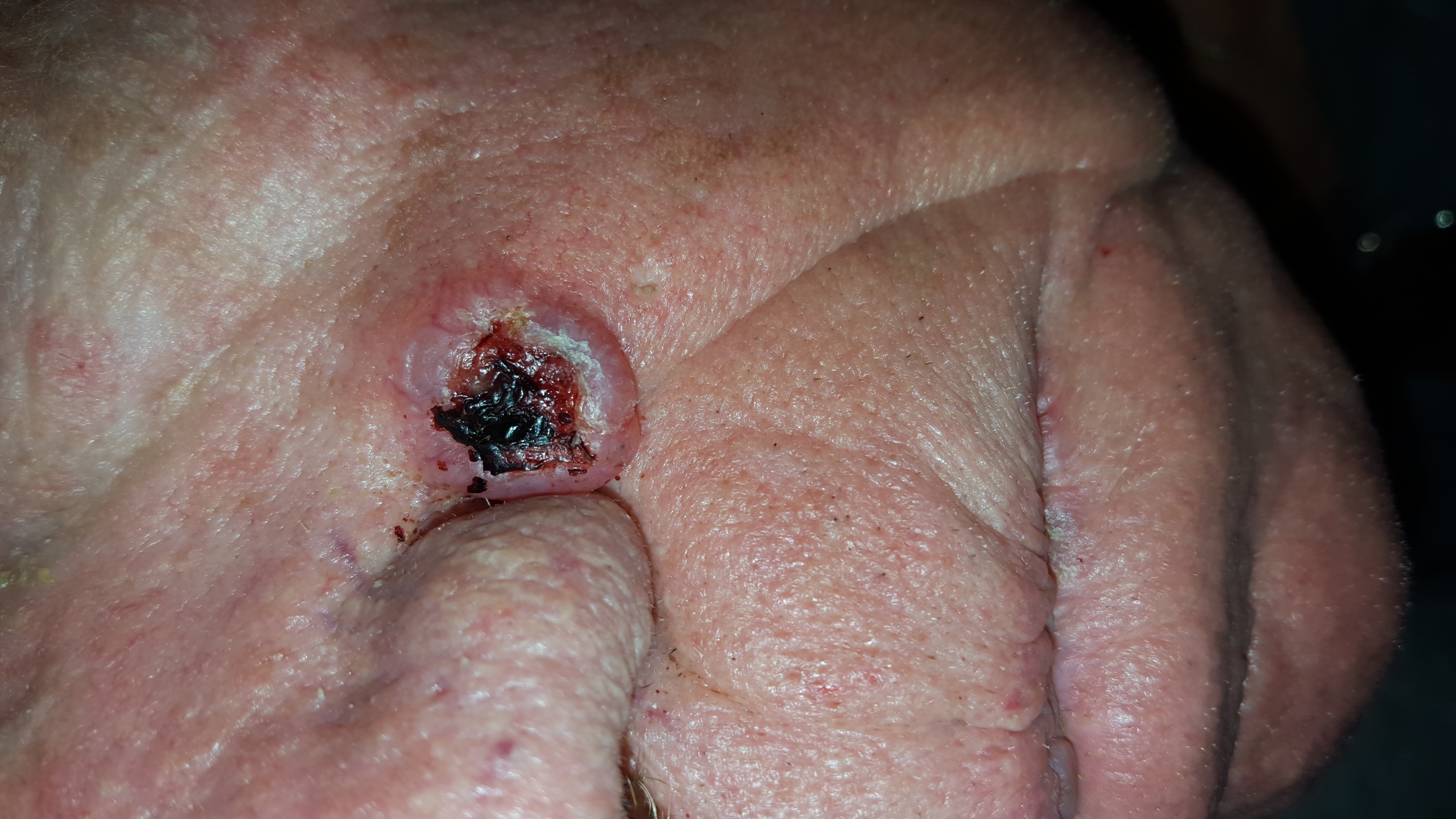
The lesion is an impressive, round, 1.2-cm nodule with extensive central ulceration. It has a rolled, translucent border and is tucked into the upper left nasolabial fold, directly adjacent to the alar bulb. It appears in the context of his fair, sun-damaged skin; he has several scars consistent with his history of skin cancer removal.

His skin elsewhere, while quite sun-damaged, is free of worrisome lesions.
A simple shave biopsy of the lesion is performed.
What is the diagnosis?
DISCUSSION
The biopsy showed changes consistent with basal cell carcinoma (BCC). It would be difficult to find a more typical example of BCC, the most common of all the sun-caused skin cancers (which include squamous cell, melanoma, and Merkel cell carcinoma). And yet, as this case illustrates, classic signs of BCC are often missed or overlooked, leading to “infection” or “pyoderma” diagnoses by providers who are unfamiliar with BCC.
I often tell medical providers that “it will be a rare day when you don’t see a BCC” because they are that common. But what I probably should say is “it will be a rare day when you don’t have a chance to see a BCC.” By that, I mean that you do have to look for them—not all are as obvious as this man’s—and you do need to have an understanding of who is susceptible and what the lesion will look like.
Older, sun-damaged individuals (mostly men) are most at risk for BBCs. Fair skin, freckles, red hair, and blue eyes are additional high-risk characteristics. This varies from melanoma patients, who tend to be much younger and whose lesions look nothing like a basal cell.
For BCC identification, focus on the areas directly and chronically exposed to the sun, such as the tops of the ears, the face, nose, chest, back, and arms.
BCCs can take on many appearances; they look as though they would hurt, but rarely do—a fact that should be noted as significant. Typically, they break down, scab, and bleed as they grow larger. They are usually very slow-growing and often take years to become noticeable. In other words, you have to think of it first, and look for it next. The fact that almost 1.5 million new BCCs are diagnosed each year in the US should heighten your suspicions considerably.
In this case, the patient will need extensive surgery, which may be deforming. Given his history of sun exposure, he will likely develop more skin cancers in the future.
TAKE-HOME LEARNING POINTS
• Basal cell carcinoma (BCC) is by far the most common type of sun-caused skin cancer; it will be seen frequently if properly looked for.
• Identification of BCCs will be more successful if the search is concentrated on the patients most likely to develop one and the areas where they are most likely to appear.
• This patient’s noduloulcerative BCC is the most common type and developed in a classic location. There is no type of infection that presents in this fashion.
• A simple shave biopsy confirms the diagnosis of BCC.
A 68-year-old man is seen for evaluation of a facial sore, which has been present for “at least a month.” It concerns him because, while the lesion is not painful, it won’t heal. The patient denies any preceding trauma to the area but does admit to having several skin cancers removed in his lifetime.
His lesion has been previously diagnosed as “pyoderma” and treated unsuccessfully with topical mupirocin and oral cephalexin.
He has held a number of jobs throughout his life, all of which involved being out in the sun all day—often seven days a week. And it wasn’t until he turned 45 that he finally started wearing a hat.
EXAMINATION
The lesion is an impressive, round, 1.2-cm nodule with extensive central ulceration. It has a rolled, translucent border and is tucked into the upper left nasolabial fold, directly adjacent to the alar bulb. It appears in the context of his fair, sun-damaged skin; he has several scars consistent with his history of skin cancer removal.
His skin elsewhere, while quite sun-damaged, is free of worrisome lesions.
A simple shave biopsy of the lesion is performed.
What is the diagnosis?
DISCUSSION
The biopsy showed changes consistent with basal cell carcinoma (BCC). It would be difficult to find a more typical example of BCC, the most common of all the sun-caused skin cancers (which include squamous cell, melanoma, and Merkel cell carcinoma). And yet, as this case illustrates, classic signs of BCC are often missed or overlooked, leading to “infection” or “pyoderma” diagnoses by providers who are unfamiliar with BCC.
I often tell medical providers that “it will be a rare day when you don’t see a BCC” because they are that common. But what I probably should say is “it will be a rare day when you don’t have a chance to see a BCC.” By that, I mean that you do have to look for them—not all are as obvious as this man’s—and you do need to have an understanding of who is susceptible and what the lesion will look like.
Older, sun-damaged individuals (mostly men) are most at risk for BBCs. Fair skin, freckles, red hair, and blue eyes are additional high-risk characteristics. This varies from melanoma patients, who tend to be much younger and whose lesions look nothing like a basal cell.
For BCC identification, focus on the areas directly and chronically exposed to the sun, such as the tops of the ears, the face, nose, chest, back, and arms.
BCCs can take on many appearances; they look as though they would hurt, but rarely do—a fact that should be noted as significant. Typically, they break down, scab, and bleed as they grow larger. They are usually very slow-growing and often take years to become noticeable. In other words, you have to think of it first, and look for it next. The fact that almost 1.5 million new BCCs are diagnosed each year in the US should heighten your suspicions considerably.
In this case, the patient will need extensive surgery, which may be deforming. Given his history of sun exposure, he will likely develop more skin cancers in the future.
TAKE-HOME LEARNING POINTS
• Basal cell carcinoma (BCC) is by far the most common type of sun-caused skin cancer; it will be seen frequently if properly looked for.
• Identification of BCCs will be more successful if the search is concentrated on the patients most likely to develop one and the areas where they are most likely to appear.
• This patient’s noduloulcerative BCC is the most common type and developed in a classic location. There is no type of infection that presents in this fashion.
• A simple shave biopsy confirms the diagnosis of BCC.
Acne: Maybe She's Born With It?
A recent treatment regimen worsened this 13-year-old Native American girl’s “acne.” Within hours of beginning the treatment (application of benzoyl peroxide–containing gel and twice-daily use of a benzoyl peroxide–based cleanser), she reported intense burning and redness in the treated areas. She stopped this regimen after a week, and her primary care provider referred her for evaluation.
The lesions have been on her face since age 2 and are not usually problematic.
The patient has a notable history of atopy, with seasonal allergies and occasional bouts of asthma. She claims to have no other health problems but does admit to being overweight for most of her life; this is an issue for most of her family members, as well. Several of her relatives developed diabetes as adults.
EXAMINATION
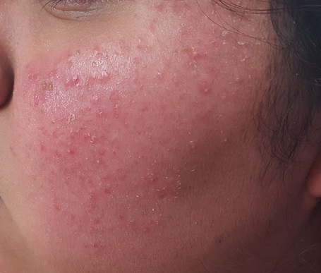
The patient is 5 ft tall and weighs 250 lb. She has type IV skin, consistent with her Native American heritage. The posterior aspects of both cheeks are quite red and covered with hyperkeratotic, rough, pinpoint papules. There are no comedones or pustules there or elsewhere on her body. 
No other blemishes or lesions are seen on her face, but there are hundreds of hyperkeratotic papules on her bilateral triceps. The skin in all her intertriginous (skin-on-skin) areas is quite dark and has a faintly papular, velvety appearance.
What is the diagnosis?
DISCUSSION
This girl’s facial and arm condition is keratosis pilaris (KP), an extremely common and harmless problem that affects up to 70% of newborns, though it may not fully express until age 1 or 2. Caused by an overproduction of perifollicular keratin, KP is inherited in an autosomal dominant pattern and is often seen in conjunction with atopic dermatitis and related conditions (eg, eczema, xerosis, asthma, icthyosis). KP can affect skin anywhere on the body except glabrous skin (palms and soles).
Variants of KP are also common; the one affecting this patient is keratosis pilaris rubra facei. This condition is often confused with acne, but treating it as such worsens irritation—especially in the wintertime, when humidity levels are low.
The presence of the patient’s condition from such an early age, the lack of comedones or pustules, and the discovery of more typical KP lesions on her arms all served to rule out acne and rosacea (the other item in the differential). The intertriginous hyperpigmentation represents acanthosis nigricans, reflecting her obese and (probable) prediabetic state, and is unrelated to her KP.
She will be treated with a one-week course of 2.5% hydrocortisone cream (bid application) and cessation of benzoyl peroxide–containing products. The condition should improve rapidly and become less prominent over time. Given the nature of KP, I have also found it helpful to encourage parents of affected children to research the condition online, to reinforce the information I’ve provided in the clinic.
TAKE-HOME LEARNING POINTS
• Keratosis pilaris (KP) is extremely common, affecting up to 70% of all newborns.
• Keratosis pilaris rubra facei, a common variant, affects the posterior two-thirds of the cheeks. Virtually all patients with this form will also exhibit KP on more typical areas (eg, the triceps).
• While there is no cure, moisturization helps to ease symptoms, as do other products (eg, salicylic acid–containing creams).
• KP is often mistaken for acne; unfortunately, treatment as such is counterproductive.
A recent treatment regimen worsened this 13-year-old Native American girl’s “acne.” Within hours of beginning the treatment (application of benzoyl peroxide–containing gel and twice-daily use of a benzoyl peroxide–based cleanser), she reported intense burning and redness in the treated areas. She stopped this regimen after a week, and her primary care provider referred her for evaluation.
The lesions have been on her face since age 2 and are not usually problematic.
The patient has a notable history of atopy, with seasonal allergies and occasional bouts of asthma. She claims to have no other health problems but does admit to being overweight for most of her life; this is an issue for most of her family members, as well. Several of her relatives developed diabetes as adults.
EXAMINATION
The patient is 5 ft tall and weighs 250 lb. She has type IV skin, consistent with her Native American heritage. The posterior aspects of both cheeks are quite red and covered with hyperkeratotic, rough, pinpoint papules. There are no comedones or pustules there or elsewhere on her body.
No other blemishes or lesions are seen on her face, but there are hundreds of hyperkeratotic papules on her bilateral triceps. The skin in all her intertriginous (skin-on-skin) areas is quite dark and has a faintly papular, velvety appearance.
What is the diagnosis?
DISCUSSION
This girl’s facial and arm condition is keratosis pilaris (KP), an extremely common and harmless problem that affects up to 70% of newborns, though it may not fully express until age 1 or 2. Caused by an overproduction of perifollicular keratin, KP is inherited in an autosomal dominant pattern and is often seen in conjunction with atopic dermatitis and related conditions (eg, eczema, xerosis, asthma, icthyosis). KP can affect skin anywhere on the body except glabrous skin (palms and soles).
Variants of KP are also common; the one affecting this patient is keratosis pilaris rubra facei. This condition is often confused with acne, but treating it as such worsens irritation—especially in the wintertime, when humidity levels are low.
The presence of the patient’s condition from such an early age, the lack of comedones or pustules, and the discovery of more typical KP lesions on her arms all served to rule out acne and rosacea (the other item in the differential). The intertriginous hyperpigmentation represents acanthosis nigricans, reflecting her obese and (probable) prediabetic state, and is unrelated to her KP.
She will be treated with a one-week course of 2.5% hydrocortisone cream (bid application) and cessation of benzoyl peroxide–containing products. The condition should improve rapidly and become less prominent over time. Given the nature of KP, I have also found it helpful to encourage parents of affected children to research the condition online, to reinforce the information I’ve provided in the clinic.
TAKE-HOME LEARNING POINTS
• Keratosis pilaris (KP) is extremely common, affecting up to 70% of all newborns.
• Keratosis pilaris rubra facei, a common variant, affects the posterior two-thirds of the cheeks. Virtually all patients with this form will also exhibit KP on more typical areas (eg, the triceps).
• While there is no cure, moisturization helps to ease symptoms, as do other products (eg, salicylic acid–containing creams).
• KP is often mistaken for acne; unfortunately, treatment as such is counterproductive.
A Case of Cold Feet Hands
A 51-year-old woman is referred to dermatology for multiple problems she has had for several months—and, in some cases, years. The most immediate is a new nodule on the side of one thumb. Although it is asymptomatic, the fact that it simply “appeared” worries her, because her favorite uncle recently died of melanoma.
Several new lesions also materialized on her finger pads and chest. These do not cause symptoms either, but their newness makes them worrisome.
When asked about her health in general, the patient initially states it is “fine”—until her husband corrects her. She then recalls that she is being seen by various specialists, including a gastroenterologist for dysphagia-like symptoms and a rheumatologist for vague joint pains and what sounds like Raynaud disease.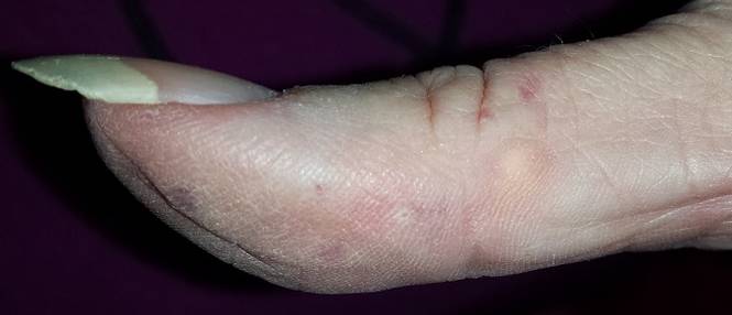
EXAMINATION
On the lateral right thumb is a shallow, intradermal, white lesion. It is firm and round and measures 5 mm. No overlying skin changes (eg, redness or disruption of the skin surface) are observed. Under local anesthesia, a small incision is made in the lesion’s surface, allowing for exploration with small forceps; granular, gritty, white material is extracted.
On the affected thumb and two other fingers, telangiectasias can be seen. There are additional patches on the patient’s chest, but none around her mouth.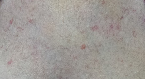
No sign of Raynaud disease is seen. However, the patient is adamant that this only appears when her hands are quite cold.
What is the diagnosis?
DISCUSSION
Connecting these dots leads to the likelihood of CREST syndrome, a variant of systemic sclerosis (SS); the acronym stands for calcinosis, Raynaud disease, esophageal dysmotility, sclerodactyly, and telangiectasia. The classification of CREST in the spectrum of SS is still open to discussion, but it is widely recognized as a real and distinct condition.
SS shares with CREST the involvement of anticentromere antibodies, but there are significant differences in the clinical course of each condition. Raynaud disease is often the first sign of CREST and can precede the other findings by years. Unlike SS, CREST typically spares the kidneys, and if it affects the lungs at all, it is through pulmonary artery hypertension and not fibrosis (the latter of which is seen in SS). Both conditions, however, can present with swelling of the hands and dysphagia.
Interpretation of these disparate findings requires the skill of a rheumatologist, who often shares management with other specialists.
As for differential diagnoses: Any one of these components can be a stand-alone diagnosis or can manifest with other diseases or syndromes. But when found together, they are highly suggestive of CREST. In this patient’s case, more testing needs to be done before a definitive diagnosis can be made.
TAKE-HOME LEARNING POINTS
• CREST syndrome is considered a variant of systemic sclerosis but has significantly different features and clinical course.
• The components of CREST (calcinosis, Raynaud disease, esophageal dysmotility, sclerodactyly, and telangiectasia) are often viewed as isolated phenomena by those unfamiliar with the condition.
• Raynaud disease is often the initial sign of CREST, preceding the rest by years.
• The telangiectasias seen with CREST often manifest on the hands and chest but can also be found on mucosal surfaces.

Itchy Lesions, Picking Patient
A 59-year-old woman presents urgently to dermatology for evaluation of very itchy lesions that manifested at least 20 years ago. They have been previously diagnosed as staph infection, fungal infection, psoriasis, and scabies but have not responded to a variety of topical steroid creams and topical antibacterials (eg, triple-antibiotic cream, mupirocin, and clindamycin solution). The patient has also been prescribed numerous oral antibiotics, the most recent of which was a 10-day course of clindamycin. This induced a raging case of diarrhea; her primary care provider subsequently referred her to dermatology.
She denies any family history of skin problems until her sister, who has accompanied her to this appointment, mentions that she and her mother have had similar problems for decades. More questioning reveals that all three women also have extensive psychiatric histories of chronic anxiety and depression.
When pressed, the patient and her sister admit to picking at their skin “constantly,” but especially when under stress.
EXAMINATION
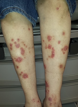
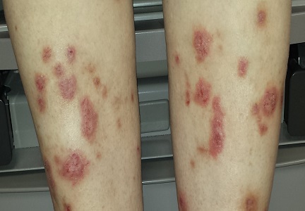
Most of the patient’s lesions—round to oval, orangish red, scaly plaques that range from 2 to 5 cm—are on her legs, below her knees. There are about 15 lesions total. They look and feel very thick but have no increased warmth or tenderness. The erythema is confined to the lesions and does not extend into the surrounding areas.
Punch biopsy of one leg lesion shows a hypertrophic epidermis with focal parakeratosis, elongation of rete ridges, and perhaps most importantly, no signs of other items in the differential (such as psoriasis or skin cancer).
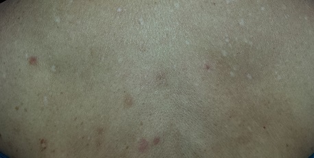
Elsewhere on her skin, extensive punctate disciform scarring extends across her upper back, stopping abruptly at the T2 level. The skin below remains totally clear.
What is the diagnosis?
DISCUSSION
The correct diagnosis is prurigo nodularis (PN), which was first described in 1909 and has been extensively studied since. Though much remains unknown about PN, we know that, typically, the patient plays a major role in the genesis and perpetuation of the condition. In other words, these patients are pickers—but as they’ll tell you, they “wouldn’t pick if the lesions didn’t itch.” It does appear that they have a much lower threshold for itching than the general population.
Personal and family history of serious, ongoing bouts of depression and anxiety, as seen in this patient, are also typical among PN patients. Women appear to be far more likely to be diagnosed with PN, though there is no documentation of this in the literature.
These observations, while helpful for diagnosis, still don’t fully explain exactly why one person has PN while another doesn’t.
Besides the biopsy results, other keys to distinguishing PN include the characteristic lesions themselves, as well as the sparing of sites out of the patient’s reach. As is often the case, multiple old scars were seen on this patient’s upper back, confirming the chronicity of the condition.
In addition to educating patients about their role in the creation and perpetuation of PN, I find the most useful treatment to be intralesional triamcinolone in a 10-mg/cc strength (1/4 cc stock triamcinolone diluted in ¾ cc lidocaine), delivered directly into the lesion by 30-gauge needle Alternatively, a class 1 topical steroid cream (eg, clobetasol 0.05%) can be applied twice daily and then covered with a bandage. In cooler weather, when patients tend to bundle up, additional layers of clothing help to create a barrier that prevents patients from picking as much.
New cases of “picking” suggest the possibility of other diagnoses, including renal or hepatic failure, occult cancer, or other skin disease (eg, mastocytosis).
TAKE-HOME LEARNING POINTS
• Prurigo nodularis (PN) often presents with multiple papules, nodules, and/or plaques that develop primarily as a result of picking, rubbing, or scratching the area.
• PN patients appear to have a lowered threshold for itching, which may play a role in their disease.
• The differential for PN includes squamous cell carcinoma, psoriasis, atypical mycobacteria infection, and metastatic cancer.
• Biopsy plays a key role in establishing the correct diagnosis.
• Intralesional steroid injection with 10 mg/cc triamcinolone stops the itching and shrinks the lesion.
A 59-year-old woman presents urgently to dermatology for evaluation of very itchy lesions that manifested at least 20 years ago. They have been previously diagnosed as staph infection, fungal infection, psoriasis, and scabies but have not responded to a variety of topical steroid creams and topical antibacterials (eg, triple-antibiotic cream, mupirocin, and clindamycin solution). The patient has also been prescribed numerous oral antibiotics, the most recent of which was a 10-day course of clindamycin. This induced a raging case of diarrhea; her primary care provider subsequently referred her to dermatology.
She denies any family history of skin problems until her sister, who has accompanied her to this appointment, mentions that she and her mother have had similar problems for decades. More questioning reveals that all three women also have extensive psychiatric histories of chronic anxiety and depression.
When pressed, the patient and her sister admit to picking at their skin “constantly,” but especially when under stress.
EXAMINATION
Most of the patient’s lesions—round to oval, orangish red, scaly plaques that range from 2 to 5 cm—are on her legs, below her knees. There are about 15 lesions total. They look and feel very thick but have no increased warmth or tenderness. The erythema is confined to the lesions and does not extend into the surrounding areas.
Punch biopsy of one leg lesion shows a hypertrophic epidermis with focal parakeratosis, elongation of rete ridges, and perhaps most importantly, no signs of other items in the differential (such as psoriasis or skin cancer).
Elsewhere on her skin, extensive punctate disciform scarring extends across her upper back, stopping abruptly at the T2 level. The skin below remains totally clear.
What is the diagnosis?
DISCUSSION
The correct diagnosis is prurigo nodularis (PN), which was first described in 1909 and has been extensively studied since. Though much remains unknown about PN, we know that, typically, the patient plays a major role in the genesis and perpetuation of the condition. In other words, these patients are pickers—but as they’ll tell you, they “wouldn’t pick if the lesions didn’t itch.” It does appear that they have a much lower threshold for itching than the general population.
Personal and family history of serious, ongoing bouts of depression and anxiety, as seen in this patient, are also typical among PN patients. Women appear to be far more likely to be diagnosed with PN, though there is no documentation of this in the literature.
These observations, while helpful for diagnosis, still don’t fully explain exactly why one person has PN while another doesn’t.
Besides the biopsy results, other keys to distinguishing PN include the characteristic lesions themselves, as well as the sparing of sites out of the patient’s reach. As is often the case, multiple old scars were seen on this patient’s upper back, confirming the chronicity of the condition.
In addition to educating patients about their role in the creation and perpetuation of PN, I find the most useful treatment to be intralesional triamcinolone in a 10-mg/cc strength (1/4 cc stock triamcinolone diluted in ¾ cc lidocaine), delivered directly into the lesion by 30-gauge needle Alternatively, a class 1 topical steroid cream (eg, clobetasol 0.05%) can be applied twice daily and then covered with a bandage. In cooler weather, when patients tend to bundle up, additional layers of clothing help to create a barrier that prevents patients from picking as much.
New cases of “picking” suggest the possibility of other diagnoses, including renal or hepatic failure, occult cancer, or other skin disease (eg, mastocytosis).
TAKE-HOME LEARNING POINTS
• Prurigo nodularis (PN) often presents with multiple papules, nodules, and/or plaques that develop primarily as a result of picking, rubbing, or scratching the area.
• PN patients appear to have a lowered threshold for itching, which may play a role in their disease.
• The differential for PN includes squamous cell carcinoma, psoriasis, atypical mycobacteria infection, and metastatic cancer.
• Biopsy plays a key role in establishing the correct diagnosis.
• Intralesional steroid injection with 10 mg/cc triamcinolone stops the itching and shrinks the lesion.
Red Facial Lesions Are No Day at the Beach
A 70-year-old man is seen for “broken blood vessels” on his face. The lesions were recently noted by a relative who had not seen him in years.
The patient has a pronounced history of sun overexposure as a result of his job and his hobbies, which include fishing and hunting. As a younger man, he enjoyed waterskiing and going to the lake almost every weekend during the warmer months.
He is in decent health apart from the lesions and has no other skin complaints.

EXAMINATION
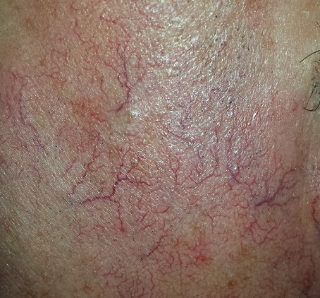
The sides of the patient’s face are covered with linear, red, dilated blood vessels running in jagged patterns. The lesions are asymptomatic and range from 0.5 to 1.0 mm in diameter; some are several centimeters long. They are readily blanchable with digital pressure, though none are palpable.
Far more blood vessels are seen on the patient’s left side than on his right. The underlying skin is quite fair and thin. In some locations (eg, the lateral forehead), there are patches of white skin free of surface adnexa (ie pores, skin lines, or hair follicles).
His dorsal arms are very rough, with hundreds of solar lentigines, while the skin on his volar arms is relatively unaffected.
What is the diagnosis?
DISCUSSION
This collection of findings is called dermatoheliosis (DHe), literally “sun-skin condition,” caused by overexposure to UV light. Telangiectasias, one manifestation, have many potential causes, but sun exposure is the most common—especially in those whose skin burns easily and frequently.
These blood vessels are simply normal surface vasculature made more obvious by two factors: First, chronic sun exposure overheats the skin for prolonged periods, leading to persistent vasodilatation that eventually becomes fixed. Second, solar atrophy may occur when the skin thins from sun overexposure, making typically invisible structures apparent.
DHe can also manifest as actinic keratosis, weathering of the skin, solar elastosis, and sun-caused skin cancers (basal and squamous cell carcinomas, melanoma, etc). The condition is incredibly common.
Many causes of telangiectasias are unrelated to sun exposure. Other processes that thin the skin—such as radiation therapy or injudicious use of topical steroids—may produce telangiectasias. Further examples include spider vessels on legs due to venous insufficiency and newly acquired vascular lesions resulting from pregnancy or liver cirrhosis. There are also two unusual but significant conditions that involve telangiectasias: a variant of systemic sclerosis called CREST syndrome (calcinosis, Raynaud’s, esophageal dysmotility, sclerodactyly, and telangiectasias) and hereditary hemorrhagic telangiectasias (also known eponymically as Osler-Weber-Rendu syndrome).
Treatment for sun-caused telangiectasias includes laser and electrodessication.
TAKE-HOME LEARNING POINTS
• Telangiectasias are permanently dilated surface vasculature usually seen on the most prominently sun-exposed areas of the skin. The left face and arm are often more heavily affected than the right, since they are closer to the car window during driving.
• Telangiectasias are only one of a number of sun-caused skin changes that are collectively termed dermatoheliosis (DHe).
• Telangiectasias are important because they identify patients at risk for sun-caused skin cancers, such as basal or squamous cell carcinomas and melanoma.
• Other potential causes of telangiectasias include injudicious use of topical steroids, focal radiation therapy, pregnancy, liver disease, and rare conditions such as CREST syndrome and hereditary hemorrhagic telangiectasias.
A 70-year-old man is seen for “broken blood vessels” on his face. The lesions were recently noted by a relative who had not seen him in years.
The patient has a pronounced history of sun overexposure as a result of his job and his hobbies, which include fishing and hunting. As a younger man, he enjoyed waterskiing and going to the lake almost every weekend during the warmer months.
He is in decent health apart from the lesions and has no other skin complaints.
EXAMINATION
The sides of the patient’s face are covered with linear, red, dilated blood vessels running in jagged patterns. The lesions are asymptomatic and range from 0.5 to 1.0 mm in diameter; some are several centimeters long. They are readily blanchable with digital pressure, though none are palpable.
Far more blood vessels are seen on the patient’s left side than on his right. The underlying skin is quite fair and thin. In some locations (eg, the lateral forehead), there are patches of white skin free of surface adnexa (ie pores, skin lines, or hair follicles).
His dorsal arms are very rough, with hundreds of solar lentigines, while the skin on his volar arms is relatively unaffected.
What is the diagnosis?
DISCUSSION
This collection of findings is called dermatoheliosis (DHe), literally “sun-skin condition,” caused by overexposure to UV light. Telangiectasias, one manifestation, have many potential causes, but sun exposure is the most common—especially in those whose skin burns easily and frequently.
These blood vessels are simply normal surface vasculature made more obvious by two factors: First, chronic sun exposure overheats the skin for prolonged periods, leading to persistent vasodilatation that eventually becomes fixed. Second, solar atrophy may occur when the skin thins from sun overexposure, making typically invisible structures apparent.
DHe can also manifest as actinic keratosis, weathering of the skin, solar elastosis, and sun-caused skin cancers (basal and squamous cell carcinomas, melanoma, etc). The condition is incredibly common.
Many causes of telangiectasias are unrelated to sun exposure. Other processes that thin the skin—such as radiation therapy or injudicious use of topical steroids—may produce telangiectasias. Further examples include spider vessels on legs due to venous insufficiency and newly acquired vascular lesions resulting from pregnancy or liver cirrhosis. There are also two unusual but significant conditions that involve telangiectasias: a variant of systemic sclerosis called CREST syndrome (calcinosis, Raynaud’s, esophageal dysmotility, sclerodactyly, and telangiectasias) and hereditary hemorrhagic telangiectasias (also known eponymically as Osler-Weber-Rendu syndrome).
Treatment for sun-caused telangiectasias includes laser and electrodessication.
TAKE-HOME LEARNING POINTS
• Telangiectasias are permanently dilated surface vasculature usually seen on the most prominently sun-exposed areas of the skin. The left face and arm are often more heavily affected than the right, since they are closer to the car window during driving.
• Telangiectasias are only one of a number of sun-caused skin changes that are collectively termed dermatoheliosis (DHe).
• Telangiectasias are important because they identify patients at risk for sun-caused skin cancers, such as basal or squamous cell carcinomas and melanoma.
• Other potential causes of telangiectasias include injudicious use of topical steroids, focal radiation therapy, pregnancy, liver disease, and rare conditions such as CREST syndrome and hereditary hemorrhagic telangiectasias.
The Things You Do (but Don’t) See Every Day …
Fifty-year-old twin sisters are seen for what appears to be the same problem: changes in the color and texture of their neck skin, recently noted by a family member. Both deny ever noticing it before and further deny experiencing any associated symptoms.
The sisters are accompanying their mother, who is being treated for basal cell carcinoma. All three women grew up living and working on the family farm, spending a great deal of time in the sun from an early age.

EXAMINATION
Both patients have type III skin, meaning that they seldom burn, tan fairly easily, and are able to keep that tan. The patients have identical, obvious hypo- and hyperpigmentation covering the sides of their faces and necks, sparing a well-defined oval area on the submental anterior neck.
The affected areas appear hyperemic, displaying a reddish brown tinge, but fail to blanch with digital pressure. The changes are completely macular.
What is the diagnosis?
DISCUSSION
The changes seen with poikiloderma of Civatte (PC) are obvious to the observer (see photograph) but appear so gradually that the patient often fails to notice them until they are pointed out. Both patients in this case almost certainly have had the condition for years.
PC was first described by Achille Civatte in France in 1923, around the same time that sunbathing became popular. Prior to that time, most women carefully protected their skin from the sun.
Civatte and others found that virtually all PC patients had a history of chronic overexposure to the sun, and that the clinical features of the condition—especially the characteristic sparing of the anterior neck skin (due to shading by the chin)—supported this hypothesis. They were also able to detect histologic changes (eg, solar elastosis) on biopsy, which further corroborated this theory.
There is little doubt that chronic overexposure to UV radiation is the cause, though other factors appear to be involved, as this case illustrates. For example, PC affects far more women than men, which suggests a possible role for hormones. It also appears that, in some cases, the tendency to develop PC is due to the genetic inheritance of an increased susceptibility to UV light. The mother of the two women in this case also had PC.
The range of presentations of PC includes involvement of the chest, face, and posterior neck. It should also be noted that somewhat similar patterns can be seen in other diseases, such as lupus, dermatomyositis, and early cutaneous T-cell lymphoma. Careful history-taking and biopsy, when indicated, will serve to distinguish between these diagnoses.
PC is usually left untreated, although lasers have been successfully used on motivated patients.
TAKE-HOME LEARNING POINTS
• Poikiloderma of Civatte (PC) is quite common and is seen more in women than men.
• Chronic overexposure to UV light is the primary cause of PC, but other factors—such as gender and heredity—appear to be involved.
• The term poikiloderma refers to the variability of the red, brown, and white color changes, as well as the solar atrophy seen with this condition.
• The differential for PC includes lupus, dermatomyositis, and early cutaneous T-cell lymphoma.
Fifty-year-old twin sisters are seen for what appears to be the same problem: changes in the color and texture of their neck skin, recently noted by a family member. Both deny ever noticing it before and further deny experiencing any associated symptoms.
The sisters are accompanying their mother, who is being treated for basal cell carcinoma. All three women grew up living and working on the family farm, spending a great deal of time in the sun from an early age.
EXAMINATION
Both patients have type III skin, meaning that they seldom burn, tan fairly easily, and are able to keep that tan. The patients have identical, obvious hypo- and hyperpigmentation covering the sides of their faces and necks, sparing a well-defined oval area on the submental anterior neck.
The affected areas appear hyperemic, displaying a reddish brown tinge, but fail to blanch with digital pressure. The changes are completely macular.
What is the diagnosis?
DISCUSSION
The changes seen with poikiloderma of Civatte (PC) are obvious to the observer (see photograph) but appear so gradually that the patient often fails to notice them until they are pointed out. Both patients in this case almost certainly have had the condition for years.
PC was first described by Achille Civatte in France in 1923, around the same time that sunbathing became popular. Prior to that time, most women carefully protected their skin from the sun.
Civatte and others found that virtually all PC patients had a history of chronic overexposure to the sun, and that the clinical features of the condition—especially the characteristic sparing of the anterior neck skin (due to shading by the chin)—supported this hypothesis. They were also able to detect histologic changes (eg, solar elastosis) on biopsy, which further corroborated this theory.
There is little doubt that chronic overexposure to UV radiation is the cause, though other factors appear to be involved, as this case illustrates. For example, PC affects far more women than men, which suggests a possible role for hormones. It also appears that, in some cases, the tendency to develop PC is due to the genetic inheritance of an increased susceptibility to UV light. The mother of the two women in this case also had PC.
The range of presentations of PC includes involvement of the chest, face, and posterior neck. It should also be noted that somewhat similar patterns can be seen in other diseases, such as lupus, dermatomyositis, and early cutaneous T-cell lymphoma. Careful history-taking and biopsy, when indicated, will serve to distinguish between these diagnoses.
PC is usually left untreated, although lasers have been successfully used on motivated patients.
TAKE-HOME LEARNING POINTS
• Poikiloderma of Civatte (PC) is quite common and is seen more in women than men.
• Chronic overexposure to UV light is the primary cause of PC, but other factors—such as gender and heredity—appear to be involved.
• The term poikiloderma refers to the variability of the red, brown, and white color changes, as well as the solar atrophy seen with this condition.
• The differential for PC includes lupus, dermatomyositis, and early cutaneous T-cell lymphoma.

Obesity, Scales, and a Sweaty Situation
A 67-year-old Asian woman presents with changes under her breasts that manifested several years ago and are slowly becoming more pronounced. Occasionally itchy, the skin is always hot and sweaty. This problem has been treated numerous times over the years with oral and topical antiyeast preparations, to no good effect. She is currently on fluconazole oral.
Aside from obesity, she claims that her only other health problem is “skin rashes” on her arms and scalp, which have persisted for years. Cursory examination of her chart, however, reveals that she also has type 2 diabetes and dementia.
EXAMINATION
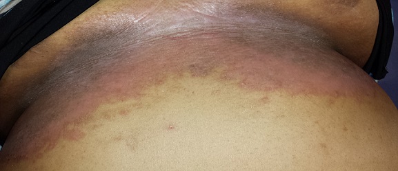
The patient is 5 feet tall and weighs 240 lb. She has type II skin with classic Asian coloring. The skin under her breasts is bright pink, with focal areas of dark brown. The affected area extends downward onto her upper abdomen. The surface of the rash is totally macular, with no palpable component.
On both extensor forearms, a scaly, pink rash is seen, covered with thick, white scales. Likewise, on her scalp, particularly above and behind the ears, there are large patches of white scaling with underlying pink skin.
What is the diagnosis?
DISCUSSION
While it is uncommon in those of Asian ancestry, this patient has psoriasis. (The involvement of her scalp and arms helps to corroborate this diagnosis.) But she has other issues that serve to confuse the picture: Her extreme obesity and pendulous breasts trap heat and sweat, which induces a condition called intertrigo. This chronic inflammatory process has become “koebnerized,” a phenomenon in which psoriasis appears in a traumatized area, such as a surgical wound or burn. To make matters even worse, the skin immediately under her breasts has darkened as a result of postinflammatory hyperpigmentation.
So this patient actually has three overlapping problems: intertrigo, koebnerized psoriasis, and postinflammatory hyperpigmentation. Her obesity, which is unlikely to improve, is a major contributor to each. Luckily, the inframammary skin changes are relatively asymptomatic.
All three problems can be alleviated by reducing heat and sweat (with antiperspirants, talcum-based powder, weight loss, and use of cotton cloth to separate the breasts from the underlying chest skin), and through use of topical class III steroid creams. We can stop treating it as a yeast infection, because yeast plays little to no part in the process.
TAKE-HOME LEARNING POINTS
• Skin injury—whether from surgery, burn, excoriation, or chronic maceration—can cause psoriasis to flare, a process known as the Koebner phenomenon.
• Intertrigo refers to an inflammatory condition affecting intertriginous areas such as the groin, axilla, abdominal or other folds, and inframammary skin.
• Intertrigo is caused by chronic maceration related to heat and perspiration and is especially common under the breasts.
• The same chronic inflammation that causes intertrigo and the Koebner phenomenon can also result in postinflammatory hyperpigmentation in those with darker skin.
• Bacteria and yeast organisms play only a small role, if any, in these processes.
A 67-year-old Asian woman presents with changes under her breasts that manifested several years ago and are slowly becoming more pronounced. Occasionally itchy, the skin is always hot and sweaty. This problem has been treated numerous times over the years with oral and topical antiyeast preparations, to no good effect. She is currently on fluconazole oral.
Aside from obesity, she claims that her only other health problem is “skin rashes” on her arms and scalp, which have persisted for years. Cursory examination of her chart, however, reveals that she also has type 2 diabetes and dementia.
EXAMINATION
The patient is 5 feet tall and weighs 240 lb. She has type II skin with classic Asian coloring. The skin under her breasts is bright pink, with focal areas of dark brown. The affected area extends downward onto her upper abdomen. The surface of the rash is totally macular, with no palpable component.
On both extensor forearms, a scaly, pink rash is seen, covered with thick, white scales. Likewise, on her scalp, particularly above and behind the ears, there are large patches of white scaling with underlying pink skin.
What is the diagnosis?
DISCUSSION
While it is uncommon in those of Asian ancestry, this patient has psoriasis. (The involvement of her scalp and arms helps to corroborate this diagnosis.) But she has other issues that serve to confuse the picture: Her extreme obesity and pendulous breasts trap heat and sweat, which induces a condition called intertrigo. This chronic inflammatory process has become “koebnerized,” a phenomenon in which psoriasis appears in a traumatized area, such as a surgical wound or burn. To make matters even worse, the skin immediately under her breasts has darkened as a result of postinflammatory hyperpigmentation.
So this patient actually has three overlapping problems: intertrigo, koebnerized psoriasis, and postinflammatory hyperpigmentation. Her obesity, which is unlikely to improve, is a major contributor to each. Luckily, the inframammary skin changes are relatively asymptomatic.
All three problems can be alleviated by reducing heat and sweat (with antiperspirants, talcum-based powder, weight loss, and use of cotton cloth to separate the breasts from the underlying chest skin), and through use of topical class III steroid creams. We can stop treating it as a yeast infection, because yeast plays little to no part in the process.
TAKE-HOME LEARNING POINTS
• Skin injury—whether from surgery, burn, excoriation, or chronic maceration—can cause psoriasis to flare, a process known as the Koebner phenomenon.
• Intertrigo refers to an inflammatory condition affecting intertriginous areas such as the groin, axilla, abdominal or other folds, and inframammary skin.
• Intertrigo is caused by chronic maceration related to heat and perspiration and is especially common under the breasts.
• The same chronic inflammation that causes intertrigo and the Koebner phenomenon can also result in postinflammatory hyperpigmentation in those with darker skin.
• Bacteria and yeast organisms play only a small role, if any, in these processes.

Morphing Lesion = Worried Patient
A 25-year-old woman is concerned about lesions on the dorsum of her right foot that first appeared about four months ago. Initially, there was one red lesion; several similarly colored lesions manifested within a few weeks. The original lesion darkened from red to brown before gradually fading to its current appearance. All the lesions are asymptomatic.
The patient denies injury to the foot. She denies any history of skin problems, including any similar lesions elsewhere on her body. She describes her general health as “quite good,” aside from mild seasonal allergies. She is taking no medications of any kind.
EXAMINATION
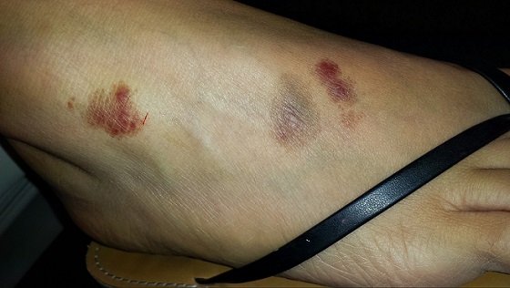
Three macular patches—each about 2.4 cm and roughly round, with sharp margins—are seen on the dorsum of her right foot. There is no palpable component to any of them, nor increased warmth or tenderness. Two are dark reddish brown, with nonblanchable redness. The middle lesion looks quite different—brownish gray—but it too is totally macular. Elsewhere, her type IV Hispanic skin is free of any notable lesions.
A shave biopsy is taken from one of the active (red) lesions. The pathology results indicate capillaritis.
What is the diagnosis?
DISCUSSION
When red inflammatory processes fail to blanch with digital pressure, we know that red blood cells (RBCs) have likely leaked from damaged blood vessels and stained the interstitial spaces. Generically, we call this purpura. On occasion, purpura can be an ominous sign, indicating a vasculitis such as leukocytoclastic vasculitis (LCV), in which an immune complex damages the walls of blood vessels that supply vital organs. This can occur in the context of a connective tissue disease, such as lupus, or an allergic reaction to an ingestant (food, drink, or medicine).
But there are specific histologic criteria (eg, nuclear dust from white blood cells that have expended themselves in attacking blood vessel walls and extravasated RBCs from these same leaky walls) for the diagnosis of LCV that were missing in this case. Significantly, the affected blood vessels in this case were capillaries, which are not affected by LCV.
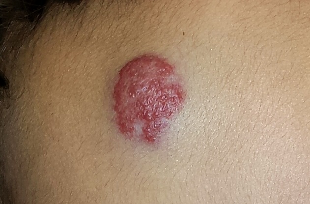
The histologic and clinical findings put us squarely into a relatively benign group of conditions called capillaritis. Collectively, these are known as pigmented purpuris dermatoses; the most common is Schamberg disease (progressive pigmentary dermatosis). These dermatoses are characterized by extravasation of RBCs with marked hemosiderin deposition.
In this case, however, we’re looking at another common form, lichen aureus (LA). The lesions of LA are round to polygonal, sometimes shiny, and brownish red (which can look darker in patients with darker skin).
Schamberg lesions are said to resemble sprinkled cayenne pepper. They typically start on the lower legs and move upward to just below the knee, then slowly descend and resolve. This entire process will occur within a period of several months.
The cause of this family of dermatoses is unknown, but, thankfully, they are all asymptomatic and self-limited. Topical and systemic treatment does not help.
The differential also includes granulomatous disease (eg, granuloma annulare) and fixed drug eruption. Biopsy, as in this case, is often warranted.
TAKE-HOME LEARNING POINTS
• Lichen aureus and Schamberg disease are the most common forms of capillaritis, a benign condition usually affecting the legs and feet.
• These forms of capillaritis are caused by extravasation of red blood cells (RBCs) from damaged capillaries.
• They typically manifest with brownish red macules and patches that do not blanch with digital pressure.
• The cause of capillaritis is unknown, although it is clear that it results from damage to capillary vessel walls that allows RBCs to leak into the interstitial tissues.
• The main item in the differential is leukocytoclastic vasculitis (LCV), which is of more acute onset and usually has a petichial appearance. Since it has systemic implications, suspected LCV must be confirmed or ruled out with biopsy.
A 25-year-old woman is concerned about lesions on the dorsum of her right foot that first appeared about four months ago. Initially, there was one red lesion; several similarly colored lesions manifested within a few weeks. The original lesion darkened from red to brown before gradually fading to its current appearance. All the lesions are asymptomatic.
The patient denies injury to the foot. She denies any history of skin problems, including any similar lesions elsewhere on her body. She describes her general health as “quite good,” aside from mild seasonal allergies. She is taking no medications of any kind.
EXAMINATION
Three macular patches—each about 2.4 cm and roughly round, with sharp margins—are seen on the dorsum of her right foot. There is no palpable component to any of them, nor increased warmth or tenderness. Two are dark reddish brown, with nonblanchable redness. The middle lesion looks quite different—brownish gray—but it too is totally macular. Elsewhere, her type IV Hispanic skin is free of any notable lesions.
A shave biopsy is taken from one of the active (red) lesions. The pathology results indicate capillaritis.
What is the diagnosis?
DISCUSSION
When red inflammatory processes fail to blanch with digital pressure, we know that red blood cells (RBCs) have likely leaked from damaged blood vessels and stained the interstitial spaces. Generically, we call this purpura. On occasion, purpura can be an ominous sign, indicating a vasculitis such as leukocytoclastic vasculitis (LCV), in which an immune complex damages the walls of blood vessels that supply vital organs. This can occur in the context of a connective tissue disease, such as lupus, or an allergic reaction to an ingestant (food, drink, or medicine).
But there are specific histologic criteria (eg, nuclear dust from white blood cells that have expended themselves in attacking blood vessel walls and extravasated RBCs from these same leaky walls) for the diagnosis of LCV that were missing in this case. Significantly, the affected blood vessels in this case were capillaries, which are not affected by LCV.
The histologic and clinical findings put us squarely into a relatively benign group of conditions called capillaritis. Collectively, these are known as pigmented purpuris dermatoses; the most common is Schamberg disease (progressive pigmentary dermatosis). These dermatoses are characterized by extravasation of RBCs with marked hemosiderin deposition.
In this case, however, we’re looking at another common form, lichen aureus (LA). The lesions of LA are round to polygonal, sometimes shiny, and brownish red (which can look darker in patients with darker skin).
Schamberg lesions are said to resemble sprinkled cayenne pepper. They typically start on the lower legs and move upward to just below the knee, then slowly descend and resolve. This entire process will occur within a period of several months.
The cause of this family of dermatoses is unknown, but, thankfully, they are all asymptomatic and self-limited. Topical and systemic treatment does not help.
The differential also includes granulomatous disease (eg, granuloma annulare) and fixed drug eruption. Biopsy, as in this case, is often warranted.
TAKE-HOME LEARNING POINTS
• Lichen aureus and Schamberg disease are the most common forms of capillaritis, a benign condition usually affecting the legs and feet.
• These forms of capillaritis are caused by extravasation of red blood cells (RBCs) from damaged capillaries.
• They typically manifest with brownish red macules and patches that do not blanch with digital pressure.
• The cause of capillaritis is unknown, although it is clear that it results from damage to capillary vessel walls that allows RBCs to leak into the interstitial tissues.
• The main item in the differential is leukocytoclastic vasculitis (LCV), which is of more acute onset and usually has a petichial appearance. Since it has systemic implications, suspected LCV must be confirmed or ruled out with biopsy.

The Pimple That Wasn’t
At first, this 70-year-old man thought the lesion on his nose was a pimple, although it seemed oddly resilient. When he attempted to pop it, nothing came out.
When it not only failed to heal but also grew over a six-month period, he finally decided to consult his primary care provider (PCP). The PCP told him not to worry about it but offered to refer him to dermatology—mostly because the patient’s wife was concerned.
The patient gives a history of extensive sun exposure in childhood and young adulthood. In the 1960s, he was drafted into the military and served two tours of duty in the jungles of Vietnam, where he acquired an almost permanent sunburn.

EXAMINATION
The patient is quite fair, with modest facial sun damage manifesting as telangiectasias and solar elastosis. The lesion in question is a glassy, scarlike, concave papule with a scab at one end. It is located in the left alar groove and measures about 5 mm in diameter.
Under local anesthesia, the lesion is biopsied and sent to pathology.
What is the diagnosis?
DISCUSSION
The pathology report indicated basal cell carcinoma (BCC). Given the location, the patient was referred to a Mohs surgeon, who needed two passes to clear the site of cancer. Closure of the resulting 2.5-cm defect required a skin graft, using the postauricular neck as the donor site. Afterward, the PCP, the patient, and his wife all expressed astonishment that such a tiny lesion had required so complex a procedure.
BCC is, by far, the most common of the sun-caused skin cancers, with more than a million new cases diagnosed in the US each year. While rarely dangerous, BCCs can be highly destructive of local structures (eyelids, noses, ears, faces) if left untreated for extended periods. Virtually all BCCs grow relentlessly, but some are more aggressive than others—a trait confirmed not only by the behavior of the cancer, but also, in some cases, by characteristic histologic findings.
Location has a major impact on the behavior of a given BCC. The posterior portion of the nose (alar groove) is particularly prone to harbor more extensive and aggressive BCCs.
Given the additional cosmetic implications of removing cancers from this area, these often require Mohs surgery to ensure complete, mircroscopically controlled removal of the cancer, as well as closure of the wound in a cosmetically acceptable manner.
Some aggressive types of BCC will require radiation therapy even after Mohs or conventional surgical removal. Fortunately, deaths from BCCs usually result from local invasion and not from metastasis, which is extremely rare.
For providers, the key is a low threshold for the necessity of biopsy. Simple shave biopsy is usually adequate. When PCPs are uncomfortable performing such biopsies, expedited referral to dermatology is called for.
TAKE-HOME LEARNING POINTS
• BCC is the most common of the sun-caused skin cancers, with more than a million new cases diagnosed every year in the US.
• While they rarely metastasize, BCCs can result in extensive invasion of local structures (eg, bone, cartilage, and even brain).
• Some BCCs are more aggressive than others in terms of demonstrated biologic activity.
• Some anatomical locations are themselves predictors of increased biologic activity; the nose is a prime example of this phenomenon.
• Mohs surgery—named after Frederic Mohs, MD, who pioneered the concept—ensures complete removal of the cancer and provides cosmetically acceptable closure as well.
At first, this 70-year-old man thought the lesion on his nose was a pimple, although it seemed oddly resilient. When he attempted to pop it, nothing came out.
When it not only failed to heal but also grew over a six-month period, he finally decided to consult his primary care provider (PCP). The PCP told him not to worry about it but offered to refer him to dermatology—mostly because the patient’s wife was concerned.
The patient gives a history of extensive sun exposure in childhood and young adulthood. In the 1960s, he was drafted into the military and served two tours of duty in the jungles of Vietnam, where he acquired an almost permanent sunburn.
EXAMINATION
The patient is quite fair, with modest facial sun damage manifesting as telangiectasias and solar elastosis. The lesion in question is a glassy, scarlike, concave papule with a scab at one end. It is located in the left alar groove and measures about 5 mm in diameter.
Under local anesthesia, the lesion is biopsied and sent to pathology.
What is the diagnosis?
DISCUSSION
The pathology report indicated basal cell carcinoma (BCC). Given the location, the patient was referred to a Mohs surgeon, who needed two passes to clear the site of cancer. Closure of the resulting 2.5-cm defect required a skin graft, using the postauricular neck as the donor site. Afterward, the PCP, the patient, and his wife all expressed astonishment that such a tiny lesion had required so complex a procedure.
BCC is, by far, the most common of the sun-caused skin cancers, with more than a million new cases diagnosed in the US each year. While rarely dangerous, BCCs can be highly destructive of local structures (eyelids, noses, ears, faces) if left untreated for extended periods. Virtually all BCCs grow relentlessly, but some are more aggressive than others—a trait confirmed not only by the behavior of the cancer, but also, in some cases, by characteristic histologic findings.
Location has a major impact on the behavior of a given BCC. The posterior portion of the nose (alar groove) is particularly prone to harbor more extensive and aggressive BCCs.
Given the additional cosmetic implications of removing cancers from this area, these often require Mohs surgery to ensure complete, mircroscopically controlled removal of the cancer, as well as closure of the wound in a cosmetically acceptable manner.
Some aggressive types of BCC will require radiation therapy even after Mohs or conventional surgical removal. Fortunately, deaths from BCCs usually result from local invasion and not from metastasis, which is extremely rare.
For providers, the key is a low threshold for the necessity of biopsy. Simple shave biopsy is usually adequate. When PCPs are uncomfortable performing such biopsies, expedited referral to dermatology is called for.
TAKE-HOME LEARNING POINTS
• BCC is the most common of the sun-caused skin cancers, with more than a million new cases diagnosed every year in the US.
• While they rarely metastasize, BCCs can result in extensive invasion of local structures (eg, bone, cartilage, and even brain).
• Some BCCs are more aggressive than others in terms of demonstrated biologic activity.
• Some anatomical locations are themselves predictors of increased biologic activity; the nose is a prime example of this phenomenon.
• Mohs surgery—named after Frederic Mohs, MD, who pioneered the concept—ensures complete removal of the cancer and provides cosmetically acceptable closure as well.
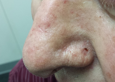
A New Approach to “Birthmarks”
A 4-month-old boy is brought in by his mother for evaluation of a “birthmark” that has become more prominent with time. The child complains a bit when the lesion is touched.
His mother gives a history of a normal full-term pregnancy, with an uneventful delivery. Other than the skin lesion, there have been no other known problems with the child’s health.

EXAMINATION
The lesion in question is a 2-cm bright red nodule in the middle of the child’s forehead. There is a faint blue halo around it, but no other abnormalities are seen in the area or elsewhere on the child’s body. The patient otherwise appears well developed and well nourished; he is quite alert and reactive to verbal stimuli.
What is the diagnosis?
DISCUSSION
Most first-year medical students could tell you this was a case of an infantile hemangioma—but within the past five years, the categorization and treatment of hemangiomas have changed rapidly.
Hemangiomas are benign and usually self-involuting vascular tumors; they are distinct from the family of permanent congenital vascular lesions, such as port wine stains. About 80% occur on the face or neck, and they more commonly occur in female patients. Those that occur near the skin’s surface tend to be bright red, while those of deeper origin are more bluish. Hemangiomas can also occur in extracutaneous areas (eg, the liver); these are usually detected via imaging.
Most infantile hemangiomas appear in the first few weeks of life but begin to involute shortly after they reach maximal size (usually by age 12 months). Involution is complete by age 5 in about 50% of cases and by age 7 in 70% of them. The remainder usually resolve by age 12.
Until recently, the parents of an affected child would be told that the lesions were benign and would likely disappear on their own by the time the child was ages 7 to 10—all true enough. These lesions were treated only when they were large or symptomatic, or if their location blocked or otherwise interfered with the function of the eyes, ears, mouth, anus, or vagina. Unfortunately, this approach ignored the psychosocial aspects of having a prominent and fire-engine red lesion, which often becomes the object of unwanted attention, and even ridicule, at a crucial developmental age.
In the past, the main treatment options were destructive in nature (laser, excision) and often caused as many problems as they solved. Pharmacologic treatment with oral prednisone, and later with interferon, was also tried with mixed success.
In 2008, in a serendipitous observation, a French team came up with a groundbreaking approach: giving systemic propranolol multiple times over a 12 to 18–month period. This method proved to be both safe and effective when used in the right setting by specialists (pediatric dermatologists and/or pediatricians) experienced in this modality. It was found that the lesions responded best if treated before age 6 months.
This has worked so well and so safely that it has quickly become routine. But it will take time before it is sufficiently well known to become the actual standard of care for these common lesions.
TAKE-HOME LEARNING POINTS
• Infantile hemangiomas (IH) usually resolve (involute) by age 12 at the latest.
• IH, by definition, is different from permanent congenital vascular malformations, such as port wine stains.
• About 80% of IHs occur on the head and neck; they are often the object of unwanted attention or even ridicule.
• A new treatment approach, systemic propranolol, is now being used routinely and successfully. It works best when the patient is 6 months old or younger.
• Ideally, this new treatment approach should be overseen by the relevant specialist, be it in a pediatric or dermatologic setting.
A 4-month-old boy is brought in by his mother for evaluation of a “birthmark” that has become more prominent with time. The child complains a bit when the lesion is touched.
His mother gives a history of a normal full-term pregnancy, with an uneventful delivery. Other than the skin lesion, there have been no other known problems with the child’s health.
EXAMINATION
The lesion in question is a 2-cm bright red nodule in the middle of the child’s forehead. There is a faint blue halo around it, but no other abnormalities are seen in the area or elsewhere on the child’s body. The patient otherwise appears well developed and well nourished; he is quite alert and reactive to verbal stimuli.
What is the diagnosis?
DISCUSSION
Most first-year medical students could tell you this was a case of an infantile hemangioma—but within the past five years, the categorization and treatment of hemangiomas have changed rapidly.
Hemangiomas are benign and usually self-involuting vascular tumors; they are distinct from the family of permanent congenital vascular lesions, such as port wine stains. About 80% occur on the face or neck, and they more commonly occur in female patients. Those that occur near the skin’s surface tend to be bright red, while those of deeper origin are more bluish. Hemangiomas can also occur in extracutaneous areas (eg, the liver); these are usually detected via imaging.
Most infantile hemangiomas appear in the first few weeks of life but begin to involute shortly after they reach maximal size (usually by age 12 months). Involution is complete by age 5 in about 50% of cases and by age 7 in 70% of them. The remainder usually resolve by age 12.
Until recently, the parents of an affected child would be told that the lesions were benign and would likely disappear on their own by the time the child was ages 7 to 10—all true enough. These lesions were treated only when they were large or symptomatic, or if their location blocked or otherwise interfered with the function of the eyes, ears, mouth, anus, or vagina. Unfortunately, this approach ignored the psychosocial aspects of having a prominent and fire-engine red lesion, which often becomes the object of unwanted attention, and even ridicule, at a crucial developmental age.
In the past, the main treatment options were destructive in nature (laser, excision) and often caused as many problems as they solved. Pharmacologic treatment with oral prednisone, and later with interferon, was also tried with mixed success.
In 2008, in a serendipitous observation, a French team came up with a groundbreaking approach: giving systemic propranolol multiple times over a 12 to 18–month period. This method proved to be both safe and effective when used in the right setting by specialists (pediatric dermatologists and/or pediatricians) experienced in this modality. It was found that the lesions responded best if treated before age 6 months.
This has worked so well and so safely that it has quickly become routine. But it will take time before it is sufficiently well known to become the actual standard of care for these common lesions.
TAKE-HOME LEARNING POINTS
• Infantile hemangiomas (IH) usually resolve (involute) by age 12 at the latest.
• IH, by definition, is different from permanent congenital vascular malformations, such as port wine stains.
• About 80% of IHs occur on the head and neck; they are often the object of unwanted attention or even ridicule.
• A new treatment approach, systemic propranolol, is now being used routinely and successfully. It works best when the patient is 6 months old or younger.
• Ideally, this new treatment approach should be overseen by the relevant specialist, be it in a pediatric or dermatologic setting.